User login
WASHINGTON – The presence of the antisynthetase antibody Jo-1, as well as Mi-2 autoantibodies, strongly predicted improvement in rituximab-treated myositis patients.
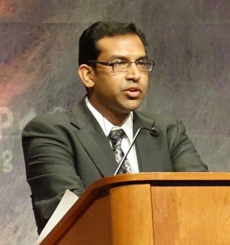
On the other hand, global damage is a poor indicator of good response to rituximab in multivariate analysis, meaning that further research into these biomarkers – especially Jo-1 – could have a major impact on treatment in this population, reported Dr. Rohit Aggarwal at the annual meeting of the American College of Rheumatology.
Dr. Aggarwal of the myositis center of the University of Pittsburgh and his colleagues looked at data from the RIM study (Rituximab in Myositis), which evaluated 200 myositis patients treated with the B-cell depleting agent, 83% of whom met the "definition of improvement" (DOI).
The DOI included a 20% or greater improvement in three of six measures: manual muscle testing, muscle enzymes, health assessment questionnaire, patient/parent global assessment, physician global disease activity assessment, and extramuscular disease activity, with not more than 25% worsening of greater than two measures.
These improvements also had to be noted at two consecutive visits to count as meeting the DOI.
Overall, 76 of the 200 myositis patients were more specifically classified as having adult polymyositis; 76 had adult dermatomyositis; and the remaining 48 patients had juvenile dermatomyositis.
In univariate analysis, several factors were associated with more rapid achievement of the DOI. These included lower baseline muscle damage, a higher white blood cell count, lower global damage, and the absence of muscle atrophy.
However, "the primary and strongest predictor of improvement on univariate analysis was autoantibodies," said Dr. Aggarwal – specifically, Jo-1 and Mi-2 – but also other myositis-associated antibodies, including SRP.
Dr. Aggarwal then conducted a multivariate analysis looking only at those variables with P value less than .1 in univariate assessment.
This time, the only significant predictors of improvement were the presence of the antisynthetase autoantibody Jo-1, but only in adults (hazard ratio, 3.08; 95% confidence interval, 1.80-5.28) as well as Mi-2, but only in juvenile and adult dermatomyositis – not adult polymyositis (HR 2.49; 95% CI, 1.42-4.41).
Other antibodies, including those to the signal recognition particle, did not confer a significantly increased association with improvement.
On the other hand, having less global damage at baseline was associated with improvement at 8 weeks, but the effect was washed out by week 20.
"Our findings regarding anti–Jo-1 are even more interesting when these results are coupled with other data that were presented in a concurrent session yesterday," said Dr. Aggarwal, citing a study he also led looking at the relation between anti–Jo-1 serum levels and the six improvement measures assessed in the DOI.
In that study, "After start of the treatment [with rituximab], autoantibody levels in Jo-1 subjects decreased by approximately nine units per week," he wrote.
"Anti–Jo-1 levels longitudinally correlated with all core set measures [of improvement] univariately and after adjusting for IgG levels."
He added, "These data suggest that anti–Jo-1 is also a disease biomarker."
"Further study of B-cell depletion in autoantibody positive, low-damage and juvenile dermatomyositis is warranted."
The RIM study was funded by Genentech, maker of rituximab; several investigators reported relationships to Genentech as well as other pharmaceutical companies.
AT THE ANNUAL MEETING OF THE AMERICAN COLLEGE OF RHEUMATOLOGY
Major Finding: Presence of the anti-synthetase autoantibody Jo-1 at baseline strongly predicted improvement in myositis patients taking rituximab (hazard ratio 3.08; 95% confidence interval, 1.80-5.28).
Data Source: A This findings is based on data from the RIM (Rituximab in Myositis) study.
Disclosures: The RIM study was funded by Genentech, maker of rituximab; several investigators reported relationships to Genentech as well as other pharmaceutical companies.

