User login
Technology makes many things possible, but not without imposing new responsibilities. When it comes to viral infections, diagnosis through serology testing, antigen assays, or amplification techniques such as polymerase chain reaction (PCR) now is possible for a number of diseases, including:
- herpes simplex virus (HSV),
- cytomegalovirus (CMV),
- hepatitis B and C viruses (HBV, HCV),
- parvovirus B19 (B19), and
- human immunodeficiency virus (HIV).
The responsibilities that come along with this ability: keeping up to date and selecting the most sensitive and specific test possible. This article reviews the latest tests and offers advice on their use in detecting 6 viruses.
How to pick the right test
When a patient’s presentation suggests viral infection, when something in her history raises a red flag, or when she reports possible exposure to a virus, the right test is critical to the diagnosis. The right test also helps prevent false positives and avoid confusion—but which test is best?
Which immunoglobulins matter?
Many of us have been taught that immunoglobulin M (IgM) correlates with acute infection, but that is not necessarily the case. Because of its high molecular weight, IgM is found most commonly in the intravascular compartment and is not transported to the fetus. IgM usually becomes apparent early during the course of infection. It has a half-life of 10 days and usually—but not always—regresses to undetectable levels over a few months.
The misconception that IgM is found only in acute infection and disappears within 3 months causes many clinicians to test for it and to misinterpret the results. In many cases, IgM fails to develop after acute infection. In others, it may persist for as long as 2 years after primary infection. It also can be detected with recurrent or reactivated infections.1
Immunoglobulin G (IgG) has a longer half-life (21 days) and is the most common immunoglobulin in humans. It is found in tissues and serum and readily crosses the placenta. It can be detected shortly after acute infection, exhibiting a steep rise and fall over several weeks after primary infection. IgG also is a sign of past infection.1
Telltale sign of acute infection
It now seems clear that an IgG antibody produced within the first months after primary infection binds to its antigen poorly. After this initial period, the IgG binds with greater intensity (ie, higher avidity) to that specific antigen (virus). Assays that measure this binding intensity are called avidity assays and are expressed as a percentage of IgG bound to the antigen after treatment with denaturing agents.
Avidity assays have been developed and studied for a variety of viruses.2 The detection of low-avidity IgG can be considered a more reliable sign of acute infection than IgM.
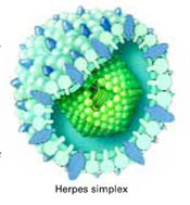
Herpes simplex virus
SERENA’S CASE
Monthly irritation and a vulvar lesion
Serena, 22, complains of irritation and pruritus that precede her period each month. She has tried treating herself with over-the-counter and prescription antifungal medication, without much relief. She presents to your office as an add-on patient and reports that the irritation started 1 day ago and usually lasts 7 days. Physical examination reveals a small area on the left vulva that is inflamed, with 2 small fissures. You obtain a vaginal pH, but it is normal, and there are negative findings on the wet prep.
In view of Serena’s history of recurrent symptoms and atypical lesions, recurrent herpes is a likely diagnosis, so you culture the lesion and order IgG type-specific serology for HSV 1 and 2. The culture is negative, but serology is positive for the HSV-2 antibody. Thus, serology confirms genital herpes.
Although Serena’s culture was negative, false-negative cultures are common with HSV, and serology testing usually is necessary to make the diagnosis of recurrent genital herpes.
HSV-2 is widespread: About 1 in 4 adults is infected. Of these, fewer than 1 in 10 is aware he or she has the virus. Thus, it makes sense to test for HSV-2 when physical findings suggest that it may be present.
Thanks to type-specific serology tests, HSV 1 and/or 2 infection can be diagnosed with confidence.3 Type-specific tests determine the IgG antibody response to an envelope glycoprotein (gG) of HSV. In the diagnosis of primary infection, the test detects seroconversion from a negative to a positive titer. The earliest time for antibody production is 3 weeks after infection—but 8 to 12 weeks should elapse prior to testing for seroconversion.4
The US Food and Drug Administration (FDA) has approved an office test for HSV-2 antibodies. The biokitHSV-2 rapid test (biokit USA, Lexington, Mass) requires blood or serum—though statistical analysis indicates that serum demonstrates higher sensitivity with this assay than does capillary whole blood—and can be performed in any outpatient setting, with excellent sensitivity and specificity.
Flaws of IgM testing and viral cultures
IgM testing for HSV is not advised, as the presence of IgM does not correlate with acute disease. As for cultures, laboratories have been slow to change from HSV cultures to PCR testing from suspected HSV lesions. Because viral cultures for HSV have extremely low sensitivity, only positive results are clinically useful.
Primary infection yields a higher positive culture rate because of increased viral shedding. In contrast, recurrent disease, with its low levels of viral shedding, yields significantly lower positive culture rates.5
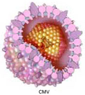
Cytomegalovirus
CATHY’S CASE
Flu-like symptoms and plans to conceive
When Cathy, 28, comes in for her annual visit, she reports that she has been experiencing exhaustion, chills, and body aches for several weeks and wonders whether she might have mononucleosis. She also mentions that she and her husband are trying to have a child. She recently started a new job in a day care center, working directly with young children. You decide to test her for the Epstein-Barr virus. Since she is planning to conceive, you also test her for CMV, and the test is positive.
How do you counsel Cathy?
CMV infection usually is diagnosed after clinical findings in the individual or fetus (by ultrasound examination) suggest this infection, or when the clinician suspects it for other reasons.
In young adults, primary CMV infection can cause flu- or mononucleosis-like symptoms such as extreme fatigue, fever, chills, and/or body aches, though they generally resolve within several weeks without further morbidity. However, CMV can cause grave illness—even death—in immunocompromised patients, who may continue to experience the disease intermittently after the first outbreak. CMV also can seriously impair development in infants who are infected in utero. The vast majority of these infants appear normal at birth and only later exhibit problems.
Cathy should be counseled to postpone conception until the virus clears, to avoid jeopardizing her infant’s health.
Testing for CMV during pregnancy
Diagnosing primary infection in pregnancy can be difficult. In the United States, we do not screen for CMV prior to pregnancy, except for special circumstances such as Cathy’s, because the virus is widespread and causes few problems in healthy, nonpregnant individuals. This lack of screening renders the best diagnostic method (ie, seroconversion, or finding CMV IgG antibody in a previously seronegative woman) impossible in most cases. Rather, the clinician usually orders IgG and IgM antibody tests, hoping they will help determine whether a primary infection is present.
In these situations, detection of the IgM antibody during pregnancy is vital to diagnose primary infection.
IgG avidity test clarifies IgM findings
The enzyme-linked immunosorbent assay (ELISA) and the capture ELISA assay are used to detect CMV IgG and IgM antibodies. However, the ELISA for IgM can yield false-positive results due to the presence of:
- IgG (especially at high titers),
- rheumatoid factor of the IgM class (IgM-RF),
- reaction between IgM antibodies and cellular antigens, and
- primary Epstein-Barr viral infection, which can stimulate production of CMV IgM antibody in CMV-immune individuals.
Thus, a positive IgM finding in the serum of a pregnant woman can be caused by acute primary CMV infection, the convalescent phase of a primary CMV infection, or simply persistent IgM antibodies.6,7
That’s where the IgG avidity test comes in. It can help confirm and clarify the clinical significance of finding the IgM antibody and distinguish between primary and recurrent CMV infection. Using the IgM antibody test together with the IgG avidity test increases the likelihood of a correct diagnosis.
A positive IgM with a low avidity index is highly suggestive of a recent (less than 3 months) primary CMV infection.
Standardization of the IgG and IgM assays and the IgG avidity assay is urgently needed to reduce the likelihood of incorrect diagnosis and unwanted intervention in otherwise normal pregnancies.
Further testing can confirm fetal transmission
If primary CMV infection is detected in a pregnant woman, accurate prenatal diagnosis is possible to determine whether the infection has been transmitted to the fetus.
Sophisticated tests such as PCR make in utero diagnosis possible. In fact, PCR remains the most accurate means of diagnosing CMV, with sensitivities ranging from 80% to 100% in prospective studies of gravidas infected with cytomegalovirus.8
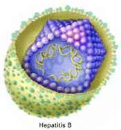
Hepatitis B
LIAN’S CASE
Pregnant, with no history of vaccination
Lian, 25, was born in Taiwan and has been in the United States 6 years. She presents for her first prenatal visit at 12 weeks’ gestation and reports no medical history, including no vaccination against hepatitis. This is her first pregnancy. Because testing for hepatitis B is routine in pregnancy, she undergoes a hepatitis B surface antigen test (HBsAg), which is positive.
What is your next step?
Lian’s positive test suggests she is a carrier for HBV. Additional testing can clarify her status and direct treatment and prevention.
Unlike some of the other viruses covered in this article, HBV has been well studied, and the identifying proteins have been described. This information has made it possible to produce sensitive and specific tests that can determine whether a patient is infected, a carrier, or immune to HBV.
Screening for carrier status is universal among gravidas in the United States. Standard ELISA assays have been developed for several HBV proteins as well as the antibody.
Signs of a carrier
The carrier state for HBV is determined by measuring HBsAg, the envelope protein of the virus, which circulates freely in the serum after acute infection (1–10 weeks) and in carrier patients. A woman is a carrier if HBsAg persists 6 months after primary infection.
The nucleocapsid core protein of the hepatitis B core antigen (HBcAg) and its secretory product e antigen (HBeAg) are produced after viral multiplication. There is no assay for HBcAg, but the hepatitis B surface antibody (HBsAb) develops after acute HBV infection. In the carrier state, no HBsAb is detected. However, HBcAb is detected in virtually all infected individuals, usually 4 to 8 weeks after infection.
HBeAg production is a sign of active production of the virus in the liver and is associated with more severe disease. It is usually cleared by 16 weeks post-infection. The antibody to the e protein (HBeAb) occurs more than 16 weeks after infection, and is a finding that suggests less severe disease.
The only finding in the serum of patients successfully vaccinated against HBV is HBsAb.9,10
How to qualify infection status
In Lian’s case, further testing should include liver function and other markers of HBV infection. The presence of hepatitis B e antibody (HBeAb) and e antigen (HBeAg) can define infection and the risk of transmitting the virus to the newborn. For example, when HBeAg is present, the patient has a higher risk of transmitting the virus to other people, including her fetus. When HBeAb is present, the patient has a lower risk of transmitting the virus to other people, including her fetus.
In addition, the viral load can be measured via PCR; if the viral titers are elevated, appropriate antiviral therapy can be given during and after pregnancy.
HBV DNA testing is more sensitive than HBeAg to detect viruses in the blood. When it is performed, DNA testing is generally in addition to regular serologic tests. It also is used to monitor therapy in individuals with chronic HBV.
Lian should also be referred for gastrointestinal (GI) consultation for management during her pregnancy, as well as long-term follow-up after delivery. Vaccination of the partner may be required to prevent his infection.
Hepatitis C
JESSICA’S CASE
Partner had hepatitis
Jessica is a 20-year-old college student home for the summer. She schedules an appointment to discuss contraception, but during her visit she also asks to be screened for sexually transmitted diseases (STDs) and wants to know if there is any way to determine whether she has had a hepatitis B vaccination. In further discussion, she tells you that she was sexually involved with a male who had hepatitis.
You order testing for STDs, including antibody testing for HBV and HCV. The results suggest that Jessica was vaccinated for HBV but is HCV-positive.
How should you proceed?
HCV infection in women is diagnosed by ELISA assays sensitive enough to detect HCV antibody. However, the US Centers for Disease Control and Prevention recommend that a person be diagnosed with HCV only after a positive test for HCV antibodies is confirmed by a more specific test, such as a nucleic amplification test or recombinant immunoblot assay.11,1 2 Thus, Jessica should undergo further testing, including liver function tests and measurement of her viral load. Supplemental testing also is recommended for patients with a borderline result on their initial ELISA.
Referral for GI consultation is indicated for Jessica’s management. The finding of HCV virus also influences which contraceptives she should be offered: Avoid any hormonal contraceptive metabolized by the liver.
Parvovirus B19
GRETCHEN’S CASE
Pregnant and occupationally exposed
Gretchen is a 31-year-old elementary school teacher who is 20 weeks pregnant with her second child. She schedules an appointment with you because she is concerned about cases of B19, or Fifth disease, reported at her school. She has been teaching in the school for 7 years and has never been tested for B19 infection. She reports no symptoms and observes that there have been no cases of B19 among the students in her classroom.
You order IgG antibody testing, which reveals that Gretchen has immunity.
In light of this finding, how do you counsel her?
Gretchen can be reassured that, though she was obviously infected in the past, B19 poses no risk to this pregnancy. Roughly 50% of women are, like Gretchen, already immune to B19. Even if a woman is exposed to the virus during pregnancy, both she and the fetus are usually only mildly affected. However, B19 infection can cause severe anemia in the fetus and trigger spontaneous abortion—although this occurs in less than 5% of pregnancies infected with B19 and is more likely to occur during the first half of a pregnancy. Fetal exposure to B19 appears to cause no birth defects or mental retardation.
In general, testing for B19 in pregnancy is warranted after exposure to the virus to assess immunity or susceptibility. The best way to do so is by testing for IgM and IgG antibodies using an ELISA technique. The IgM antibody is produced within a few days of primary infection and persists for 2 to 3 months. The IgG antibody can be found 1 week after acute infection and persists perhaps for life.
The finding of the IgG antibody in an immunocompetent patient with no IgM antibody demonstrates immunity.13,14 In some cases, additional testing using an IgG avidity assay and/or B19 DNA PCR may be necessary.15
TABLE
Sensitivity and specificity of viral screening tests
| TESTS | SENSITIVITY (%) | SPECIFICITY (%) | COMMENT |
|---|---|---|---|
| Genital herpes | |||
| IgG (ELISA) | Must be type-specific based on gG protein | ||
| HSV-1 | 91–96 | 96–97 | |
| HSV-2 | 96–100 | 92–95 | |
| Point-of-care rapid test (biokitHSV-2) | 99–100 | 96–97 | Only for HSV-2 |
| IgM | DO NOT USE | ||
| Culture of lesion in mother | 50 | 90 | Positive result clinically useful |
| PCR | >90 | >90 | Usually reserved for testing cerebrospinal fluid for encephalitis (in newborns, children, and adults) |
| Cytomegalovirus | |||
| IgG | >90 | 80–92 | |
| IgM | 65–78 | 65–82 | |
| Culture of amniotic fluid | 70 (newborns with CMV) | 100 | Culture of the amniotic fluid may be negative even with infection |
| PCR of amniotic fluid | 77 | 100 | |
| Hepatitis B | |||
| HBcAb | 95 | 95 | Marker of past infection |
| HBsAb | 95 | 95 | Only marker post-vaccination |
| HBsAg | 95 | 95 | Carrier |
| HBeAb | Marker of past infection | ||
| HBeAg | Only in carrier state. Carrier who also demonstrates HBeAg has a high risk of transmission to the newborn | ||
| Hepatitis C | |||
| HCVAb (ELISA) | 99 | 99 | 3rd-generation ELISA |
| HCV RNA-PCR | >95 | >95 | Correlates with infectivity |
| Parvovirus B19 | |||
| IgG (ELISA) | 97 | 94 | |
| IgM (antibody capture enzyme immunoassay) | 89 | 99 | Must use a reliable lab for determination of IgM because of the high incidence of false positives |
| PCR in fetus | 92 | 94 | Currently, reference lab can perform this test. Use on amniotic fluid to confirm in utero infection after primary infection in the mother is diagnosed |
| Human immunodeficiency virus | |||
| ELISA | 99 | 99 | Screening test, repeated if positive |
| Western blot | 99 | 99 | Confirmatory test |
| p24 antigen | May be positive early in acute infection before antibody response | ||
| HIV-1 RNA by PCR | May be positive early in acute infection before antibody response | ||
| ELISA=Enzyme-linked immunosorbent assay; HBcAb=hepatitis B core antibody; HBeAb=hepatitis B early antibody; HBeAg=hepatitis B early antigen; HBsAb=hepatitis B surface antibody; HBsAg=hepatitis B surface antigen; HCV=hepatitis C virus; HSV=herpes simplex virus; Ig=immunoglobulin; PCR=polymerase chain reaction. | |||
ANGELA’S CASE
Recurrent vaginitis and numerous sexual partners
After 2 years of recurrent yeast infections, 41-year-old Angela comes to your office seeking more definitive therapy. She says she is tired of frequent infections that respond to oral and topical therapy but recur less than a week after therapy ends. She reports that she was divorced 8 years ago and has had numerous sexual partners since then. One in particular had a history of drug abuse. In response to your questions, Angela reports no history of diabetes or drug abuse herself.
Physical examination reveals severe external vulvar erythema and edema, and severe vaginal overgrowth of a “cottage-cheese” discharge consistent with Candida vaginitis. Given Angela’s history and physical findings, you order an HIV test, obtaining written permission for it. The test is reported as positive.
Does Angela have HIV?
It is impossible to tell without a repeat ELISA test and confirmation by Western blot, because false-positive results do occur.
Testing for HIV is now common in ObGyn offices. All women who are sexually active, diagnosed with another STD, or are pregnant are encouraged to undergo HIV screening. Testing may need to be repeated within 6 months in consideration of the incubation period of the virus. If the woman has a new relationship or additional history of high-risk sexual practices, testing should also be repeated.
Highly sensitive and specific ELISA assays have been developed for both HIV-1 and HIV-2. Both types of HIV have the same mode of transmission and incur opportunistic infections and other conditions. However, HIV-2 develops more slowly, is generally milder, and appears to be less transmissible than HIV-1.
HIV-2 occurs primarily in West Africa, although cases have been reported in Western Europe and India.
Several generations of assays have been approved for HIV-1 and HIV-2 over the past 2 decades. Today, third-generation assays using enzyme-coupled HIV antigens are standard. These tests are able to detect HIV antibody within 22 days of acute infection.16
Rapid tests (<30 minutes) and saliva tests are available for HIV screening. The IgG antibody is found as a transudate in oral secretions. Therefore, a technique that absorbs and concentrates IgG antibody from the oral cavity can be used to assay for HIV antibodies. An oral assay has been approved, but only for HIV-1 testing.17
Beware of false-positive and false-negative HIV results
Because of the enormous impact of an HIV diagnosis, it is critical to counsel patients that HIV screening assays can be falsely positive or falsely negative. However, compared with tests for the other viral infections covered in this article, HIV testing is perhaps the most sensitive and specific of FDA-approved tests.
False-positive rates range from 1 in 1,000 to 1 in 2,000 tests.
False negatives are less common and usually occur because of the time it takes for a patient to seroconvert after acute infection, which can be up to 1 year after infection.16
Confirm positive results via Western blot
The Western blot test is the gold standard for confirming ELISA-positive HIV results. For this highly sensitive (>99%) test, specific HIV proteins are obtained, separated by electrophoresis, and incubated with the serum along with control specimens. The CDC recognizes 3 results: positive, negative, or indeterminate.
A positive Western blot reacts to 2 bands (from p24, gp41, and gp120/160). A negative result reacts to no bands, and an indeterminate test reacts to only 1 band. Several chronic autoimmune diseases such as systemic lupus erythematosus and Hashimoto’s thyroiditis are associated with p24 reactivity.18
Other HIV tests
In addition to antibody tests, antigen and RNA tests can diagnose and help monitor the course of the disease and response to therapy. The most widely used are the p24 antigen detection test and the PCR assay for HIV-1 RNA.
Finding p24 antigen in the serum precedes the antibody response and may diagnose HIV earlier than ELISA. PCR testing for HIV viral RNA may be positive earlier than the p24 and ELISA antigen tests.19
Dr. Baker reports that he receives grant/research support from GlaxoSmithKline and Merck, is a consultant to GlaxoSmithKline, and is on the speakers bureaus of 3M, GlaxoSmithKline, and Pfizer.
1. Heyman B. Regulation of antibody responses via antibodies, complement and Fc receptors. Annu Rev Immunol. 2000;18:709-737.
2. Ashley RL. Sorting out the new HSV type specific antibody tests. Sex Transm Infect. 2001;77:232-237.
3. Bodeus M, Van Ranst M, Bernard P, Hubinont C, Goubau P. Anticytomegalovirus IgG avidity in pregnancy: a 2-year prospective study. Fetal Diagn Ther. 2002;17:362-366.
4. Ashley-Morrow R, Krantz E, Wald A. Time course of seroconversion by HerpeSelect ELISA after acquisition of genital herpes simplex virus type 1 (HSV-1) or HSV-2. Sex Transm Dis. 2003;30:310-314.
5. Wald A, Huang ML, Carrell D, Selke S, Corey L. Polymerase chain reaction for detection of herpes simplex virus (HSV) DNA on mucosal surfaces: comparison with HSV isolation in cell culture. J Infect Dis. 2003;188:1345-1351.
6. Eggers M, Metzger C, Enders G. Differentiation between acute primary and recurrent human cytomegalovirus infection in pregnancy, using a microneutralization assay. J Med Virol. 1998;56:351-358.
7. Maine GT, Lazzarotto T, Landini MP. New developments in the diagnosis of maternal and congenital CMV infection. Expert Rev Mol Diagn. 2001;1:19-29.
8. Bodeus M, Hubinont C, Bernard P, Bouckaert A, Thomas K, Goubau P. Prenatal diagnosis of human cytomegalovirus by culture and polymerase chain reaction: 98 pregnancies leading to congenital infection. Prenat Diagn. 1999;19:314-317.
9. Dinsmoor MJ. Hepatitis in the obstetric patient. Infect Dis Clin North Am. 1997;11:77-91.
10. Duff P. Hepatitis in pregnancy. Semin Perinatol. 1998;22:277-283.
11. Pawlotsky JM. Use and interpretation of virological tests for hepatitis C. Hepatology. 2002;36(suppl 1):S65-S73.
12. Dienstag JL. Sexual and perinatal transmission of hepatitis C. Hepatology. 1997;26(suppl 1):S66-S70.
13. Jordan JA. Diagnosing human parvovirus B19 infection: guidelines for test selection. Mol Diagn. 2001;6:307-312.
14. Brown KE. Parvovirus B19. In: Mandell GL, Bennett JE, Dolin R, eds. Principles and Practice of Infectious Diseases. 6th ed. Philadelphia: Elsevier; 2005.
15. Katta R. Parvovirus B19: a review. Dermatol Clin. 2002;20:333-342.
16. Beelaert G, Vercauteren G, Fransen K, et al. Comparative evaluation of eight commercial enzyme linked immunosorbent assays and 14 simple assays for detection of antibodies to HIV. J Virol Methods. 2002;105:197-206.
17. Schramm W, Angulo GB, Torres PC, Burgess-Cassler A. A simple saliva-based test for detecting antibodies to human immunodeficiency virus. Clin Diagn Lab Immunol. 1999;6:577-580.
18. Makuwa M, Souquiere S, Niangui MT, et al. Reliability of rapid diagnostic tests for HIV variant infection. J Virol Methods. 2002;103:183-190.
19. Dodd RY, Notari EP, 4th, Stramer SL. Current prevalence and incidence of infectious disease markers and estimated window-period risk in the American Red Cross blood donor population. Transfusion. 2002;42:975-979.
Technology makes many things possible, but not without imposing new responsibilities. When it comes to viral infections, diagnosis through serology testing, antigen assays, or amplification techniques such as polymerase chain reaction (PCR) now is possible for a number of diseases, including:
- herpes simplex virus (HSV),
- cytomegalovirus (CMV),
- hepatitis B and C viruses (HBV, HCV),
- parvovirus B19 (B19), and
- human immunodeficiency virus (HIV).
The responsibilities that come along with this ability: keeping up to date and selecting the most sensitive and specific test possible. This article reviews the latest tests and offers advice on their use in detecting 6 viruses.
How to pick the right test
When a patient’s presentation suggests viral infection, when something in her history raises a red flag, or when she reports possible exposure to a virus, the right test is critical to the diagnosis. The right test also helps prevent false positives and avoid confusion—but which test is best?
Which immunoglobulins matter?
Many of us have been taught that immunoglobulin M (IgM) correlates with acute infection, but that is not necessarily the case. Because of its high molecular weight, IgM is found most commonly in the intravascular compartment and is not transported to the fetus. IgM usually becomes apparent early during the course of infection. It has a half-life of 10 days and usually—but not always—regresses to undetectable levels over a few months.
The misconception that IgM is found only in acute infection and disappears within 3 months causes many clinicians to test for it and to misinterpret the results. In many cases, IgM fails to develop after acute infection. In others, it may persist for as long as 2 years after primary infection. It also can be detected with recurrent or reactivated infections.1
Immunoglobulin G (IgG) has a longer half-life (21 days) and is the most common immunoglobulin in humans. It is found in tissues and serum and readily crosses the placenta. It can be detected shortly after acute infection, exhibiting a steep rise and fall over several weeks after primary infection. IgG also is a sign of past infection.1
Telltale sign of acute infection
It now seems clear that an IgG antibody produced within the first months after primary infection binds to its antigen poorly. After this initial period, the IgG binds with greater intensity (ie, higher avidity) to that specific antigen (virus). Assays that measure this binding intensity are called avidity assays and are expressed as a percentage of IgG bound to the antigen after treatment with denaturing agents.
Avidity assays have been developed and studied for a variety of viruses.2 The detection of low-avidity IgG can be considered a more reliable sign of acute infection than IgM.
Herpes simplex virus
SERENA’S CASE
Monthly irritation and a vulvar lesion
Serena, 22, complains of irritation and pruritus that precede her period each month. She has tried treating herself with over-the-counter and prescription antifungal medication, without much relief. She presents to your office as an add-on patient and reports that the irritation started 1 day ago and usually lasts 7 days. Physical examination reveals a small area on the left vulva that is inflamed, with 2 small fissures. You obtain a vaginal pH, but it is normal, and there are negative findings on the wet prep.
In view of Serena’s history of recurrent symptoms and atypical lesions, recurrent herpes is a likely diagnosis, so you culture the lesion and order IgG type-specific serology for HSV 1 and 2. The culture is negative, but serology is positive for the HSV-2 antibody. Thus, serology confirms genital herpes.
Although Serena’s culture was negative, false-negative cultures are common with HSV, and serology testing usually is necessary to make the diagnosis of recurrent genital herpes.
HSV-2 is widespread: About 1 in 4 adults is infected. Of these, fewer than 1 in 10 is aware he or she has the virus. Thus, it makes sense to test for HSV-2 when physical findings suggest that it may be present.
Thanks to type-specific serology tests, HSV 1 and/or 2 infection can be diagnosed with confidence.3 Type-specific tests determine the IgG antibody response to an envelope glycoprotein (gG) of HSV. In the diagnosis of primary infection, the test detects seroconversion from a negative to a positive titer. The earliest time for antibody production is 3 weeks after infection—but 8 to 12 weeks should elapse prior to testing for seroconversion.4
The US Food and Drug Administration (FDA) has approved an office test for HSV-2 antibodies. The biokitHSV-2 rapid test (biokit USA, Lexington, Mass) requires blood or serum—though statistical analysis indicates that serum demonstrates higher sensitivity with this assay than does capillary whole blood—and can be performed in any outpatient setting, with excellent sensitivity and specificity.
Flaws of IgM testing and viral cultures
IgM testing for HSV is not advised, as the presence of IgM does not correlate with acute disease. As for cultures, laboratories have been slow to change from HSV cultures to PCR testing from suspected HSV lesions. Because viral cultures for HSV have extremely low sensitivity, only positive results are clinically useful.
Primary infection yields a higher positive culture rate because of increased viral shedding. In contrast, recurrent disease, with its low levels of viral shedding, yields significantly lower positive culture rates.5
Cytomegalovirus
CATHY’S CASE
Flu-like symptoms and plans to conceive
When Cathy, 28, comes in for her annual visit, she reports that she has been experiencing exhaustion, chills, and body aches for several weeks and wonders whether she might have mononucleosis. She also mentions that she and her husband are trying to have a child. She recently started a new job in a day care center, working directly with young children. You decide to test her for the Epstein-Barr virus. Since she is planning to conceive, you also test her for CMV, and the test is positive.
How do you counsel Cathy?
CMV infection usually is diagnosed after clinical findings in the individual or fetus (by ultrasound examination) suggest this infection, or when the clinician suspects it for other reasons.
In young adults, primary CMV infection can cause flu- or mononucleosis-like symptoms such as extreme fatigue, fever, chills, and/or body aches, though they generally resolve within several weeks without further morbidity. However, CMV can cause grave illness—even death—in immunocompromised patients, who may continue to experience the disease intermittently after the first outbreak. CMV also can seriously impair development in infants who are infected in utero. The vast majority of these infants appear normal at birth and only later exhibit problems.
Cathy should be counseled to postpone conception until the virus clears, to avoid jeopardizing her infant’s health.
Testing for CMV during pregnancy
Diagnosing primary infection in pregnancy can be difficult. In the United States, we do not screen for CMV prior to pregnancy, except for special circumstances such as Cathy’s, because the virus is widespread and causes few problems in healthy, nonpregnant individuals. This lack of screening renders the best diagnostic method (ie, seroconversion, or finding CMV IgG antibody in a previously seronegative woman) impossible in most cases. Rather, the clinician usually orders IgG and IgM antibody tests, hoping they will help determine whether a primary infection is present.
In these situations, detection of the IgM antibody during pregnancy is vital to diagnose primary infection.
IgG avidity test clarifies IgM findings
The enzyme-linked immunosorbent assay (ELISA) and the capture ELISA assay are used to detect CMV IgG and IgM antibodies. However, the ELISA for IgM can yield false-positive results due to the presence of:
- IgG (especially at high titers),
- rheumatoid factor of the IgM class (IgM-RF),
- reaction between IgM antibodies and cellular antigens, and
- primary Epstein-Barr viral infection, which can stimulate production of CMV IgM antibody in CMV-immune individuals.
Thus, a positive IgM finding in the serum of a pregnant woman can be caused by acute primary CMV infection, the convalescent phase of a primary CMV infection, or simply persistent IgM antibodies.6,7
That’s where the IgG avidity test comes in. It can help confirm and clarify the clinical significance of finding the IgM antibody and distinguish between primary and recurrent CMV infection. Using the IgM antibody test together with the IgG avidity test increases the likelihood of a correct diagnosis.
A positive IgM with a low avidity index is highly suggestive of a recent (less than 3 months) primary CMV infection.
Standardization of the IgG and IgM assays and the IgG avidity assay is urgently needed to reduce the likelihood of incorrect diagnosis and unwanted intervention in otherwise normal pregnancies.
Further testing can confirm fetal transmission
If primary CMV infection is detected in a pregnant woman, accurate prenatal diagnosis is possible to determine whether the infection has been transmitted to the fetus.
Sophisticated tests such as PCR make in utero diagnosis possible. In fact, PCR remains the most accurate means of diagnosing CMV, with sensitivities ranging from 80% to 100% in prospective studies of gravidas infected with cytomegalovirus.8
Hepatitis B
LIAN’S CASE
Pregnant, with no history of vaccination
Lian, 25, was born in Taiwan and has been in the United States 6 years. She presents for her first prenatal visit at 12 weeks’ gestation and reports no medical history, including no vaccination against hepatitis. This is her first pregnancy. Because testing for hepatitis B is routine in pregnancy, she undergoes a hepatitis B surface antigen test (HBsAg), which is positive.
What is your next step?
Lian’s positive test suggests she is a carrier for HBV. Additional testing can clarify her status and direct treatment and prevention.
Unlike some of the other viruses covered in this article, HBV has been well studied, and the identifying proteins have been described. This information has made it possible to produce sensitive and specific tests that can determine whether a patient is infected, a carrier, or immune to HBV.
Screening for carrier status is universal among gravidas in the United States. Standard ELISA assays have been developed for several HBV proteins as well as the antibody.
Signs of a carrier
The carrier state for HBV is determined by measuring HBsAg, the envelope protein of the virus, which circulates freely in the serum after acute infection (1–10 weeks) and in carrier patients. A woman is a carrier if HBsAg persists 6 months after primary infection.
The nucleocapsid core protein of the hepatitis B core antigen (HBcAg) and its secretory product e antigen (HBeAg) are produced after viral multiplication. There is no assay for HBcAg, but the hepatitis B surface antibody (HBsAb) develops after acute HBV infection. In the carrier state, no HBsAb is detected. However, HBcAb is detected in virtually all infected individuals, usually 4 to 8 weeks after infection.
HBeAg production is a sign of active production of the virus in the liver and is associated with more severe disease. It is usually cleared by 16 weeks post-infection. The antibody to the e protein (HBeAb) occurs more than 16 weeks after infection, and is a finding that suggests less severe disease.
The only finding in the serum of patients successfully vaccinated against HBV is HBsAb.9,10
How to qualify infection status
In Lian’s case, further testing should include liver function and other markers of HBV infection. The presence of hepatitis B e antibody (HBeAb) and e antigen (HBeAg) can define infection and the risk of transmitting the virus to the newborn. For example, when HBeAg is present, the patient has a higher risk of transmitting the virus to other people, including her fetus. When HBeAb is present, the patient has a lower risk of transmitting the virus to other people, including her fetus.
In addition, the viral load can be measured via PCR; if the viral titers are elevated, appropriate antiviral therapy can be given during and after pregnancy.
HBV DNA testing is more sensitive than HBeAg to detect viruses in the blood. When it is performed, DNA testing is generally in addition to regular serologic tests. It also is used to monitor therapy in individuals with chronic HBV.
Lian should also be referred for gastrointestinal (GI) consultation for management during her pregnancy, as well as long-term follow-up after delivery. Vaccination of the partner may be required to prevent his infection.
Hepatitis C
JESSICA’S CASE
Partner had hepatitis
Jessica is a 20-year-old college student home for the summer. She schedules an appointment to discuss contraception, but during her visit she also asks to be screened for sexually transmitted diseases (STDs) and wants to know if there is any way to determine whether she has had a hepatitis B vaccination. In further discussion, she tells you that she was sexually involved with a male who had hepatitis.
You order testing for STDs, including antibody testing for HBV and HCV. The results suggest that Jessica was vaccinated for HBV but is HCV-positive.
How should you proceed?
HCV infection in women is diagnosed by ELISA assays sensitive enough to detect HCV antibody. However, the US Centers for Disease Control and Prevention recommend that a person be diagnosed with HCV only after a positive test for HCV antibodies is confirmed by a more specific test, such as a nucleic amplification test or recombinant immunoblot assay.11,1 2 Thus, Jessica should undergo further testing, including liver function tests and measurement of her viral load. Supplemental testing also is recommended for patients with a borderline result on their initial ELISA.
Referral for GI consultation is indicated for Jessica’s management. The finding of HCV virus also influences which contraceptives she should be offered: Avoid any hormonal contraceptive metabolized by the liver.
Parvovirus B19
GRETCHEN’S CASE
Pregnant and occupationally exposed
Gretchen is a 31-year-old elementary school teacher who is 20 weeks pregnant with her second child. She schedules an appointment with you because she is concerned about cases of B19, or Fifth disease, reported at her school. She has been teaching in the school for 7 years and has never been tested for B19 infection. She reports no symptoms and observes that there have been no cases of B19 among the students in her classroom.
You order IgG antibody testing, which reveals that Gretchen has immunity.
In light of this finding, how do you counsel her?
Gretchen can be reassured that, though she was obviously infected in the past, B19 poses no risk to this pregnancy. Roughly 50% of women are, like Gretchen, already immune to B19. Even if a woman is exposed to the virus during pregnancy, both she and the fetus are usually only mildly affected. However, B19 infection can cause severe anemia in the fetus and trigger spontaneous abortion—although this occurs in less than 5% of pregnancies infected with B19 and is more likely to occur during the first half of a pregnancy. Fetal exposure to B19 appears to cause no birth defects or mental retardation.
In general, testing for B19 in pregnancy is warranted after exposure to the virus to assess immunity or susceptibility. The best way to do so is by testing for IgM and IgG antibodies using an ELISA technique. The IgM antibody is produced within a few days of primary infection and persists for 2 to 3 months. The IgG antibody can be found 1 week after acute infection and persists perhaps for life.
The finding of the IgG antibody in an immunocompetent patient with no IgM antibody demonstrates immunity.13,14 In some cases, additional testing using an IgG avidity assay and/or B19 DNA PCR may be necessary.15
TABLE
Sensitivity and specificity of viral screening tests
| TESTS | SENSITIVITY (%) | SPECIFICITY (%) | COMMENT |
|---|---|---|---|
| Genital herpes | |||
| IgG (ELISA) | Must be type-specific based on gG protein | ||
| HSV-1 | 91–96 | 96–97 | |
| HSV-2 | 96–100 | 92–95 | |
| Point-of-care rapid test (biokitHSV-2) | 99–100 | 96–97 | Only for HSV-2 |
| IgM | DO NOT USE | ||
| Culture of lesion in mother | 50 | 90 | Positive result clinically useful |
| PCR | >90 | >90 | Usually reserved for testing cerebrospinal fluid for encephalitis (in newborns, children, and adults) |
| Cytomegalovirus | |||
| IgG | >90 | 80–92 | |
| IgM | 65–78 | 65–82 | |
| Culture of amniotic fluid | 70 (newborns with CMV) | 100 | Culture of the amniotic fluid may be negative even with infection |
| PCR of amniotic fluid | 77 | 100 | |
| Hepatitis B | |||
| HBcAb | 95 | 95 | Marker of past infection |
| HBsAb | 95 | 95 | Only marker post-vaccination |
| HBsAg | 95 | 95 | Carrier |
| HBeAb | Marker of past infection | ||
| HBeAg | Only in carrier state. Carrier who also demonstrates HBeAg has a high risk of transmission to the newborn | ||
| Hepatitis C | |||
| HCVAb (ELISA) | 99 | 99 | 3rd-generation ELISA |
| HCV RNA-PCR | >95 | >95 | Correlates with infectivity |
| Parvovirus B19 | |||
| IgG (ELISA) | 97 | 94 | |
| IgM (antibody capture enzyme immunoassay) | 89 | 99 | Must use a reliable lab for determination of IgM because of the high incidence of false positives |
| PCR in fetus | 92 | 94 | Currently, reference lab can perform this test. Use on amniotic fluid to confirm in utero infection after primary infection in the mother is diagnosed |
| Human immunodeficiency virus | |||
| ELISA | 99 | 99 | Screening test, repeated if positive |
| Western blot | 99 | 99 | Confirmatory test |
| p24 antigen | May be positive early in acute infection before antibody response | ||
| HIV-1 RNA by PCR | May be positive early in acute infection before antibody response | ||
| ELISA=Enzyme-linked immunosorbent assay; HBcAb=hepatitis B core antibody; HBeAb=hepatitis B early antibody; HBeAg=hepatitis B early antigen; HBsAb=hepatitis B surface antibody; HBsAg=hepatitis B surface antigen; HCV=hepatitis C virus; HSV=herpes simplex virus; Ig=immunoglobulin; PCR=polymerase chain reaction. | |||
ANGELA’S CASE
Recurrent vaginitis and numerous sexual partners
After 2 years of recurrent yeast infections, 41-year-old Angela comes to your office seeking more definitive therapy. She says she is tired of frequent infections that respond to oral and topical therapy but recur less than a week after therapy ends. She reports that she was divorced 8 years ago and has had numerous sexual partners since then. One in particular had a history of drug abuse. In response to your questions, Angela reports no history of diabetes or drug abuse herself.
Physical examination reveals severe external vulvar erythema and edema, and severe vaginal overgrowth of a “cottage-cheese” discharge consistent with Candida vaginitis. Given Angela’s history and physical findings, you order an HIV test, obtaining written permission for it. The test is reported as positive.
Does Angela have HIV?
It is impossible to tell without a repeat ELISA test and confirmation by Western blot, because false-positive results do occur.
Testing for HIV is now common in ObGyn offices. All women who are sexually active, diagnosed with another STD, or are pregnant are encouraged to undergo HIV screening. Testing may need to be repeated within 6 months in consideration of the incubation period of the virus. If the woman has a new relationship or additional history of high-risk sexual practices, testing should also be repeated.
Highly sensitive and specific ELISA assays have been developed for both HIV-1 and HIV-2. Both types of HIV have the same mode of transmission and incur opportunistic infections and other conditions. However, HIV-2 develops more slowly, is generally milder, and appears to be less transmissible than HIV-1.
HIV-2 occurs primarily in West Africa, although cases have been reported in Western Europe and India.
Several generations of assays have been approved for HIV-1 and HIV-2 over the past 2 decades. Today, third-generation assays using enzyme-coupled HIV antigens are standard. These tests are able to detect HIV antibody within 22 days of acute infection.16
Rapid tests (<30 minutes) and saliva tests are available for HIV screening. The IgG antibody is found as a transudate in oral secretions. Therefore, a technique that absorbs and concentrates IgG antibody from the oral cavity can be used to assay for HIV antibodies. An oral assay has been approved, but only for HIV-1 testing.17
Beware of false-positive and false-negative HIV results
Because of the enormous impact of an HIV diagnosis, it is critical to counsel patients that HIV screening assays can be falsely positive or falsely negative. However, compared with tests for the other viral infections covered in this article, HIV testing is perhaps the most sensitive and specific of FDA-approved tests.
False-positive rates range from 1 in 1,000 to 1 in 2,000 tests.
False negatives are less common and usually occur because of the time it takes for a patient to seroconvert after acute infection, which can be up to 1 year after infection.16
Confirm positive results via Western blot
The Western blot test is the gold standard for confirming ELISA-positive HIV results. For this highly sensitive (>99%) test, specific HIV proteins are obtained, separated by electrophoresis, and incubated with the serum along with control specimens. The CDC recognizes 3 results: positive, negative, or indeterminate.
A positive Western blot reacts to 2 bands (from p24, gp41, and gp120/160). A negative result reacts to no bands, and an indeterminate test reacts to only 1 band. Several chronic autoimmune diseases such as systemic lupus erythematosus and Hashimoto’s thyroiditis are associated with p24 reactivity.18
Other HIV tests
In addition to antibody tests, antigen and RNA tests can diagnose and help monitor the course of the disease and response to therapy. The most widely used are the p24 antigen detection test and the PCR assay for HIV-1 RNA.
Finding p24 antigen in the serum precedes the antibody response and may diagnose HIV earlier than ELISA. PCR testing for HIV viral RNA may be positive earlier than the p24 and ELISA antigen tests.19
Dr. Baker reports that he receives grant/research support from GlaxoSmithKline and Merck, is a consultant to GlaxoSmithKline, and is on the speakers bureaus of 3M, GlaxoSmithKline, and Pfizer.
1. Heyman B. Regulation of antibody responses via antibodies, complement and Fc receptors. Annu Rev Immunol. 2000;18:709-737.
2. Ashley RL. Sorting out the new HSV type specific antibody tests. Sex Transm Infect. 2001;77:232-237.
3. Bodeus M, Van Ranst M, Bernard P, Hubinont C, Goubau P. Anticytomegalovirus IgG avidity in pregnancy: a 2-year prospective study. Fetal Diagn Ther. 2002;17:362-366.
4. Ashley-Morrow R, Krantz E, Wald A. Time course of seroconversion by HerpeSelect ELISA after acquisition of genital herpes simplex virus type 1 (HSV-1) or HSV-2. Sex Transm Dis. 2003;30:310-314.
5. Wald A, Huang ML, Carrell D, Selke S, Corey L. Polymerase chain reaction for detection of herpes simplex virus (HSV) DNA on mucosal surfaces: comparison with HSV isolation in cell culture. J Infect Dis. 2003;188:1345-1351.
6. Eggers M, Metzger C, Enders G. Differentiation between acute primary and recurrent human cytomegalovirus infection in pregnancy, using a microneutralization assay. J Med Virol. 1998;56:351-358.
7. Maine GT, Lazzarotto T, Landini MP. New developments in the diagnosis of maternal and congenital CMV infection. Expert Rev Mol Diagn. 2001;1:19-29.
8. Bodeus M, Hubinont C, Bernard P, Bouckaert A, Thomas K, Goubau P. Prenatal diagnosis of human cytomegalovirus by culture and polymerase chain reaction: 98 pregnancies leading to congenital infection. Prenat Diagn. 1999;19:314-317.
9. Dinsmoor MJ. Hepatitis in the obstetric patient. Infect Dis Clin North Am. 1997;11:77-91.
10. Duff P. Hepatitis in pregnancy. Semin Perinatol. 1998;22:277-283.
11. Pawlotsky JM. Use and interpretation of virological tests for hepatitis C. Hepatology. 2002;36(suppl 1):S65-S73.
12. Dienstag JL. Sexual and perinatal transmission of hepatitis C. Hepatology. 1997;26(suppl 1):S66-S70.
13. Jordan JA. Diagnosing human parvovirus B19 infection: guidelines for test selection. Mol Diagn. 2001;6:307-312.
14. Brown KE. Parvovirus B19. In: Mandell GL, Bennett JE, Dolin R, eds. Principles and Practice of Infectious Diseases. 6th ed. Philadelphia: Elsevier; 2005.
15. Katta R. Parvovirus B19: a review. Dermatol Clin. 2002;20:333-342.
16. Beelaert G, Vercauteren G, Fransen K, et al. Comparative evaluation of eight commercial enzyme linked immunosorbent assays and 14 simple assays for detection of antibodies to HIV. J Virol Methods. 2002;105:197-206.
17. Schramm W, Angulo GB, Torres PC, Burgess-Cassler A. A simple saliva-based test for detecting antibodies to human immunodeficiency virus. Clin Diagn Lab Immunol. 1999;6:577-580.
18. Makuwa M, Souquiere S, Niangui MT, et al. Reliability of rapid diagnostic tests for HIV variant infection. J Virol Methods. 2002;103:183-190.
19. Dodd RY, Notari EP, 4th, Stramer SL. Current prevalence and incidence of infectious disease markers and estimated window-period risk in the American Red Cross blood donor population. Transfusion. 2002;42:975-979.