User login
BY JUSLEEN AHLUWALIA, MD, AND LAWRENCE F. EICHENFIELD, MD
The presence of an initial tense bulla on the vertex scalp suggests a diagnosis of aplasia cutis congenita. Given the palpable quality of the lesion, magnetic resonance imaging and angiogram (MRI/MRA) of the head with gadolinium enhancement was performed, revealing a 1.5-cm mass underlying the scalp defect, without calvarial deformations. The mass was excised with advancement flap closure. Histologic evaluation of the excised specimen revealed polypoid tissue with unremarkable epidermis and hypocellular dermis with loose connective tissue, lacking adnexal structures. Neural and glial tissue were not identified.
Aplasia cutis congenita (ACC) is a term used to describe congenital absence or defects of the skin.
According to its developmental stage in utero, scalp ACC may present at birth as a deep ulceration, superficial erosion, or a healed, alopecic scar.2 Rarely, some defects may have a cystic or bullous component that may transform into a smooth, atrophic scar with an overlying translucent membrane. This clinical subtype has been described as the “bullous” or “membranous” variant, depending on the stage of bulla maturation.4-8 Our patient displays features demonstrative of the membranous variant. Most commonly, membranous aplasia cutis is an isolated defect and, if there is no palpable component, does not require imaging.2 These usually heal spontaneously, including those with small bone defects. Larger lesions of AC (greater than 3 cm) with large bone defects require urgent imaging.2 Papules or nodules around aplasia cutis may be associated with more significant neural tube anomalies, including congenital ectopic meningeal tissue, also termed atretic or rudimentary meningoceles.7 These may be associated with defects of the skull or tracts with intracranial connections.7 Hair collars, a collarette of coarser hair surrounding bullous or membranous variant of AC, may be indicative of defective neural tube closure.4-8 MRI of the head is recommended in cases of bullous or membranous scalp ACC with a palpable nodule to evaluate for intracranial connection and ectopic neural tissue, as performed in our patient.2 Because imaging of our patient was concerning for ectopic neural tissue within the defect, excision was recommended with histologic examination for neural or glial tissue.
Small scalp ACC lesions can heal by secondary intention with supportive wound care, whereas larger defects may require bone or skin grafting.2 Cautious monitoring of the defect will help prevent the serious sequelae rarely associated with large scalp ACC, including hemorrhage, infection, sagittal sinus thrombosis, and hydrocephalus.2 Generally, most defects heal well within months and the resultant scars become relatively unnoticeable. Those that are apparent can be surgically reconstructed.2
Although most infants with ACC are otherwise healthy, several anomalies have been noted to be associated with the presence of ACC, and thus a multisystem evaluation is recommended during the initial visit. These anomalies include limb abnormalities, epidermal nevi, epidermolysis bullosa, chromosomal abnormalities, such as trisomy 13, ectodermal dysplasias, or other malformation syndromes, including Adams-Oliver syndrome.2,8 Around 30 years ago, Frieden et al. stratified ACC into nine different groups based on the location and presence of these associations.9
Differentiation of scalp ACC from other conditions may be necessary. HSV usually appears as grouped vesicles that can evolve to appear punched out.2 Iatrogenic trauma can produce erosions at the site of scalp electrode placement or forceps use, but is less likely to occur over the vertex scalp.2 Congenital triangular alopecia usually presents as an alopecic, lancet-shaped pattern on the frontal temporal scalp without evidence of scarring. Nevus sebaceous characteristically presents as a solitary yellow-orange, mammilated plaque, but may mimic an erosion given its slightly pink appearance in the neonate.2
References
1. Developmental abnormalities, in “Neonatal and infant dermatology,” 3rd ed. (Philadelphia: Saunders, 2015).
2. Dermatol Ther. 2013 Nov-Dec;26(6):439-44.
3. Br J Plast Surg. 2002 Sep;55(6):530-2.
4. J Am Acad Dermatol. 2003 May;48(5 Suppl):S95-8.
5. J Ultrasound Med. 2009 Oct;28(10):1393-6.
6. Arch Dermatol. 1995 Dec;131(12):1427-31.
7. Pediatr Dermatol. 2015 Mar-Apr;32(2):161-70.
8. Indian J Dermatol. 2011 May;56(3):337-8.
9. J Am Acad Dermatol. 1986 Apr;14(4):646-60.
Dr. Ahluwalia is with the division of pediatric and adolescent dermatology, Rady Children’s Hospital, San Diego. Dr. Eichenfield is in the departments of dermatology and pediatrics, University of California, San Diego.
BY JUSLEEN AHLUWALIA, MD, AND LAWRENCE F. EICHENFIELD, MD
The presence of an initial tense bulla on the vertex scalp suggests a diagnosis of aplasia cutis congenita. Given the palpable quality of the lesion, magnetic resonance imaging and angiogram (MRI/MRA) of the head with gadolinium enhancement was performed, revealing a 1.5-cm mass underlying the scalp defect, without calvarial deformations. The mass was excised with advancement flap closure. Histologic evaluation of the excised specimen revealed polypoid tissue with unremarkable epidermis and hypocellular dermis with loose connective tissue, lacking adnexal structures. Neural and glial tissue were not identified.
Aplasia cutis congenita (ACC) is a term used to describe congenital absence or defects of the skin.
According to its developmental stage in utero, scalp ACC may present at birth as a deep ulceration, superficial erosion, or a healed, alopecic scar.2 Rarely, some defects may have a cystic or bullous component that may transform into a smooth, atrophic scar with an overlying translucent membrane. This clinical subtype has been described as the “bullous” or “membranous” variant, depending on the stage of bulla maturation.4-8 Our patient displays features demonstrative of the membranous variant. Most commonly, membranous aplasia cutis is an isolated defect and, if there is no palpable component, does not require imaging.2 These usually heal spontaneously, including those with small bone defects. Larger lesions of AC (greater than 3 cm) with large bone defects require urgent imaging.2 Papules or nodules around aplasia cutis may be associated with more significant neural tube anomalies, including congenital ectopic meningeal tissue, also termed atretic or rudimentary meningoceles.7 These may be associated with defects of the skull or tracts with intracranial connections.7 Hair collars, a collarette of coarser hair surrounding bullous or membranous variant of AC, may be indicative of defective neural tube closure.4-8 MRI of the head is recommended in cases of bullous or membranous scalp ACC with a palpable nodule to evaluate for intracranial connection and ectopic neural tissue, as performed in our patient.2 Because imaging of our patient was concerning for ectopic neural tissue within the defect, excision was recommended with histologic examination for neural or glial tissue.
Small scalp ACC lesions can heal by secondary intention with supportive wound care, whereas larger defects may require bone or skin grafting.2 Cautious monitoring of the defect will help prevent the serious sequelae rarely associated with large scalp ACC, including hemorrhage, infection, sagittal sinus thrombosis, and hydrocephalus.2 Generally, most defects heal well within months and the resultant scars become relatively unnoticeable. Those that are apparent can be surgically reconstructed.2
Although most infants with ACC are otherwise healthy, several anomalies have been noted to be associated with the presence of ACC, and thus a multisystem evaluation is recommended during the initial visit. These anomalies include limb abnormalities, epidermal nevi, epidermolysis bullosa, chromosomal abnormalities, such as trisomy 13, ectodermal dysplasias, or other malformation syndromes, including Adams-Oliver syndrome.2,8 Around 30 years ago, Frieden et al. stratified ACC into nine different groups based on the location and presence of these associations.9
Differentiation of scalp ACC from other conditions may be necessary. HSV usually appears as grouped vesicles that can evolve to appear punched out.2 Iatrogenic trauma can produce erosions at the site of scalp electrode placement or forceps use, but is less likely to occur over the vertex scalp.2 Congenital triangular alopecia usually presents as an alopecic, lancet-shaped pattern on the frontal temporal scalp without evidence of scarring. Nevus sebaceous characteristically presents as a solitary yellow-orange, mammilated plaque, but may mimic an erosion given its slightly pink appearance in the neonate.2
References
1. Developmental abnormalities, in “Neonatal and infant dermatology,” 3rd ed. (Philadelphia: Saunders, 2015).
2. Dermatol Ther. 2013 Nov-Dec;26(6):439-44.
3. Br J Plast Surg. 2002 Sep;55(6):530-2.
4. J Am Acad Dermatol. 2003 May;48(5 Suppl):S95-8.
5. J Ultrasound Med. 2009 Oct;28(10):1393-6.
6. Arch Dermatol. 1995 Dec;131(12):1427-31.
7. Pediatr Dermatol. 2015 Mar-Apr;32(2):161-70.
8. Indian J Dermatol. 2011 May;56(3):337-8.
9. J Am Acad Dermatol. 1986 Apr;14(4):646-60.
Dr. Ahluwalia is with the division of pediatric and adolescent dermatology, Rady Children’s Hospital, San Diego. Dr. Eichenfield is in the departments of dermatology and pediatrics, University of California, San Diego.
BY JUSLEEN AHLUWALIA, MD, AND LAWRENCE F. EICHENFIELD, MD
The presence of an initial tense bulla on the vertex scalp suggests a diagnosis of aplasia cutis congenita. Given the palpable quality of the lesion, magnetic resonance imaging and angiogram (MRI/MRA) of the head with gadolinium enhancement was performed, revealing a 1.5-cm mass underlying the scalp defect, without calvarial deformations. The mass was excised with advancement flap closure. Histologic evaluation of the excised specimen revealed polypoid tissue with unremarkable epidermis and hypocellular dermis with loose connective tissue, lacking adnexal structures. Neural and glial tissue were not identified.
Aplasia cutis congenita (ACC) is a term used to describe congenital absence or defects of the skin.
According to its developmental stage in utero, scalp ACC may present at birth as a deep ulceration, superficial erosion, or a healed, alopecic scar.2 Rarely, some defects may have a cystic or bullous component that may transform into a smooth, atrophic scar with an overlying translucent membrane. This clinical subtype has been described as the “bullous” or “membranous” variant, depending on the stage of bulla maturation.4-8 Our patient displays features demonstrative of the membranous variant. Most commonly, membranous aplasia cutis is an isolated defect and, if there is no palpable component, does not require imaging.2 These usually heal spontaneously, including those with small bone defects. Larger lesions of AC (greater than 3 cm) with large bone defects require urgent imaging.2 Papules or nodules around aplasia cutis may be associated with more significant neural tube anomalies, including congenital ectopic meningeal tissue, also termed atretic or rudimentary meningoceles.7 These may be associated with defects of the skull or tracts with intracranial connections.7 Hair collars, a collarette of coarser hair surrounding bullous or membranous variant of AC, may be indicative of defective neural tube closure.4-8 MRI of the head is recommended in cases of bullous or membranous scalp ACC with a palpable nodule to evaluate for intracranial connection and ectopic neural tissue, as performed in our patient.2 Because imaging of our patient was concerning for ectopic neural tissue within the defect, excision was recommended with histologic examination for neural or glial tissue.
Small scalp ACC lesions can heal by secondary intention with supportive wound care, whereas larger defects may require bone or skin grafting.2 Cautious monitoring of the defect will help prevent the serious sequelae rarely associated with large scalp ACC, including hemorrhage, infection, sagittal sinus thrombosis, and hydrocephalus.2 Generally, most defects heal well within months and the resultant scars become relatively unnoticeable. Those that are apparent can be surgically reconstructed.2
Although most infants with ACC are otherwise healthy, several anomalies have been noted to be associated with the presence of ACC, and thus a multisystem evaluation is recommended during the initial visit. These anomalies include limb abnormalities, epidermal nevi, epidermolysis bullosa, chromosomal abnormalities, such as trisomy 13, ectodermal dysplasias, or other malformation syndromes, including Adams-Oliver syndrome.2,8 Around 30 years ago, Frieden et al. stratified ACC into nine different groups based on the location and presence of these associations.9
Differentiation of scalp ACC from other conditions may be necessary. HSV usually appears as grouped vesicles that can evolve to appear punched out.2 Iatrogenic trauma can produce erosions at the site of scalp electrode placement or forceps use, but is less likely to occur over the vertex scalp.2 Congenital triangular alopecia usually presents as an alopecic, lancet-shaped pattern on the frontal temporal scalp without evidence of scarring. Nevus sebaceous characteristically presents as a solitary yellow-orange, mammilated plaque, but may mimic an erosion given its slightly pink appearance in the neonate.2
References
1. Developmental abnormalities, in “Neonatal and infant dermatology,” 3rd ed. (Philadelphia: Saunders, 2015).
2. Dermatol Ther. 2013 Nov-Dec;26(6):439-44.
3. Br J Plast Surg. 2002 Sep;55(6):530-2.
4. J Am Acad Dermatol. 2003 May;48(5 Suppl):S95-8.
5. J Ultrasound Med. 2009 Oct;28(10):1393-6.
6. Arch Dermatol. 1995 Dec;131(12):1427-31.
7. Pediatr Dermatol. 2015 Mar-Apr;32(2):161-70.
8. Indian J Dermatol. 2011 May;56(3):337-8.
9. J Am Acad Dermatol. 1986 Apr;14(4):646-60.
Dr. Ahluwalia is with the division of pediatric and adolescent dermatology, Rady Children’s Hospital, San Diego. Dr. Eichenfield is in the departments of dermatology and pediatrics, University of California, San Diego.

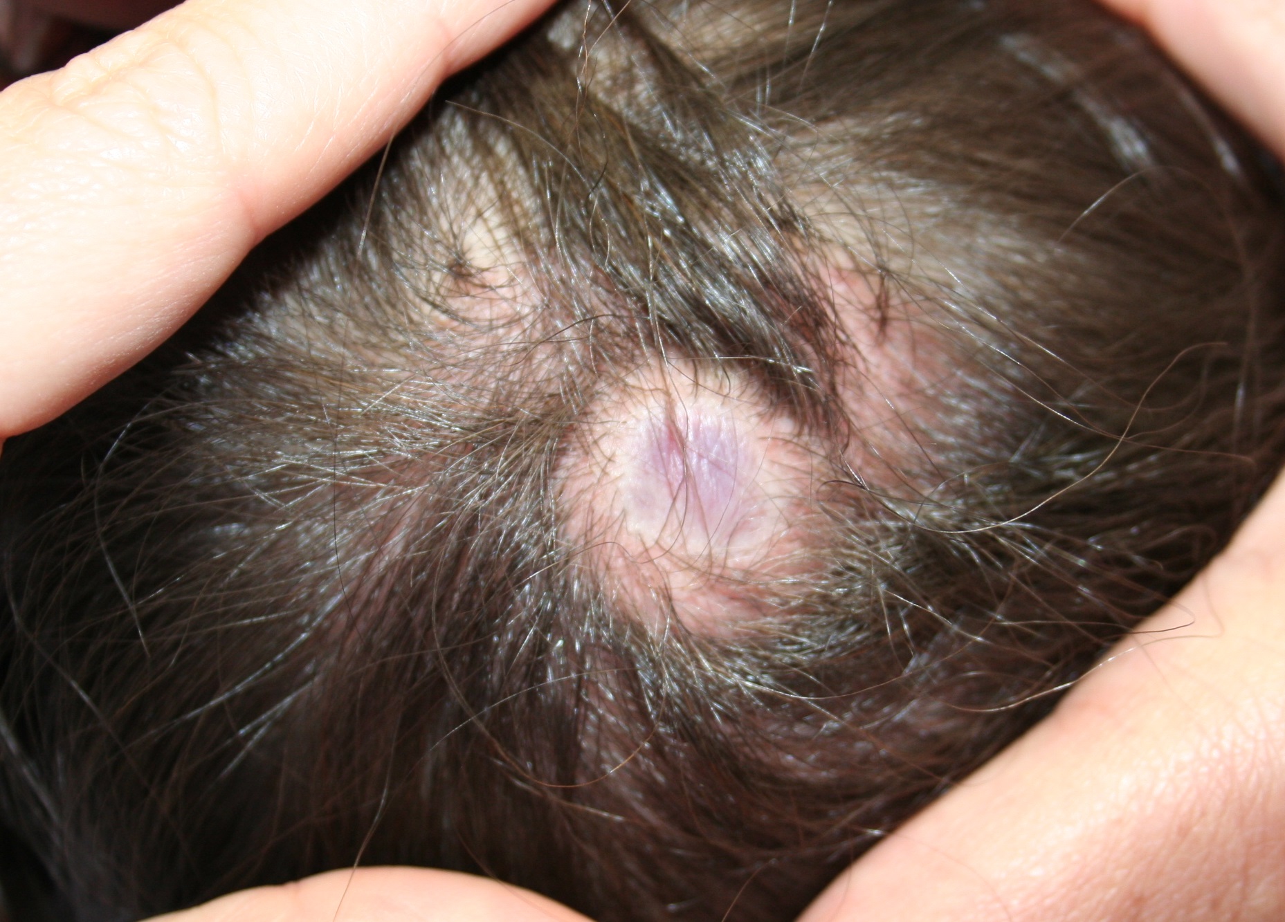
A 9-month-old previously healthy female, born at term, presents to the dermatology clinic for evaluation of a bluish nodule on the posterior vertex of the scalp (Figure 1). Initially at birth, the area was described as a hairless, tense blister that flattened over several weeks. The hairless nodule has persisted without symptoms. The family denies any history of trauma, including the use of forceps or scalp electrodes during labor. Family history was noncontributory. Review of systems was otherwise negative.
On examination, the patient is a well-developed, active female with a 2-cm, bluish nodule with an overlying atrophic, glistening membrane on the posterior parietal scalp, lateral to the midline. The lesion is surrounded by a ring of subtly coarse, terminal hair. There is no evidence of bleeding or ulceration, and no scalp defects were palpable. The lesion was unchanged with crying. The remainder of the physical examination was normal.