User login
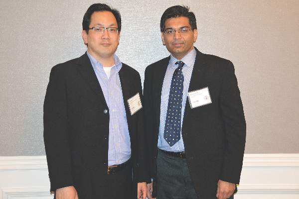
SAN FRANCISCO – At the 2015 AGA Tech Summit, which is sponsored by the AGA Center for GI Innovation and Technology, two researchers provided attendees with an update on their innovative research projects made possible thanks to grants from the AGA Research Foundation.
First, Dr. Richard Kwon updated attendees on the status of his research supported by the inaugural AGA–Covidien Research & Development Pilot Award in Technology. Announced in the fall of 2014, this award is supported by a generous grant from Covidien, a leading global provider of health care products. With this grant, Dr. Kwon and his associates at the University of Michigan are investigating how an image processing and analysis method known as analytic morphomics can be used to improve the care of patients with pancreatic cysts. To date, the inability to accurately distinguish the cyst types has led to a significant amount of unnecessary surgeries and surveillance.
“Nonmucinous cysts generally are comprised of serous cystadenomas as well as inflammatory cells and a small handful of zebra cysts,” explained Dr. Kwon, a gastroenterologist at the university. “These are generally considered benign and do not require follow-up. In contrast, mucinous cysts are made of mucinous cystic neoplasms, which are characterized by a variant stroma and IPMN [intraductal papillary mucinous neoplasm]. This is important because these are the cysts that have malignant potential and therefore require some sort of follow-up and/or surgery.”
He described analytic morphomics as “a novel method to quantitatively analyze structural data such as shape and surface contour (which are otherwise descriptive) from CT scans in order to predict disease states. In our minds, the benefit is that it overcomes some of the shortcomings of imaging curvilinear organs. It has the potential to overcome qualitative descriptions and the limitations of characterizing these cysts based on one cut or one slice of imaging. Hopefully, it will allow us to establish quantifiable criteria, it can be performed on existing studies so you don’t need a new CT scan, and there’s no specialized laboratory or equipment that you need other than a high-powered computer.”
Dr. Kwon presented results from 81 cystic lesions processed with the method. Analytic morphomics yielded a sensitivity of 78% in predicting serous cystadenomas (SCA), a specificity of 90%, a positive predictive value of 73%, and negative predictive value of 88%. The accuracy was 84%. He and his associates are in the process of validating the findings and are in the planning stages of a prospective study comparing morphomics and cyst fluid analysis. “The accuracy of the SCA prediction model appears equal to cyst fluid carcinoembryonic antigen testing, suggesting morphomic data have the potential to replace endoscopic ultrasound/fine needle aspiration in certain scenarios,” he said.
During a separate presentation, Dr. Ashish Nimgaonkar updated attendees on the status of his research, supported by the inaugural AGA–Boston Scientific Career Development Technology & Innovation Award. Bestowed in the fall of 2014, this award is supported by a grant from Boston Scientific, a leading innovator of medical solutions. Dr. Nimgaonkar’s research focuses on developing technology to manage patients with refractory ascites – a condition in which fluid builds up in the abdomen. This fluid accumulation eventually becomes resistant to medical therapy and the only definitive treatment at this stage is liver transplantation, which is limited by organ availability. The only option for them is removal of this fluid every few weeks in a hospital or clinical setting. “When we started to look at this problem, the biggest driving factor for a [solution] was a way to keep these patients out of the hospital and to be able to do this at home,” said Dr. Nimgaonkar, associate medical director at the Center for Bioengineering Innovation and Design in the department of biomedical engineering at Johns Hopkins University, Baltimore.
He and his colleagues have developed a wireless implantable shunt technology to pull the fluid from the peritoneal space into the stomach. With this approach, patients can manage their fluid drainage needs at home – significantly improving their quality of life, as well as reducing the cost of care associated with frequent hospital visits. “You can think of it as a combination of a peritoneal drain and a percutaneous gastrostomy tube, which is a feeding tube hooked but completely implanted in the subcutaneous space,” he said of the technology, which is currently being refined and tested in animal models. “It can be done under conscious sedation. The current design we are working on is a simple, nonelectromechanical solution – silicone-based – which uses the diaphragmatic movements of the patient to drive the fluid. It turns out that the pressure differential between the peritoneal space and the intragastric cavity is minimal, so you need some kind of actuation mechanism.”
Dr. Nimgaonkar and his colleagues hope to study the technology in humans “in the next year or so.”

