User login
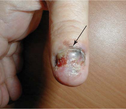
The FP suspected subungual melanoma of the thumb and referred the patient to a surgical oncologist, who confirmed the FP’s suspicions. Subungual melanoma is a type of acrolentiginous melanoma.
The arrow in the image points to hyperpigmentation on the proximal nail fold (Hutchinson’s sign), which is strongly indicative of melanoma. The ulceration suggested that the bad prognosis was going to get even worse.
The initial biopsy showed that the melanoma was greater than 1 mm in depth, so a sentinel node biopsy was planned for the time of surgery. The oncologist thought that the patient might need a partial amputation of the thumb, so a consultation with a hand surgeon was planned.
Subungual melanoma arises on the hand in 45% to 60% of cases, and most of those occur in the thumb. On the foot, subungual melanoma usually occurs in the great toe. The median age at which subungual melanoma is usually diagnosed is in the sixth and seventh decades.
Text for Photo Rounds Friday is courtesy of Richard P. Usatine, MD. Photo is courtesy of Dr. Dubin at http://www.skinatlas.com. This case was adapted from: Mayeaux EJ. Pigmented nail disorders. In: Usatine R, Smith M, Mayeaux EJ, et al, eds. The Color Atlas of Family Medicine. New York, NY: McGraw-Hill; 2009:822-825.
To learn more about The Color Atlas of Family Medicine, see:
• http://www.amazon.com/Color-Atlas-Family-Medicine/dp/0071474641
You can now get The Color Atlas of Family Medicine as an app for mobile devices including the iPhone and iPad by clicking this link: