User login
The American Gastroenterological Association recently published a Clinical Practice Update Commentary outlining surveillance strategies following endoscopic submucosal dissection (ESD) of dysplasia and early gastrointestinal cancer considered pathologically curative.
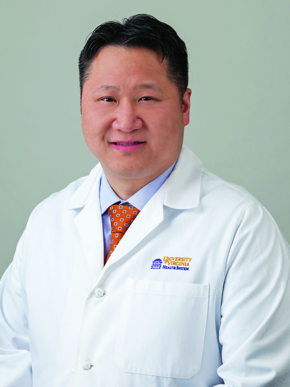
The suggested practice advice, which was put together by Andrew Y. Wang, MD, of the University of Virginia, Charlottesville, and colleagues, offers timelines and modalities of surveillance based on neoplasia type and location, with accompanying summaries of relevant literature.
“Long-term U.S. data about ESD outcomes for early GI neoplasia are only beginning to emerge,” the authors wrote in Gastroenterology. “As such, the current clinical practice regarding endoscopic surveillance intervals and the need for other testing (such as radiographic imaging) after ESD considered curative by histopathology is extrapolated from data derived from Asia and other countries, from concepts learned from polypectomy and piecemeal endoscopic mucosal resection (EMR), and from guideline recommendations after local surgical resection.”
The authors went on to suggest that current recommendations for post-ESD surveillance, including international guidelines “are based more so on expert opinion than rigorous evidence.”
The present update was written to offer additional clarity in this area by providing “a reasonable framework for clinical care and launch points for future research to refine and standardize optimal post-ESD surveillance strategies.”
Foremost, Dr. Wang and colleagues suggested that post-ESD surveillance is necessary because of a lack of standardization concerning the definition of complete resection, along with variable standards of pathological assessment in Western countries, compared with Japan, where pathologists use 2-3 mm serial sectioning and special stains to detect lymphovascular invasion, “which is essential to accurate histopathologic diagnosis and determination of curative resection.”
According to the authors, surveillance endoscopy should be performed with a high-definition endoscope augmented with dye-based or electronic chromoendoscopy, and ideally with optimal magnification.
“Although no supporting data are available at this time, it is prudent and may be reasonable to obtain central and peripheral biopsies of the post-ESD scar,” the authors wrote, noting that relevant mucosa should be checked for metachronous lesions.
Esophageal dysplasia and esophageal squamous cell carcinoma
Following curative resection of low-grade or high-grade esophageal squamous dysplasia, the authors suggested follow-up esophagogastroduodenoscopy (EGD) initially at intervals of 6-12 months, while advising against endoscopic ultrasonography and radiographic surveillance.
In contrast, Dr. Wang and colleagues suggested that superficial esophageal squamous cell carcinoma removed by ESD may benefit from a shorter interval of endoscopic surveillance, with a range of 3-6 months for first and second follow-up EGDs. Clinicians may also consider endoscopic ultrasonography with each EGD, plus an annual CT scan of the abdomen and chest, for 3-5 years.
“A limitation of ESD is that the at-risk esophagus is left in place, and there is a possibility of developing local recurrence or metachronous neoplasia,” the authors wrote. “Although local recurrence after ESD deemed pathologically curative of esophageal squamous cell carcinoma is infrequent, the development of metachronous lesions is not.”
Barrett’s dysplasia and esophageal adenocarcinoma
For all patients, curative removal of Barrett’s dysplasia or esophageal adenocarcinoma should be followed by endoscopy with mucosal ablative therapy at 2-3 months, with treatments every 2-3 months until complete eradication of intestinal metaplasia is achieved, according to Dr. Wang and colleagues.
After complete eradication, patients should be endoscopically screened from 3-12 months, depending on the degree of dysplasia or T-stage of adenocarcinoma, followed by screening procedures ranging from 6 months to 3 years, again depending on disease type.
“Endoscopic resection of visible Barrett’s neoplasia without treatment of Barrett’s esophagus has been associated with significant recurrence rates, so the objective of treatment should be endoscopic resection of visible or nodular dysplasia, followed by complete ablation of any remaining Barrett’s esophagus and associated (flat and/or invisible) dysplasia,” the authors wrote.
Gastric dysplasia and gastric adenocarcinoma
According to the update, after curative resection of gastric dysplasia, first follow-up endoscopy should be conducted at 6-12 months. Second follow-up should be conducted at 12 months for low-grade dysplasia versus 6-12 months for high-grade dysplasia, with annual exams thereafter.
For T1a early gastric cancer, the first two follow-up endoscopies should be performed at 6-month intervals, followed by annual exams. T1b Sm1 disease should be screened more aggressively, with 3-6 months intervals for first and second follow-up EGDs, plus CT scans of the abdomen and chest and/or endoscopic ultrasound every 6-12 months for 3-5 years.
“For lesions where a curative resection was achieved based on clinical criteria and histopathologic examination, surveillance is performed primarily to detect metachronous gastric cancers,” the authors wrote.
Colonic dysplasia and adenocarcinoma
According to the authors, adenomas with low-grade dysplasia or serrated sessile lesions without dysplasia removed by ESD should be rechecked by colonoscopy at 1 year and then 3 years, followed by adherence to U.S. Multi-Society Task Force recommendations.
For traditional serrated adenomas, serrated sessile lesions with dysplasia, adenomas with high-grade dysplasia, carcinoma in situ, intramucosal carcinoma, or dysplasia in the setting of inflammatory bowel disease, first follow-up colonoscopy should be conducted at 6-12 months, 1 year later, then 3 years after that, followed by reversion to USMSTF recommendations, although patients with IBD may benefit from annual colonoscopy.
Finally, patients with superficial T1 colonic adenocarcinoma should be screened more frequently, with colonoscopies at 3-6 months, 6 months, and 1 year, followed by adherence to USMSTF recommendations.
“The current Japanese guideline suggests that recurrence or metastasis after endoscopic resection of T1 (Sm) colonic carcinomas occurs mainly within 3-5 years,” the authors noted.
Rectal dysplasia and adenocarcinoma
Best practice advice suggestions for rectal dysplasia and adenocarcinoma are grouped similarly to the above advice for colonic lesions.
For lower-grade lesions, first follow-up with flexible sigmoidoscopy is suggested after 1 year, then 3 years, followed by reversion to USMSTF recommendations. Higher-grade dysplastic lesions should be checked after 6-12 months, 1 year, then 3 years, followed by adherence to USMSTF guidance, again excluding patients with IBD, who may benefit from annual exams.
Patients with superficial T1 rectal adenocarcinoma removed by ESD deemed pathologically curative should be checked with flexible sigmoidoscopy at 3-6 months, again at 3-6 months after first sigmoidoscopy, then every 6 months for a total of 5 years from the time of ESD, followed by adherence to USMSTF recommendations. At 1 year following ESD, patients should undergo colonoscopy, which can take the place of one of the follow-up flexible sigmoidoscopy exams; if an advanced adenoma is found, colonoscopy should be repeated after 1 year, versus 3 years if no advanced adenomas are found, followed by adherence to USMSTF recommendations. Patients with superficial T1 rectal adenocarcinoma should also undergo endoscopic ultrasound or pelvic MRI with contrast every 3-6 months for 2 years, followed by intervals of 6 months for a total of 5 years. Annual CT of the chest and abdomen may also be considered for a duration of 3-5 years.
Call for research
Dr. Wang and colleagues concluded their update with a call for research.
“We acknowledge that the level of evidence currently available to support much of our surveillance advice is generally low,” they wrote. “The intent of this clinical practice update was to propose surveillance strategies after potentially curative ESD for various GI neoplasms, which might also serve as reference points to stimulate research that will refine future clinical best practice advice.”
The article was supported by the AGA. The authors disclosed relationships with MicroTech, Olympus, Lumendi, U.S. Endoscopy, Boston Scientific, Steris and others.
This article was updated Dec. 15, 2021.
FROM GASTROENTEROLOGY

