User login
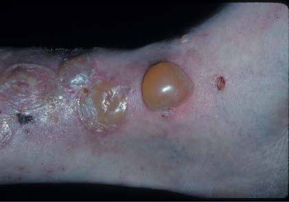
The family physician suspected bullous pemphigoid (BP) based on the collection of tense bullae on the leg; the diagnosis was confirmed with a punch biopsy at the edge of the intact bulla.
BP is a chronic autoimmune disorder of the skin with an average age of onset of 65 years. The generalized bullous form is the most common. One atypical aspect of this case was the localized nature of the bullae. Tense bullae occur on both erythematous and normal appearing skin surfaces, which usually heal without scarring. Drug-induced BP has been reported with drugs containing sulfhydryl groups, including penicillamine, furosemide, captopril, and sulfasalazine.
Biopsy is required to establish the diagnosis. A 4-mm punch biopsy from the edge of an early blister including part of the normal-appearing skin shows a subepidermal blister and an eosinophil-rich mixed dermal inflammatory infiltrate. (A scoop shave biopsy under a small intact blister is also a good method of making a diagnosis of BP or other bullous diseases.)
One can start with a standard biopsy and perform another biopsy for direct immunofluorescence (DIF) if needed. This can be done with a second 4-mm punch biopsy or a shave from perilesional skin. This biopsy for DIF should be transported in Michel’s solution. DIF demonstrates linear IgG and complement C3 deposits at the dermal–epidermal junction.
The family physician started the patient on prednisone 60 mg daily on the day of the biopsy and referred him to a dermatologist for long-term management. The patient began to heal dramatically within days.
Photo courtesy of Eric Kraus, MD. Text for Photo Rounds Friday courtesy of Richard P. Usatine, MD. This case was adapted from: Mohmand A. Bullous pemphigoid. In: Usatine R, Smith M, Mayeaux EJ, et al, eds. The Color Atlas of Family Medicine. New York, NY: McGraw-Hill; 2009:790-796.
To learn more about The Color Atlas of Family Medicine, see:
• http://www.amazon.com/Color-Atlas-Family-Medicine/dp/0071474641
The Color Atlas of Family Medicine is also available as an app for mobile devices. See