Article Type
Changed
Tue, 08/01/2017 - 12:25
Display Headline
Clinical Challenges - August 2017 What's your diagnosis?

The diagnosis

Answer to “What’s your diagnosis?” on page 2: Lemmel’s syndrome

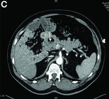
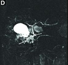
Figure C shows a 30 × 22-mm juxtapapillary diverticulum (JPD) containing air, fluid, and food debris (white arrow). Figure D shows the JPD (solid white arrow) causing distortion of a normal-caliber distal common bile duct (dashed white arrow) and pancreatic duct (PD; black arrow), with no gallbladder or ductal stones demonstrated. The final diagnosis was cholangitis and pancreatitis associated with a JPD without choledocholithiasis (Lemmel’s syndrome). He remains well 2 months later on follow-up, with normal blood tests.

Duodenal diverticuli occur commonly (15%–20% of autopsies), especially in the elderly, are usually discovered incidentally, and mostly asymptomatic. The majority (75%) are periampullary; those located within 2–3 cm of the ampulla of Vater are called JPD. Patients may rarely present with symptoms including abdominal pain, steatorrhea, gastrointestinal bleeding, perforation, intestinal obstruction, diverticulitis, and cholangiopancreatic disease (obstructive jaundice, cholangitis, and pancreatitis).1 Primary choledocholithiasis occurs commonly (incidence 20%–40%) in patients with periampullary duodenal diverticuli, owing to the production of beta-glucuronidase producing microorganisms. However, many patients (up to 41%) with JPD and cholangiopancreatic symptoms have normal biliary and pancreatic ducts on cholangiography.1,2 Lemmel’s syndrome is the combination of JPD and cholangiopancreatic disease without choledocholithiasis.3 It is postulated to arise from the reflux of duodenal contents with intestinal bacteria into the common bile duct and pancreatic duct, mechanical compression, or distortion of the distal common bile duct and pancreatic duct arising from an impacted enterolith or food debris in the diverticulum, or sphincter of Oddi dysfunction, with resultant bile stasis.1 Patients characteristically describe postprandial epigastric pain or fullness. The mainstay of treatment in the majority of patients is conservative, although endoscopic removal of impacted food debris may occasionally be necessary. Operative intervention, preferably duodenojejunostomy, may be indicated for persistent/recurrent symptoms or severe complications (such as recurrent cholangitis/pancreatitis, bleeding or perforation).


References

1. Egawa, N., Anjiki, H., Takuma, K., et al. Juxtapapillary duodenal diverticula and pancreatobiliary disease. Dig Surg. 2010;27:105-9.

2. Lobo, D.N., Balfour, T.W., Iftikhar, S.Y. Periampullary diverticula: consequences of failed ERCP. Ann Royal Coll Surg. 1998;80:326-31.

3. Lemmel, G. Die klinische Bedeutung der Duodenaldivertikel. Archiv fur Verdauungskrankheiten. 1934;56:59-70.


Publications
Sections

The diagnosis

Answer to “What’s your diagnosis?” on page 2: Lemmel’s syndrome

Figure C shows a 30 × 22-mm juxtapapillary diverticulum (JPD) containing air, fluid, and food debris (white arrow). Figure D shows the JPD (solid white arrow) causing distortion of a normal-caliber distal common bile duct (dashed white arrow) and pancreatic duct (PD; black arrow), with no gallbladder or ductal stones demonstrated. The final diagnosis was cholangitis and pancreatitis associated with a JPD without choledocholithiasis (Lemmel’s syndrome). He remains well 2 months later on follow-up, with normal blood tests.

Duodenal diverticuli occur commonly (15%–20% of autopsies), especially in the elderly, are usually discovered incidentally, and mostly asymptomatic. The majority (75%) are periampullary; those located within 2–3 cm of the ampulla of Vater are called JPD. Patients may rarely present with symptoms including abdominal pain, steatorrhea, gastrointestinal bleeding, perforation, intestinal obstruction, diverticulitis, and cholangiopancreatic disease (obstructive jaundice, cholangitis, and pancreatitis).1 Primary choledocholithiasis occurs commonly (incidence 20%–40%) in patients with periampullary duodenal diverticuli, owing to the production of beta-glucuronidase producing microorganisms. However, many patients (up to 41%) with JPD and cholangiopancreatic symptoms have normal biliary and pancreatic ducts on cholangiography.1,2 Lemmel’s syndrome is the combination of JPD and cholangiopancreatic disease without choledocholithiasis.3 It is postulated to arise from the reflux of duodenal contents with intestinal bacteria into the common bile duct and pancreatic duct, mechanical compression, or distortion of the distal common bile duct and pancreatic duct arising from an impacted enterolith or food debris in the diverticulum, or sphincter of Oddi dysfunction, with resultant bile stasis.1 Patients characteristically describe postprandial epigastric pain or fullness. The mainstay of treatment in the majority of patients is conservative, although endoscopic removal of impacted food debris may occasionally be necessary. Operative intervention, preferably duodenojejunostomy, may be indicated for persistent/recurrent symptoms or severe complications (such as recurrent cholangitis/pancreatitis, bleeding or perforation).


References

1. Egawa, N., Anjiki, H., Takuma, K., et al. Juxtapapillary duodenal diverticula and pancreatobiliary disease. Dig Surg. 2010;27:105-9.

2. Lobo, D.N., Balfour, T.W., Iftikhar, S.Y. Periampullary diverticula: consequences of failed ERCP. Ann Royal Coll Surg. 1998;80:326-31.

3. Lemmel, G. Die klinische Bedeutung der Duodenaldivertikel. Archiv fur Verdauungskrankheiten. 1934;56:59-70.


Publications
Publications
Article Type
Display Headline
Clinical Challenges - August 2017 What's your diagnosis?
Display Headline
Clinical Challenges - August 2017 What's your diagnosis?
Sections
Questionnaire Body

By Crispin Musumba, MBChB, PhD, Edward Britton, MBBS, MRCP, and Howard Smart, MBBS, DM. Published previously in Gastroenterology (2013;144:274, 468-469).
A 50-year-old man presented with a 2-week history of intermittent epigastric pain. His pain typically started 1-2 hours after a meal and lasted for 10 minutes, with associated nausea and vomiting. On the day of admission, the pain had become severe and continuous (lasting 2 hours), exacerbated by lying flat, with accompanying jaundice and rigors. On examination, he was icteric and pyrexial (39.2 °C), with tenderness in the epigastrium and normal bowel sounds. Bloods tests revealed a hemoglobin count of 13.0 g/dL, leukocytosis (13 × 109/L), neutrophilia (13.0 × 10⁹/L), elevated C-reactive protein (161 mg/L), alanine transaminase (424 U/L), alkaline phosphatase (230 U/L), gamma-glutamyl transpeptidase (579 U/L), bilirubin (77 mmol/L), albumin (32 g/L), glucose (7.4 mmol/L), amylase (375 U/L; normal, less than 150), and a normal lipid profile and calcium levels. Liver ultrasonography was unremarkable.

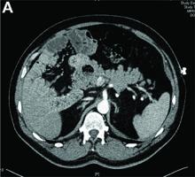
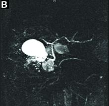
An abdominal computed tomography (CT) scan was done (Figure A). He was treated conservatively initially with intravenous morphine for pain relief and started on a course of intravenous antibiotics, with good clinical improvement. After improvement of his blood tests, he was discharged a week after admission, and had a magnetic resonance cholangiopancreatography (Figure B) performed as an outpatient. What is the likely diagnosis and pathogenetic mechanisms?

Disallow All Ads
Content Gating
No Gating (article Unlocked/Free)
Alternative CME
Disqus Comments
Default