User login
|
|
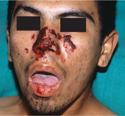
A rush biopsy ordered after the family physician (FP) consulted with a dermatologist confirmed a diagnosis of pemphigus vulgaris.
Pemphigus vulgaris should be considered in any bullous disease involving the oral mucosa. As this is a life-threatening illness, treatment should begin with oral prednisone at the same time that biopsies are performed.
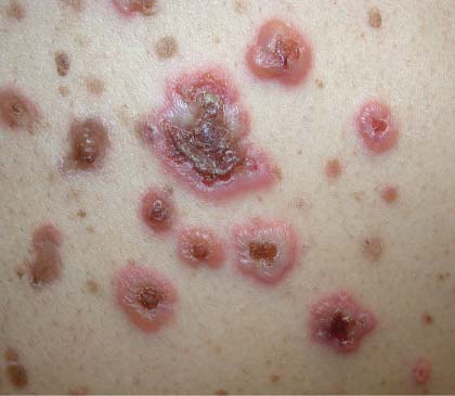
Pemphigus vulgaris presents with flaccid bullae, which rupture easily, creating erosions. Since bullae are short-lived, erosions are the more common presenting physical finding. Lesions are typically tender and heal with post-inflammatory hyperpigmentation, which resolves without scarring. A Nikolsky sign may be present, but is not diagnostic. This is elicited by applying lateral pressure to a bulla or adjacent normal skin, resulting in a separation of the epidermis from deeper layers.
The most common mucosal site is the oral mucosa. Mucosal lesions may be followed by skin lesions on the scalp, face, and upper torso. Pemphigus vulgaris should be suspected if an oral ulcer persists beyond a month.
Two skin biopsies are preferred, but even a single biopsy can be used to make the diagnosis. The first biopsy is sent in formalin for routine histopathology. This biopsy should be of the freshest lesion with an intact bulla if possible. Perform a shave of the edge of the bulla to include the epidermis. The second is taken from perilesional normal skin, and is sent on a gauze pad soaked in normal saline or Michel’s solution for direct immunofluorescence.
The skin lesions are treated with topical high-potency steroids, such as clobetasol ointment. The facial lesions are treated with topical triamcinolone ointment. Oral prednisone is needed in relatively high doses.
Oral hygiene is crucial. Mouthwashes such as chlorhexidine 0.2% or 1:4 hydrogen peroxide may be used. Topical anesthetics may be used for pain. Prednisone should be started at 60 mg a day and titrated as needed. The dose may be increased by 50% every 1 to 2 weeks until disease activity is controlled. However, some experts will not go beyond 1 mg/kg/day. Once remission is induced, the dose is tapered by 25% every 1 to 2 weeks to the lowest dose needed to suppress recurrence of new lesions.
High dose and prolonged treatment with steroids can have serious side effects. Therefore, it is advisable to start adjuvant steroid-sparing therapy within 2 to 4 weeks of treatment. Adjuvant agents have a lag period of 4 to 6 weeks before they become effective, so starting them sooner allows for an earlier steroid taper. Adjuvant agents include azathioprine, cyclophosphamide, methotrexate, and mycophenolate. They may be used alone to maintain remission after steroid withdrawal.
Photos and text for Photo Rounds Friday courtesy of Richard P. Usatine, MD. This case was adapted from: Mittal S. Pemphigus. In: Usatine R, Smith M, Mayeaux EJ, et al, eds. The Color Atlas of Family Medicine. New York, NY: McGraw-Hill; 2009:794-798.
To learn more about The Color Atlas of Family Medicine, see:
• http://www.amazon.com/Color-Atlas-Family-Medicine/dp/0071474641
The Color Atlas of Family Medicine is also available as an app for mobile devices. See
|
|
|
|
A rush biopsy ordered after the family physician (FP) consulted with a dermatologist confirmed a diagnosis of pemphigus vulgaris.
Pemphigus vulgaris should be considered in any bullous disease involving the oral mucosa. As this is a life-threatening illness, treatment should begin with oral prednisone at the same time that biopsies are performed.
Pemphigus vulgaris presents with flaccid bullae, which rupture easily, creating erosions. Since bullae are short-lived, erosions are the more common presenting physical finding. Lesions are typically tender and heal with post-inflammatory hyperpigmentation, which resolves without scarring. A Nikolsky sign may be present, but is not diagnostic. This is elicited by applying lateral pressure to a bulla or adjacent normal skin, resulting in a separation of the epidermis from deeper layers.
The most common mucosal site is the oral mucosa. Mucosal lesions may be followed by skin lesions on the scalp, face, and upper torso. Pemphigus vulgaris should be suspected if an oral ulcer persists beyond a month.
Two skin biopsies are preferred, but even a single biopsy can be used to make the diagnosis. The first biopsy is sent in formalin for routine histopathology. This biopsy should be of the freshest lesion with an intact bulla if possible. Perform a shave of the edge of the bulla to include the epidermis. The second is taken from perilesional normal skin, and is sent on a gauze pad soaked in normal saline or Michel’s solution for direct immunofluorescence.
The skin lesions are treated with topical high-potency steroids, such as clobetasol ointment. The facial lesions are treated with topical triamcinolone ointment. Oral prednisone is needed in relatively high doses.
Oral hygiene is crucial. Mouthwashes such as chlorhexidine 0.2% or 1:4 hydrogen peroxide may be used. Topical anesthetics may be used for pain. Prednisone should be started at 60 mg a day and titrated as needed. The dose may be increased by 50% every 1 to 2 weeks until disease activity is controlled. However, some experts will not go beyond 1 mg/kg/day. Once remission is induced, the dose is tapered by 25% every 1 to 2 weeks to the lowest dose needed to suppress recurrence of new lesions.
High dose and prolonged treatment with steroids can have serious side effects. Therefore, it is advisable to start adjuvant steroid-sparing therapy within 2 to 4 weeks of treatment. Adjuvant agents have a lag period of 4 to 6 weeks before they become effective, so starting them sooner allows for an earlier steroid taper. Adjuvant agents include azathioprine, cyclophosphamide, methotrexate, and mycophenolate. They may be used alone to maintain remission after steroid withdrawal.
Photos and text for Photo Rounds Friday courtesy of Richard P. Usatine, MD. This case was adapted from: Mittal S. Pemphigus. In: Usatine R, Smith M, Mayeaux EJ, et al, eds. The Color Atlas of Family Medicine. New York, NY: McGraw-Hill; 2009:794-798.
To learn more about The Color Atlas of Family Medicine, see:
• http://www.amazon.com/Color-Atlas-Family-Medicine/dp/0071474641
The Color Atlas of Family Medicine is also available as an app for mobile devices. See