User login

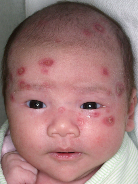
The physician diagnosed neonatal lupus erythematosus, a rare syndrome in which maternal autoantibodies are passively transferred to the baby and cause cutaneous lesions or isolated congenital heart block. The skin rash generally appears a few days to weeks after birth, typically after sun exposure, and shows well-demarcated erythematous scaling patches that are often annular and predominately on the scalp, neck, or face. It is self-limited and generally resolves without scarring by 6 to 7 months of age.
Diagnosis is based on physical findings in an infant <6 months of age and detection of autoantibodies against Ro (SSA), La (SSB), and/or U1-ribonucleoprotein (U1-RNP) in the child or mother. All infants diagnosed with neonatal lupus should have an EKG to detect heart block. If the EKG is abnormal, referral to a pediatric cardiologist is warranted.
Infants with neonatal lupus should also be protected from the sun through avoidance and protective clothing. Mild topical steroids may be helpful. Children with neonatal lupus may be at higher risk of developing autoimmune disorders or rheumatic disease later in life.
The infant described here had abnormal anti-Ro, anti-La, and anti-RNP levels. Her mother’s autoantibody levels were similarly abnormal. The infant’s initial nursery stay was complicated by a transient and otherwise asymptomatic bradycardia. An EKG detected a heart rate of 96 bpm with no heart block, but there were changes suggestive of left ventricular hypertrophy. An echocardiogram was normal. The patient was referred to a pediatric cardiologist and a second echocardiogram and EKG were normal.
At 7 weeks, the infant’s cutaneous lesions were present, but were resolving on their own. The skin lesions were fully resolved by 6 months of age.
Text for Photo Rounds Friday courtesy of Richard P. Usatine, MD. This case was adapted from: Mayeaux, EJ. Lupus erythematosus (systemic and cutaneous). In: Usatine R, Smith M, Mayeaux EJ, et al, eds. The Color Atlas of Family Medicine. New York, NY: McGraw-Hill; 2009:766-771.
Photo courtesy of: Warner AM, Frey KA, Connolly S. Photo Rounds: Annular rash on a newborn. J Fam Pract. 2006;55:128–129.
To learn more about The Color Atlas of Family Medicine, see:
* http://www.amazon.com/Color-Atlas-Family-Medicine/dp/0071474641