User login
CHICAGO – Fluorescence in situ hybridization testing for aneuploidy and loss of the tumor suppressor gene P16 can be useful in predicting progression to high-grade dysplasia and adenocarcinoma in patients with nondysplastic Barrett’s esophagus.
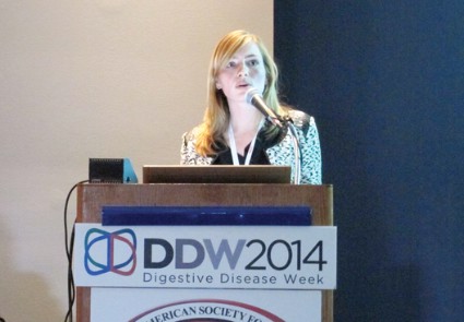
Adding these genetic biomarkers to the clinical risk factors of age and Barrett’s esophagus (BE) segment length provides for an even more robust prediction tool, Dr. Margriet R. Timmer said at the annual Digestive Disease Week.
This conclusion is based on a prospective, multicenter cohort study involving 428 patients with nondysplastic BE. The average age of the patients was 59 years and maximum BE length 3 cm.
Fluorescence in situ hybridization (FISH) analysis was used to detect genetic abnormalities on endoscopic cytology brushes from all patients. The FISH panel included probes for six candidate biomarkers: the tumor suppressor genes P16 and P53, the oncogenes MYC, HER-2/neu, and 20q, as well as aneuploidy.
After a median follow-up of 45 months, 22 patients had histologic progression after review by two expert pathologists: 13 cases of high-grade dysplasia (HGD) and 9 cases of esophageal adenocarcinoma (EAC).
The rate of progression to HGD/EAC was 1.09% per patient-year and 0.65% per patient-year for EAC, said Dr. Timmer of the Center for Experimental and Molecular Medicine, Academic Medical Center, Amsterdam, the Netherlands.
Univariable analysis revealed that P16 loss (hazard ratio, 2.56; P = .03) and aneuploidy (HR, 2.66; P = .04) significantly predicted progression, as did increasing age (HR, 1.06; P = .008) and longer BE length (HR, 1.15; P = .017).
None of the other biomarkers or clinical factors such as male sex, body mass index, tobacco use, family history of BE or EAC, or years of BE were significant.
A prediction model was then created using the significant predictors. The C statistic, which takes into account specificity and sensitivity, was 0.68 for the basic model that included only age and BE length, but this improved significantly to 0.73 when the biomarkers were added, she said.
Based on the prediction model, 236 patients were considered high risk and 192 patients were low risk.
Importantly, 5-year progression-free survival was significantly lower in the high-risk group than in the low-risk group (93.6% vs. 98.4%; P = .001), Dr. Timmer noted.
Session cochair Roy Wong of Walter Reed Army Medical Center, Washington, said in an interview that "intellectually, the concept is terrific," but that clinicians will need a specific risk cutoff to apply the findings to an individual patient in the community setting and that overall survival data would strengthen the results.
The study authors acknowledged that FISH testing is still costly and that they have yet to use it in the clinical setting. One major advantage of FISH, however, is that it can be applied to cytology specimens that can be easily obtained during upper endoscopy by brushing the entire Barrett’s segment, thereby overcoming the potential problem of biopsy sampling error, Dr. Timmer said.
The study was funded by grants from KWF Kanderbestrijding (the Dutch Cancer Society), NOW (De Nederlandse Organisatie voor Wetenschappelijk Onderzoek), Fonds NutsOhra, Gut Club, and Abbott Molecular. Dr. Timmer and her coauthors reported no financial disclosures.
AT DDW 2014
Key clinical point: Assessing aneuploidy and P16 as well as age and BE length can be useful in stratifying patients with Barrett’s esophagus into high- and low-risk disease categories to improve the efficacy of surveillance programs.
Major finding: P16 loss (hazard ratio, 2.56; P = .03) and aneuploidy (HR, 2.66; P = .04), increasing age (HR, 1.06; P = .008), and longer BE length (HR, 1.15; P = .017) predicted progression.
Data source: A prospective study of 428 patients with nondysplastic Barrett’s esophagus.
Disclosures: The study was funded by grants from KWF Kanderbestrijding (the Dutch Cancer Society), NOW (De Nederlandse Organisatie voor Wetenschappelijk Onderzoek), Fonds NutsOhra, Gut Club, and Abbott Molecular. Dr. Timmer and her coauthors reported no financial disclosures.

