User login
Digestive Disease Week (DDW 2014)
Hepatology section
The hepatology session at the AGA Postgraduate Course in Chicago had a range of excellent topics for the practicing gastroenterologist/hepatologist. Perhaps the area that is changing most rapidly is that of treatment of hepatitis C. Dr. Jordan Feld from the University of Toronto presented “Choosing the optimal HCV regimen.” Dr. Feld’s presentation highlighted the recent approval of simeprevir and sofosbuvir both with pegylated interferon and ribavirin for treatment of chronic hepatitis C genotype 1. HCV cure rates of 80% to 90% were described. He also told us about exciting developments with interferon-free regimens that will be available in October/November of 2014.
The combination of sofosbuvir and ledipasvir and the combination of three direct-acting antiviral agents with or without ribavirin will revolutionize our care of hepatitis C patients from now on. Many clinicians have already been using interferon-free regimens by combining simeprevir and sofosbuvir for 12 weeks with excellent results. Dr. Feld finished by highlighting the fact that the American Association for the Study of Liver Diseases/Infectious Diseases Society of America guidelines are now available and provide a useful resource for treatment for your hepatitis C patients.
Dr. Michael Lucey from the University of Wisconsin School of Medicine and Public Health talked about the management of alcoholic hepatitis. Dr. Stephen Harrison from Brooke Army Medical Center presented impressive data on the huge number of patients that we will be seeing with nonalcoholic fatty liver disease/nonalcoholic steatohepatitis. By careful estimates, he predicts that there may be as many as 25 million patients in the United States with NASH. New treatments are being developed, but we are still a long way from having reliable treatments for this disease. Thus, lifestyle modifications with judicious weight loss and exercise regimens remain the most important treatments available for NASH/NAFLD. Dr. Harrison also highlighted the recent studies that demonstrate that regular coffee consumption may be helpful for reducing fibrosis in patients with NAFLD.
Dr. Jorge Marrero from the University of Texas Southwestern Medical School presented data on kidney injury in cirrhosis and how to differentiate acute kidney injury from hepatorenal syndrome. Finally, Dr. Sam Lee from the University of Calgary presented “Management of coagulation disorders in cirrhosis.” He discussed the use of anticoagulation and taught us to recognize that cirrhosis is not a hypocoagulable state and that there are situations where anticoagulant therapy in cirrhosis is definitely a benefit. The idea of using anticoagulation in patients with cirrhosis is counterintuitive to many of us, but Dr. Lee presented data to endorse this approach. I think all of will have to think hard in our practices on how we proceed with the use of anticoagulation in patients with portal vein thrombosis.
Dr. Bacon is the James F. King MD Endowed Chair in Gastroenterology, professor of internal medicine, and director of abdominal transplantation at Saint Louis University School of Medicine, division of gastroenterology and hepatology, St. Louis.
The pancreatic-biliary section
The first presentation was on benign biliary strictures, given by Dr. Greg Cote of Indiana University. Dr. Cote divided etiologies into extrinsic vs. intrinsic and noted which ones require only bridging plastic stents while treating the underlying cause. The etiologies that require endoscopic therapy include chronic pancreatitis, postoperative (both post cholecystectomy and post transplant), primary sclerosing cholangitis, and the rare common bile duct stone–induced stricture. For primary sclerosing cholangitis, dilation therapy alone is usually sufficient and safer than dilation plus stenting. Strictures that are postoperative or due to chronic pancreatitis require aggressive endo-therapy combining dilation and placement of the maximal number of plastic stents possible vs. fully covered metal stent placement.
Dr. David Lichtenstein gave the second talk on indeterminate and malignant bile duct strictures. He discussed the multiple diagnostic methods needed to determine whether a stricture is benign or malignant. At endoscopic retrograde cholangiopancreatography it is useful to combine both brush cytology (+/– FISH) and intraductal biopsy. Even higher sensitivities are obtained by utilizing endoscopic ultrasound (EUS)/fine-needle aspiration (FNA), although if the patient is a resection or transplant candidate, some surgeons are fearful of tumor seeding with FNA. Other techniques discussed included cholangioscopy and probe-based confocal microscopy. The pros and cons of various palliative stents were also thoroughly discussed, as were experimental endoscopic therapies including photodynamic therapy and endobiliary radiofrequency ablation.
Dr. Robert Hawes discussed the management of chronic pancreatitis pain with medical, endoscopic, and surgical therapy. He reviewed the causes of pain in chronic pancreatitis, the variable clinical presentations (main pancreatic duct obstruction or not, and ongoing inflammation or not), and the fact that placebo response rates are high. Medical therapies, including pancreatic enzymes, antioxidants, and octreotide are not well proven although often tried. Celiac axis blockade has at best a 50% chance of benefit and is not long lasting. Endoscopic therapy (often combined with extracorporeal shock wave lithotripsy has some efficacy for obstructive disease. Surgery is most effective for obstructive disease and surgical resection carries the risk of insulin dependence.
Dr. Martin Freeman discussed acute idiopathic recurrent pancreatitis and its likely multifactorial causes with interaction of genetic, anatomic, and environmental factors. He pointed out the frequent overlap presentation of acute pancreatitis versus chronic pancreatitis. He also pointed out the key diagnostic role of endoscopic ultrasound and the value of secretin-stimulated magnetic resonance cholangiopancreatography. The controversial role of endoscopic therapy was discussed.
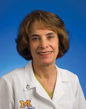
Dr. Elta is professor of internal medicine, University of Michigan, Ann Arbor. She moderated the course at the 2014 Digestive Diseases Week.
Esophagus/Upper GI section
One highlight of the AGA Postgraduate Course was the esophageal disease session. The presentation by Dr. Michael B. Wallace summarized recent studies using advanced imaging modalities in patients with Barrett’s esophagus. Studies using chromoscopy and virtual chromoscopy techniques such as narrow-band imaging have increased the detection of dysplasia in BE patients. These are so-called red flag techniques that image large areas of mucosa to detect mucosal abnormalities suspicious for the presence of dysplasia or neoplasia.
Endomicroscopy describes the use of real-time, targeted endoscopic imaging modalities that are capable of producing histologic-like images of mucosa at depths up to 200 microns. Confocal laser endomicroscopy (CLE) uses a blue light laser (405 nm) and collimated light detection and analysis to produce 1000-fold magnified images. When used with a fluorescent contrast agent such as fluorescein or acriflavin dye, these systems produce cellular level images that are comparable to those images seen with optical microscopy. A recent study from Canto et al found that the use of CLE detected BE dysplasia at rates similar to targeted plus random biopsy protocols. Further, a multicenter study will soon begin using a tethered-capsule (nonendoscopic) form of volumetric laser endomicroscopy as a method to screen for BE.
Dr. Amitabh Chak expanded on these issues and reviewed the issues surrounding screening and surveillance of BE patients for the early detection and treatment of esophageal adenocarcinoma. This presentation suggested that necessary future improvements include cost-effective advanced imaging techniques optimized for use in clinical practice, molecular biomarker panels for prediction of which patients may progress to dysplasia and neoplasia, and high-quality intensive endoscopic surveillance for high risk BE patients.
Dr. Joe Murray’s comprehensive presentation of celiac disease described the protean clinical presentations of this disease as well as optimal use of serologic and endoscopic testing. Celiac disease is increasingly identified in middle-aged patients (median 45 years) without diarrhea. Classic malabsorption symptoms of diarrhea, weight loss, steatorrhea, and nutritional deficiencies are found in 25% of patients. Half of celiac patients will have only one symptom such as anemia, diarrhea, lactose intolerance, or weight loss. Nongastrointestinal symptoms are present in another 25% of patients such as infertility, bone disease, chronic fatigue, or abnormal liver enzyme test results.
Optimal use of serologic and endoscopic testing was reviewed, including the differential diagnosis of lymphocytic duodenosis including use of nonsteroidal anti-inflammatory agents (NSAIDs), Helicobacter pylori infection, Crohn’s disease, and Sjogren’s syndrome. Proper duodenal biopsy technique was emphasized with two forceps biopsy samples obtained from the duodenal bulb and four biopsy samples obtained from the second portion of the duodenum. Also discussed was the utility of HLA typing for DQ2/8 in patients currently using a gluten free diet, patients with negative serology results but abnormal duodenal biopsy findings, and those with negative serology results who are at increased genetic risk.
Dr. James Scheiman discussed management of the complex interaction and risks associated with the use of NSAIDs, aspirin, clopidogrel, and proton pump inhibitors in the setting of previous ulcer disease, gastrointestinal bleeding, and Helicobacter pylori infection. Results from randomized controlled studies and observational studies were the basis for the Consensus Group to recommend the use of proton pump inhibitor therapy as the GI bleeding protective strategy of choice. PPI therapy was also recommended as cost-effective treatment for aspirin-using patients, although the risks and benefits of long-term PPI treatment require patient education and individualization.
Finally, Dr. Rhonda Souza discussed eosinophilic esophagitis (EoE), a chronic immune/antigen-mediated esophageal disease characterized clinically by symptoms related to esophageal dysfunction associated with eosinophil-predominant inflammation such as dysphagia, food impaction, chest pain, heartburn, abdominal pain, and refractory reflux dyspepsia. Endoscopic features include the ringed esophagus, white specks, linear furrows and stricture. Histologic features of EoE are eosinophilia (more than 15 intraepithelial eosinophils per high power field), basal zone hyperplasia, and dilated intercellular spaces. These eosinophils are activated via T-helper 2 immune system via interleukins-4, -5 and -13. This inflammation is mediated by the dramatic upregulation involving the eotaxin-3 gene that produces a potent chemoattractant for eosinophils. Treatment of EoE usually requires the use of proton pump inhibitors based on their acid suppression, anti-oxidant and anti-inflammatory effects. The use of topical corticosteroids and endoscopic dilation for symptomatic strictures may also be necessary. Nondrug treatment approaches such as the six food elimination diet (SFED) of the most common food allergens such as milk, soy, eggs, wheat, nuts and seafood have also been successful.
Dr. Wolfsen is in the division of gastroenterology and hepatology, Mayo Clinic, Jacksonville, Fla. He moderated this session during the 2014 Digestive Diseases Week.
The colon course
The colon course brought us numerous pearls on a broad range of topics, from how to perform quality endoscopy safely, to providing updates on genetic screening for colon cancer syndromes and fecal microbiota transplant.
Quality colonoscopy is no longer just a personal effort to provide the best care possible to our patients, it will become a barometer for not only government and private payers but also patients looking for top-notch care. Adenoma detection rate is an objective parameter that we should all monitor regularly and strive to meet or exceed national benchmarks.
We learned from Dr. Thomas Imperiale that advances in colonoscopy preparation have led to marked improvements in quality bowel preparation, with resultant improvements in adenoma detection rate (ADR). We now know that split preparations are most effective, and minimizing the time between prep completion and performance of colonoscopy improves bowel prep quality. Dr. Sameer Saini reviewed additional system-level factors to improve ADRs, such as high-definition and wide-angle colonoscopes, high-quality withdrawal techniques, and colonoscope adjuncts such as caps, chromoendoscopy, and narrow-band imaging.
A quality endoscopy is also a safe endoscopy, and Dr. Neena Abraham provided numerous “cardiogastroenterology” pearls to help guide our care of patients with gastrointestinal diseases requiring endoscopy in the setting of anticoagulant and antiplatelet therapy. If you remember one key point from her talk, it should be: Do not stop aspirin for endoscopy.
A major goal of colonoscopy is adenoma detection and removal, and Dr. Joseph Elmunzer guided us through techniques to tackle the “defiant polyp,” including submucosal injection, proper colonoscope positioning, clip closure of defects, and the importance of complete adenoma resection.
When we find adenomas and cancers, we should be vigilant in inquiring about patients’ family history, and be mindful of patterns suggestive of a hereditary cancer syndrome such as a large number of adenomas, right-sided sessile serrated adenomas, and family members with colorectal neoplasia at a young age. Dr. Elena Stoffel reviewed clinical features of common hereditary syndromes and emphasized that genetic testing is ideally performed in conjunction with genetic counseling.
In the evolving field of fecal microbiota transplant, Dr. Lawrence Brandt reviewed the history and current status of treatment of refractory or recurrent Clostridium difficile infection. He discussed therapy with fidaxomicin, which has been shown to be superior to vancomycin for C. difficile recurrence, and provided a thorough overview of the rationale, data, and process of fecal microbiota transplant.
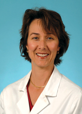
Dr. Early, professor of medicine, Washington University, St. Louis, moderated this presentation during the 2014 Digestive Diseases Week.
J. Edward Berk, M.D., D.Sc., FASGE Lecture: Endoscopic treatment of obesity in 2014
It was an honor to be chosen to deliver the 2014 J. Edward Berk Endowed Lecture. Dr. Berk was a gifted physician and teacher who made monumental contributions to medicine. He was also an insightful leader in the field of gastroenterology during a critical formative period. I am humbled to have joined distinguished past recipients of this lectureship, including Prateek Sharma, Kenneth Chang, Robert Hawes, Robert Schoen, Glenn Eisen, Douglas Rex, and Paul Fockens. I am also delighted to have been asked to speak about bariatric endoscopy.
Obesity and its associated metabolic comorbidities represent a pandemic that requires a multidisciplinary approach, to which gastroenterologists are integral. More than one third of U.S. adults are obese, accounting for over 85 million people. This contrasts with the 1.4 million Americans with inflammatory bowel disease, or the 500,000 patients that require ERCP each year, conditions for which fellows commonly seek additional years of training. Unfortunately, although nutrition is briefly covered in most gastroenterology fellowship programs, little time is devoted to obesity or the complications of bariatric surgery.
Obesity has been formally recognized as a disease by the American Medical Association and was recently the focus of the United Nations General Assembly on the Prevention and Control of Noncommunicable Diseases. Obesity is now considered the fifth leading risk for global deaths and is causally related to diabetes, ischemic heart disease, esophageal reflux, liver disease, and certain orthopedic and cancer burdens. Nevertheless, less than 2% of obese patients seek surgical treatment for a variety of reasons, including fear of complications.
Several less invasive endoscopic devices are available for the treatment of obesity, outside of the US. These devices include a variety of balloons, gastric remodeling platforms, endoluminal sleeves, and anastomotic devices. These devices focus on different mechanisms of action and offer a range of therapeutic benefits. The Food and Drug Administration has recently taken the position that risk-benefit ratio is an important concept in the treatment of obesity, allowing therapies with different risk profiles distinct therapeutic goals. As a result, many of these technologies are now being investigated in formal U.S. clinical trials.
Endoscopy currently offers solutions to many patients with complications of bariatric surgery. Additionally, there are several endoscopic devices that show promise and may be of help to this struggling patient population. This lecture focuses on these newer technologies and the current state of U.S. clinical trials.
Dr. Christopher C. Thompson, FASGE, FACG, is director of therapeutic endoscopy at Brigham and Women’s Hospital, and associate professor of medicine, Harvard Medical School, Boston. His comments were made during the ASGE and AGA joint Presidential Plenary at the annual Digestive Disease Week.
Point/Counterpoint: Is ‘resect and discard’ ready for prime time?
Yes
"Resect and discard" is a policy whereby colorectal polyps smaller than a certain size have their pathology estimated during colonoscopy by an endoscopist using imaging criteria, followed by endoscopic resection of the polyps, after which the polyps are discarded and not sent to the pathology lab. In this country, target polyps are less than 5 mm. In the U.K.: less than 9 mm.
For the target polyps in the U.S. the prevalence of villous elements and high-grade dysplasia is very low, and identification of these elements in small polyps is subject to extreme interobserver variation among pathologists (Dig. Liver Dis. 2013;45:1049-55). The prevalence of cancer in polyps less than 5 mm in size is nearly nil (Aliment. Pharmacol. Ther. 2009; 31:210-7; Gastrointest. Endosc. 2012;75:1022-30). Cancer should be endoscopically recognizable in polyps of this size by criteria such as ulceration and disruption of the surface vascular pattern.
Recommendations that CT colonographers ignore polyps less than 5 mm without reporting them (Radiology 2005;236:3-9) are accepted without controversy, while paradoxically, we argue about the wisdom of not sending diminutive polyps to pathology after their pathology has been endoscopically estimated with high confidence and the polyps have then been resected.
Adenoma detection rates (ADRs) are increasing and the cost of pathology evaluation of diminutive polyps is high. Several endoscopic modalities allow accurate estimation of pathology based on image analysis (Lancet Oncol. 2013;14:1337-47). The American Society of Gastrointestinal Endoscopy (ASGE) set a threshold of 90% agreement between postpolypectomy surveillance intervals determined by endoscopists using image analysis and intervals determined by pathology evaluation of all polyps (Gastrointest. Endosc. 2011;73:419-22) to establish resect and discard as an appropriate paradigm for clinical practice. Narrow-band imaging allows endoscopists to achieve the threshold set by ASGE (Gut 2013;62:1704-13) and only a few studies, done primarily by community-based endoscopists with limited training, have failed to meet the ASGE target (Gastroenterology 2013;144:5-8; Gut 2014;63:458-65). Cost analyses show that "resect and discard" could save more than $1 billion per year in the U.S. alone (Endoscopy 2011;43:683-91).
ASGE recommends that polyps be discarded only when pathology is interpreted with high confidence. When a diagnosis is already recognized in clinical practice with high probability, ordering a confirmatory test is considered money wasted. Money is considered well spent when the diagnosis is uncertain, and pathologic interpretation of polyps less than 5 mm in size is a good example.
"Resect and discard" will require endorsement by GI societies, re-education of patients, a credentialing process (given the recent low performance by community endoscopists), creation of training tools, and new documentation schemes based on photography for quality measures such as ADR. Documentation of ADR and medical-legal protection for endoscopists will require storage of high-quality images that record the basis for an endoscopist’s decision. Finally, institutional policies that require submission of resected tissue for pathology will need revision.
Success will depend on aligning the policy with the financial incentives of endoscopists with the introduction of a CPT code for confocal laser microscopy in the United States. Bundled payment, which includes the cost of pathology (Gastroenterology 2014;146:849-53), would create strong motivation for "resect and discard."
Dr. Douglas K. Rex, AGAF, is Distinguished Professor of Medicine, Indiana University School of Medicine, Indianapolis; Chancellor’s Professor, Indiana University–Purdue University Indianapolis; and director of endoscopy, Indiana University Hospital.
No
First, standards will be needed for a new diagnostic test. Point of care testing (POCT) is defined as medical diagnostic testing performed outside the clinical laboratory in close proximity to where the patient is receiving care. POCT is typically performed by nonlaboratory personnel and the results are used for clinical decision making. This is therefore a POCT and subject to all of the requirements of POC tests. Accreditation bodies hold departments of pathology accountable for their oversight and quality by Clinical Laboratory Improvement Amendments (CLIA) and the College of American Pathologists (CAP), while input comes from the Food and Drug Administration, Centers for Medicare & Medicaid Services, and the Centers for Disease Control and Prevention.
Second, accreditation will be required to guarantee the quality of all POC tests as well as re-accreditation. Will accreditation be for each endoscopist who will need to demonstrate diagnostic competence, or for each endoscopy unit? How will this be accomplished? At what cost? How will maintenance of competence work?
If one argues there will be no need for accreditation, there will almost certainly be opposition from CLIA, CAP, and numerous other pathologist groups. The obvious argument is that endoscopic assessment does not match up to pathology so it is a substandard test.
The notion that it can be justified as it gives an immediate diagnosis needs no further comment. There may also be opposition from gastroenterology/endoscopy groups who doubt that those who are more academic or better equipped can do this with 95%+ success or better than they or their own group can.
Are we assuming that the lower diagnostic standards of endoscopy are acceptable as they will not impact significantly on patient management? Why do we need to know precisely the pathology of polyps? Important differential diagnoses that really change follow-up include detecting the third adenoma, distinguishing adenomas from serrated polyps, hyperplastic polyps from sessile serrated polyps/adenomas (SSP/As), which can be difficult histologically, and especially dysplastic SSP/As from nondysplastic ones. Endoscopists need to demonstrate their competence at these skills for accreditation when this may not currently be possible, and identifying adenomas and SSP/As with early invasive cancer, which may not be resectable. Although uncommon, these are sometimes less than 5 mm.
Third, the issue is that it is one thing to say that a specific endoscopist can be accurate in studies based on less than 500 polyps, but when this is translated to an entire unit – after 10,000+ polyps, or a state, or the entire country. When 25 missed diagnoses become 1000, or 100,000. What gets missed that matters? Other tumors presenting as lower bowel polyps include carcinoids (low grade neuroendocrine tumors), metastatic carcinoma, metastatic melanomas, and lymphomas. While all of these are indicative of widespread disease, many are treatable.
There are intangibles at work in my argument as well. You never really know what you are dealing with when you discard the polyp. Even if accredited, are you really comfortable doing this? Would you be happy having your own polyps thrown out?
So where does this leave us? There are many issues, some of which are obvious but others less so, and there may even be some that have not yet to come to light. This summarizes the state of the art regarding the field of surveillance endoscopy and pathology. There are far too many challenges and hurdles to seriously consider implementing this policy now. It is not ready for prime time.
Dr. Robert H. Riddell is professor of laboratory medicine and pathobiology, University of Toronto, and Mount Sinai Hospital, Toronto.
Both Dr. Rex and Dr. Riddell made their comments during the ASGE and AGA joint Presidential Plenary at the annual Digestive Disease Week.
Do you think 'resect and discard' is ready for prime time? Take our Quick Poll on the GI & Hepatology News homepage.
Pancreatic cancer: progress, challenges, and hope
Over the past two decades, remarkable progress has been made in understanding the biological underpinnings of many cancers. Survival of patients with the top four leading causes of cancer deaths in the U.S., carcinomas of the lung, colon and rectum, breast, and prostate, has dramatically increased. In sharp contrast, survival in patients with pancreatic ductal adenocarcinoma (PDAC) has not changed significantly. PDAC is currently the fourth leading cause of cancer death in the U.S., but it is expected to account for more deaths than colorectal, breast, or prostate cancer by 2020, as the incidence of this cancer continues to rise and survival continues to be shockingly brief (median survival of 6 months). Nab-paclitaxel recently became the first FDA-approved drug for advanced PDAC in many years – based on an improvement of 2 months in median survival.
A better understanding of the underlying biology of PDAC is required to even consider the possibility of clinical trials yielding survival on the scale of years as opposed to months. For too long, lessons learned from the study of other cancers have been hastily applied to pancreas cancer, with lackluster results and deleterious costs to our patients. Scientists have only recently fully embraced that the biology of PDAC is unique. Under the microscope, PDAC looks different, owing to an aggressive desmoplastic response – an amalgamation of activated fibroblasts, macrophages and other inflammatory cells admixed with profuse scar tissue. Moreover, pancreas tumors are uniquely hypovascular.
Cancer cells that persist in this cauldron of hypoxia and acidity have likely evolved survival mechanisms that are particular to this disease and produce cells that are particularly resistant to therapy. One such survival mechanism is the ability to seed distant organs; this may occur surprisingly early in the course of PDAC. Furthermore, with limited blood flow within tumors, impaired delivery of systemic chemotherapy is a unique and pressing issue in PDAC.
Despite past failures, our field has recalibrated and rebooted, and we are now poised to make serious inroads against this disease. Scientists and clinicians have recently developed novel tools that will enable new discoveries that will translate to the clinic. Novel, genetically engineered mouse models have catalyzed our understanding of PDAC while also serving as a more faithful model for testing novel drugs. Vogelstein and colleagues at Johns Hopkins utilized advanced sequencing technologies to define the genomic landscape of PDAC, enabling many to identify novel drug targets and develop new assays for early diagnosis utilizing state-of-the-art microfluidic technology. Others have begun to tackle the drug delivery problem in PDAC with nanoparticle vehicles and other targeted modalities. Finally, the manipulation and engineering of the patient\'s immune system to target nascent PDAC cells is an area of intense investigation and represents perhaps one of the most promising therapeutic strategies in our armamentarium.
After many years in the dark, we are poised to enter a new age – an Age of Enlightenment for pancreatic cancer. There is no more promising time for pancreatic cancer research than the present, as we now have a plethora of new tools to understand this disease. And with better understanding, we will undoubtedly be in a better position to deliver to our patients what they sorely need most: hope for survival on the scale of years, as opposed to months.
Dr. Rhim is assistant professor of internal medicine and assistant director for translational research, division of gastroenterology, department of internal medicine, University of Michigan, Ann Arbor. His comments were made during the ASGE and AGA joint Presidential Plenary at the annual Digestive Disease Week.
Have we reached the top for therapy for IBD?
Medical therapy for the treatment of patients with inflammatory bowel disease has clearly improved the lives of patients and improved outcomes, but does not benefit all patients. Since the initial introduction of corticosteroids and mesalamine derivatives in the 1950s, medical therapy for IBD has improved significantly.
With the widespread use of antimetabolite therapy and the introduction of biologics in 1998 – infliximab, adalimumab, golimumab, certolizumab pegol, natalizumab, and finally vedolizumab – we can induce remission in steroid-dependent and steroid-refractory patients and decrease rates of surgery as well.
The goals of medical therapy have also evolved as the potency of the agents to treat patients with IBD has improved. Initially, symptom control was the desired goal of medical therapy, but soon it was recognized that this was inadequate and mucosal healing became the desired target. Now we recognize that mucosal healing is important while achieving other important endpoints – reduced hospitalization, reduction in surgery, reduced social and occupational burden, and finally doing so with improved safety and tolerability of medications.
Historically, we have used a step-up approach starting with the least effective agents and "stepping up" to more potent therapies. In this model, therapy is stepped up according to severity at presentation or failure at the prior step. We now have the ability to "profile" certain patients and treat those who will have aggressive disease with early aggressive therapy. In Crohn’s disease in particular, we are able to predict which patients will have a more virulent disease course.
Highlights of specific examples from clinical trials of current medical therapies make it very clear that there is a large unmet need for medical treatment of patients with IBD. The data presented focus on well recognized clinical studies from trials in patients with ulcerative colitis; several of these trials led to Food and Drug Administration approval.
A recent study evaluated the addition of oral mesalamine to topical mesalamine in patients with extensive ulcerative colitis, which achieved remission in 44% of patients; leaving 56% of patients with active disease.
Similarly, there were two studies evaluating patients with ulcerative colitis who received budesonide MMX (CORE I and II studies). In these studies, the rates of remission with steroid withdrawal were 17.4% and 17.9%; thus, over 80% of patients did not achieve remission.
In a recent meta-analysis the mean efficacy for azathioprine in ulcerative colitis patients was 60%. In a study using dual therapy of infliximab and azathioprine (SUCCESS trial) monotherapy with azathioprine was able to achieve remission in 24% of patients, infliximab in 22% and combination therapy in only 40% of patients; thus leaving 60% of patients not in remission.
In patients with Crohn’s disease we have similar limitations with current medical therapy. Although azathioprine has been widely used – it has recently been demonstrated to be inadequate as monotherapy for treatment of early Crohn’s disease. The rates of remission are inadequate in patients receiving infliximab (ACCENT I trial) and adalimumab (CHARM Trial) with approximately one-quarter to one-third of patients at most achieving remission. In the SONIC trial, when combination therapy with infliximab and azathioprine was used, remission with steroid-free status was achieved in 57% of patients at week 26 compared with 30% and 45%, respectively, in the azathioprine and infliximab monotherapy treatment arms.
Early aggressive therapy has been demonstrated to achieve higher rates of mucosal healing. We now recognize that it is appropriate to treat early disease, but not in all patients.
We attempt to prognosticate and treat those patients who have a poor prognosis with early aggressive therapy – specifically those patients who have fistulas, early steroid users, smo-ers, those with severe endoscopic lesions, those younger than 40, and those with extensive disease. We attempt to use combination therapy (anti-TNFs and immunomodulators) when appropriate given the added benefits: specifically, higher drug levels, fewer acute side effects, fewer infusion reactions, fewer serious infections, and also better efficacy.
Our efforts to optimize the use of treatment agents involve monitoring for antidrug antibodies and if present, switching the patient to different agents; we also monitor drug levels and when low we can increase the frequency of administration or escalate the drug dose.
We recognize that patients who have higher therapeutic trough levels within the therapeutic window are more likely to achieve clinical remission.
Several factors influence anti-TNF pharmacokinetics, including the presence of antidrug antibodies, concomitant use of immunosuppressives, low serum albumin, high baseline CRP concentration, large body size, and male gender.
Medical therapy has lessened the need for patients to undergo bowel surgery.
A recent Danish population cohort study evaluated 48,967 patients with IBD (CD, 13,185 and UC, 35,782) during 1979-2011. The 5-year cumulative probability of the first major surgery decreased from 44.7% in the early cohort (1979-1986) to 19.6% in the later cohort (2003-2011; P less than .001) for Crohn’s disease, and from 11.7% in early cohort to 7.5% in later cohort (P less than .001) for ulcerative colitis.
There are numerous drugs in development with different mechanisms of action for patients with both ulcerative colitis and Crohn’s disease. Too many IBD patients still face surgery, although there has been improvement. We are learning how to better use the medications we have and optimize therapy.
Dr. Lichtenstein is professor of medicine, department of medicine, division of gastroenterology, at the University of Pennsylvania, Philadelphia, and director of the IBD program at the university. His comments were made during the ASGE and AGA joint Presidential Plenary at the annual Digestive Disease Week.
Liver Cancer Without Cirrhosis Surprisingly Common: Is NAFLD the Cause?
CHICAGO – A full 13% of patients diagnosed with hepatocellular carcinoma did not have underlying cirrhosis in a national sample of 1,500 veterans.
Although hepatocellular carcinoma (HCC) in patients with cirrhosis typically arises against a background of alcohol abuse or hepatitis C virus infection, this entity was strongly associated with nonalcoholic fatty liver disease (NAFLD) and idiopathic HCC.
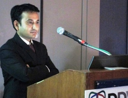
"While this represents a small proportion, this poses a logistical problem for HCC surveillance, given the large population with NAFLD and no real identifying features of who is going to develop HCC," Dr. Sahil Mittal said at the annual Digestive Disease Week.
Despite a threefold increase in HCC in the United States over the last 3 decades and emerging evidence for NAFLD presenting in the absence of cirrhosis, this is the first study to assess the prevalence and risk factors for HCC without cirrhosis in a national sample of HCC patients, he said.
The investigators, led by Dr. Hashem B. El-Serag, section chief, gastroenterology and hepatology, Baylor College of Medicine, Houston, performed a chart review of 1,500 veterans with a confirmed diagnosis of HCC randomly selected between 2005 and 2011. Of these, cirrhosis was absent in 43 (3%), highly improbable in 151 (10%), and definite in 1,201 (80%), with the diagnosis unclear because of insufficient data in 105 (7%).
As expected, HCC patients with cirrhosis were significantly more likely than those with no or probable no cirrhosis to abuse alcohol (84% vs. 67.4% vs. 63.6%; P less than .01) and to have hepatitis C infection (72.2% vs. 42% vs. 44.4%; P less than .01), said Dr. Mittal, also with Baylor’s College of Medicine.
Hepatitis B infection was similar in all three groups (5% vs. 4.7% vs. 2%).
In contrast, NAFLD was more common in patients with no or probably no cirrhosis than in those with cirrhosis (14% vs. 20.5% vs. 5.8%; P less than .01), as was idiopathic HCC (18.6% vs. 27.8% vs. 8.2%; P less than .01), he said.
"NAFLD is associated with a significantly increased risk of HCC in the absence of cirrhosis, compared with hepatitis C or alcohol," he said.
With regard to clinical factors, sex and race did not differ between groups. However, the no and probably no cirrhosis patients were significantly older than were those with HCC accompanied by cirrhosis (65.5 years, vs. 69.7 years vs. 62.6 years) and significantly more likely to have comorbidities associated with metabolic syndrome, such as hypertension (88.4% vs. 86.8% vs. 72.6%), myocardial infarction (11.6% vs. 18.5% vs. 7.5%), and peripheral vascular disease (11.6% vs. 20% vs. 9.5%), Dr. Mittal reported. The differences among groups were statistically significant with P values of less than .01.
The investigators then performed logistic regression analysis to examine predictors of HCC without cirrhosis. After adjustment for confounders, they found NAFLD was more than threefold likely (odds ratio, 3.1) and idiopathic HCC more than twofold likely (OR, 2.8) in patients with HCC without cirrhosis, compared with patients with hepatitis C–related HCC.
This entity of HCC in the absence of cirrhosis was also associated with the metabolic syndrome–related comorbidities of hypertension (OR, 1.8) and myocardial infarction (OR, 1.8).
The findings suggest that the risk of HCC in patients with NAFLD is increased not only because of progression to cirrhosis, but possibly through other alternative noncirrhosis pathways, Dr. Mittal said in an interview. This has important implications for understanding the pathogenesis of HCC, as well as for HCC screening in NAFLD patients.
"Due to the high prevalence of NAFLD in the general population, conventional screening by ultrasonography may not be a feasible strategy, especially if one cannot rely on cirrhosis as the main predisposing lesion to HCC," he said. "Studies are needed to identify risk factors and biomarkers that can identify NAFLD patients at higher risk of developing HCC. Future research is also needed to investigate the role of chemoprevention."
Dr. Mittal reported no conflicting interests. Lead author Dr. El-Serag reported consulting fees and grant/research support from Gilead Sciences.
AT DDW 2014

