User login
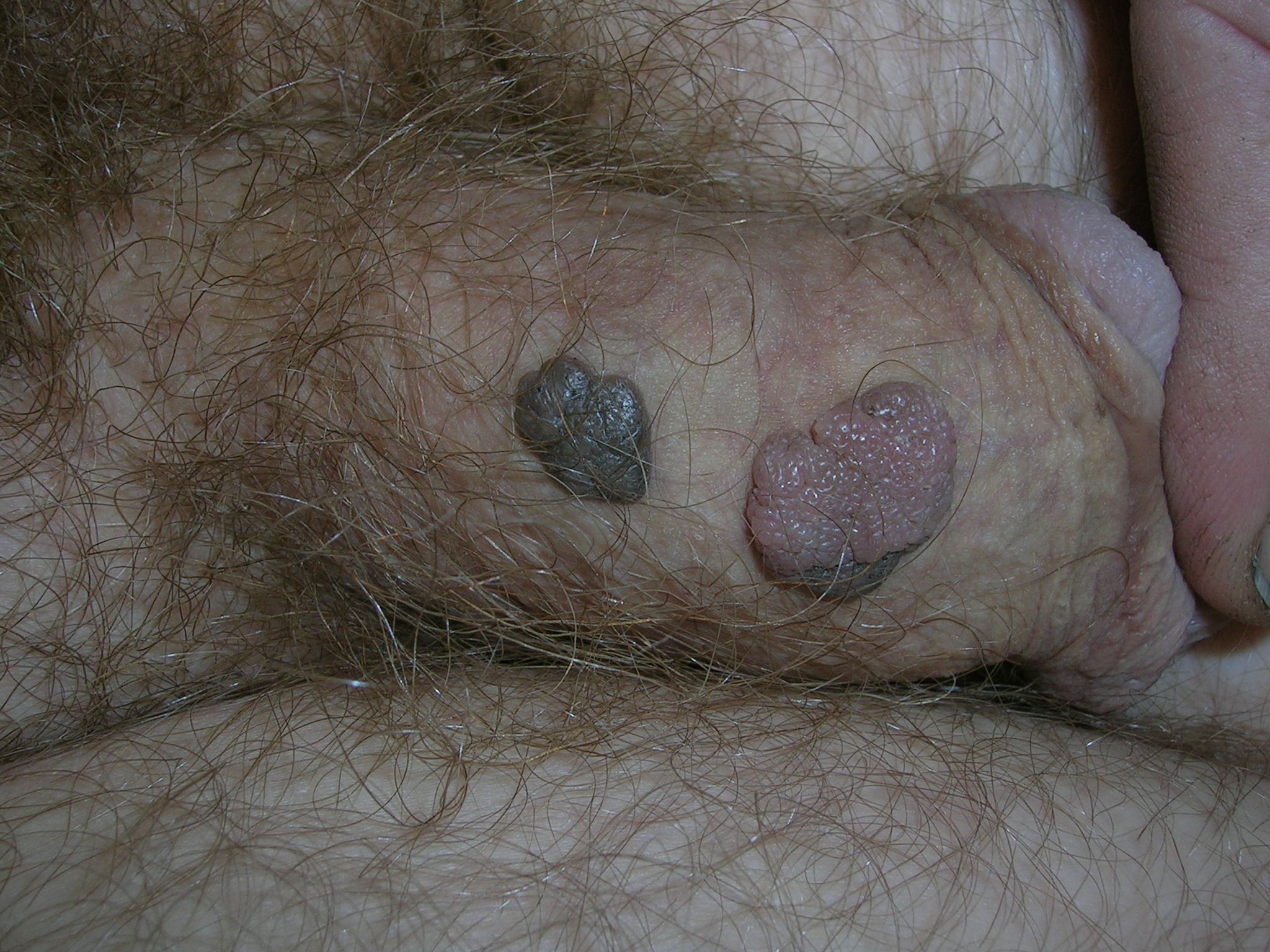
The Diagnosis
A shave biopsy of the pink lesion was positive for condylomata acuminata. (The differential diagnosis also included seborrheic keratoses.) The patient consented to blood tests for human immunodeficiency virus and syphilis; he was negative for both. The benefits and costs of urine screening for Chlamydia trachomatis and Neisseria gonorrhea were discussed and the patient decided that he would forego that test.
Condyloma acuminata is caused by human papillomavirus (HPV) infection and is the most common STD in the United States. HPV types 6 and 11 account for most external genital warts and are rarely associated with invasive carcinoma of the external genitalia. Most infections are transient and clear within 2 years, but many infections persist.
The shave biopsy that was done for diagnostic purposes also served as partial treatment. The remaining lesion was treated successfully with 2 courses of cryotherapy. Other treatment choices include topical imiquimod and podofilox.
Photos and text for Photo Rounds Friday courtesy of Richard P. Usatine, MD. This case was adapted from: Mayeaux EJ. Usatine R. Genital warts. In: Usatine R, Smith M, Mayeaux EJ, Chumley H, Tysinger J, eds. The Color Atlas of Family Medicine. New York, NY: McGraw-Hill; 2009:530-534.
To learn more about The Color Atlas of Family Medicine, see:
* http://www.amazon.com/Color-Atlas-Family-Medicine/dp/0071474641