User login
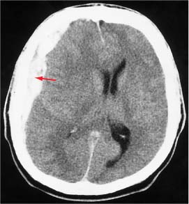
The CT scan revealed an acute subdural hematoma. The patient was hospitalized, and a neurosurgeon was consulted for surgical management.
Subdural hematomas occur at all ages. Mortality rates in treated older adults are approximately 8% for patients <65 years and 33% for patients >65 years. Most subdural hematomas are caused by trauma from a direct injury to the head or shaking injury in an infant. Motion of the brain within the skull causes a shearing force to the cortical surface and interhemispheric bridging veins. This force tears the weakest bridging veins as they cross the subdural space, resulting in an acute subdural hematoma.
Most subdural hematomas are managed surgically, and there is little evidence on conservative management. One should obtain an urgent noncontrast CT scan on any patient suspected of having a subdural hematoma. If the noncontrast CT scan is nonrevealing, obtain a contrast CT or magnetic resonance imaging scan, particularly if the traumatic event occurred 2 to 3 days earlier. Emergently refer patients with a subdural hematoma and deteriorating neurologic status or evidence of brain edema or midline shift to a hospital with neurosurgeons.
Photo courtesy of Kasper DL, Braunwald E, Fauci, AS, et al. Text for Photo Rounds Friday courtesy of Richard P. Usatine, MD. This case was adapted from: Chumley H. Subdural hematoma. In: Usatine R, Smith M, Mayeaux EJ, et al, eds. The Color Atlas of Family Medicine. New York, NY: McGraw-Hill; 2009:972-975.
To learn more about The Color Atlas of Family Medicine, see:
• http://www.amazon.com/Color-Atlas-Family-Medicine/dp/0071474641
You can now get The Color Atlas of Family Medicine as an app for mobile devices including the iPhone, iPad, and all Android devices by clicking this link: