User login
BOSTON – With art supplies and “mystery” sutures, a team of investigators has determined that immediate intraoperative inking of lumpectomy specimens appears to be a better method than suture placement for orienting specimens for pathology, a finding that could reduce re-excisions.
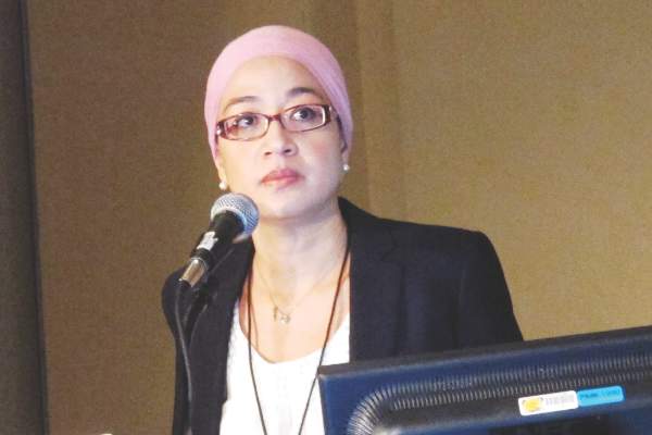
“Intraoperative specimen suturing appears to be inaccurate for margin identification, and additional means by the surgeon to improve the accuracy are needed. This could be either immediate inking or routine excision of shave margins, either at the original surgery or when you go back for re-excision, to take all the margins instead of one specific margin,” Dr. Angel Arnaout, a breast surgeon at the University of Ottawa, Ont., said at the annual Society of Surgical Oncology Cancer Symposium.
She presented results of the randomized, blinded SMART (Specimen Margin Assessment Technique) trial comparing suture placement with intraoperative inking within the same surgical specimen.
Positive resection margins during breast-conserving surgery are associated with a twofold increase in the risk of ipsilateral tumor recurrence, and require re-excision or conversion to mastectomy. Re-excisions rates range from 25% to 45%, but in the majority of cases – 53% to 65% – no additional disease is found, Dr. Arnaout said.
In addition to adding to patient distress, re-excisions diminish cosmetic results because they take additional tissue, and delay the start of adjuvant breast cancer therapy, she noted.
Surgeons typically orient specimens for pathologists by placing two or three sutures in the center of the margin surfaces or, less frequently, by intraoperative marking of the margins with ink. But in transit from the OR to the pathology lab, specimens often flatten or “pancake” under their own weight, making it challenging for the pathologist to identify the originally marked margins.
To test whether suturing or inking is better at identifying the extent of tumors during breast-conserving surgery, the investigators devised a strategy using benign breast tissue removed during prophylactic mastectomy or reduction mammoplasty.
The surgeons first performed a sham lumpectomy within the tissue. They then painted the specimen margins in the OR using a phospholuminescent paint obtained from an art supply store. The paint dries clear and colorless, but glows in different colors under black light. By using the paint, carefully selected so as not to interfere with pathology staining, the investigators were able to blind the pathologists to the actual orientation of the margins.
While the tissue was still in the OR, the surgeons then placed two or three orienting sutures in customary locations, plus a third, “mystery” suture at a location known only to the surgeon.
In the pathology lab, the assistant was asked to look at the sutures and outline in black ink the edges of the margins using the sutures for guidance.
The specimen was then examined under black light to determine the degree of discordance between surgeons and pathologist for identification of margins using the suture and painting methods (primary outcome), and to evaluate the discrepancy in the extent of margin surface areas as identified by the surgeons and the pathologists (secondary outcome).
“We asked breast surgeons ‘what would be a clinically significant discordance rate for you?’ and most of them said between 10% and 20%, so we looked for a 15% discordance rate as being clinically significant between the two margin assessment techniques,” Dr. Arnaout said.
They found that the overall discordance in the location of the mystery suture between surgeons and pathologists was similar whether the samples contained two or three location sutures, with discordance rates of 46% (34 of 75 samples) for two sutures, and 47% (41 of 88 samples) for three sutures.
For the secondary outcome of discordance in the extent of margins, they found that pathologists tended to estimate the mean surface of the anterior and posterior margins to be significantly larger than the surgeons did (P less than .01), while significantly underestimating the superior, inferior, and lateral margins (P less than .01 for superior and inferior, and P = .04 for lateral margins).
Examination under black light showed that often two or three additional margins would be included by the surgeon within what the pathologist had identified as the anterior margin.
“This has implications for the surgeon and for the patient,” Dr. Arnaout said. “For most of us that do lumpectomies that go from skin down to chest wall, if an anterior margin or posterior margin is positive, a lot of times we would fight not to go back because we would say there is no further room for re-excision.”
But if the tumor is within what is actually the superior margin but labeled by the pathologist as an anterior margin, the patient may miss an opportunity for successful re-excision, she explained.
The study was limited by the fact that the sham lumpectomy specimens did not contain skin or muscle, which would have allowed for more accurate margin orientation in an actual operative setting; by lack of compression of specimens with small nonpalpable lesions in containers, which further distorts specimens; and by the use of smaller lumpectomy specimens than normally obtained during cancer surgery, which could have resulted in overestimation of the discordance that might occur when larger specimens are taken, Dr. Arnaout said.
“The conclusion of our study is that specimen margin orientation really should be defined by the surgeon who knows the original shape and orientation of the tissue during surgery, and not to rely on the pathology to reorient based on some sutures placed in the centers of the specimens without defining the extent of the surface area of each of the margins,” she said.
The Canadian Cancer Society Research Institute and the Canadian Surgical Research Fund supported the study. Dr. Arnaout and coauthors reported no conflicts of interest.
FROM SSO 2016
Key clinical point: Intraoperative suture placement is an inaccurate method for orienting breast tumor specimens for pathology.
Major finding: Discordance in identifying tumor margin surface between surgeons and pathologists was similar whether the samples contained two or three location sutures, with discordance rates of 46% (34 of 75 samples) for two sutures, and 47% (41 of 88 samples) for three sutures.
Data source: Randomized clinical trial of 163 specimens obtained from 49 patients.
Disclosures: The Canadian Cancer Society Research Institute and the Canadian Surgical Research Fund supported the study. Dr. Arnaout and coauthors reported no conflicts of interest.

