User login

| 
|
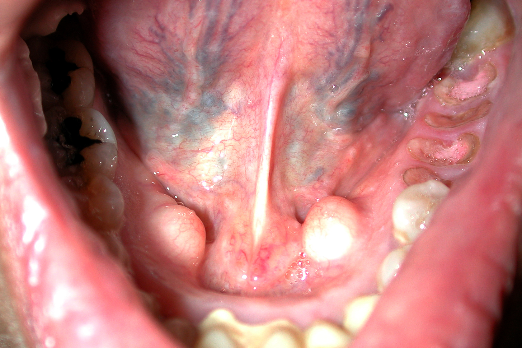
The diagnosis
The mass in the patient’s mouth was a torus palatinus (FIGURE 1) and nothing needed to be done about it.
A torus palatinus is the most common bony maxillofacial exostosis. It is usually seen in adults over 30 years of age. It is more common in women than in men. This benign bony exostosis occurs in the midline of the hard palate. It is often noticed incidentally as a hard lump protruding from the hard palate into the mouth covered with normal mucous membrane. It is important not to miss a squamous cell carcinoma in this area, but an SCC is not as hard and the mucous membranes are usually ulcerated.
Excision can be considered if the lesion interferes with function such as the fit of dentures. A variation of this is the torus mandibularis (FIGURE 2).
Photos and text for Photo Rounds Friday courtesy of Richard P. Usatine, MD. This case was adapted from: French L. Torus palatinus. In: Usatine R, Smith M, Mayeaux EJ, Chumley H, Tysinger J, eds. The Color Atlas of Family Medicine. New York, NY: McGraw-Hill; 2009:148-149.
To learn more about The Color Atlas of Family Medicine, see:
* http://www.amazon.com/Color-Atlas-Family-Medicine/dp/0071474641
|
|
|
The diagnosis
The mass in the patient’s mouth was a torus palatinus (FIGURE 1) and nothing needed to be done about it.
A torus palatinus is the most common bony maxillofacial exostosis. It is usually seen in adults over 30 years of age. It is more common in women than in men. This benign bony exostosis occurs in the midline of the hard palate. It is often noticed incidentally as a hard lump protruding from the hard palate into the mouth covered with normal mucous membrane. It is important not to miss a squamous cell carcinoma in this area, but an SCC is not as hard and the mucous membranes are usually ulcerated.
Excision can be considered if the lesion interferes with function such as the fit of dentures. A variation of this is the torus mandibularis (FIGURE 2).
Photos and text for Photo Rounds Friday courtesy of Richard P. Usatine, MD. This case was adapted from: French L. Torus palatinus. In: Usatine R, Smith M, Mayeaux EJ, Chumley H, Tysinger J, eds. The Color Atlas of Family Medicine. New York, NY: McGraw-Hill; 2009:148-149.
To learn more about The Color Atlas of Family Medicine, see:
* http://www.amazon.com/Color-Atlas-Family-Medicine/dp/0071474641