User login
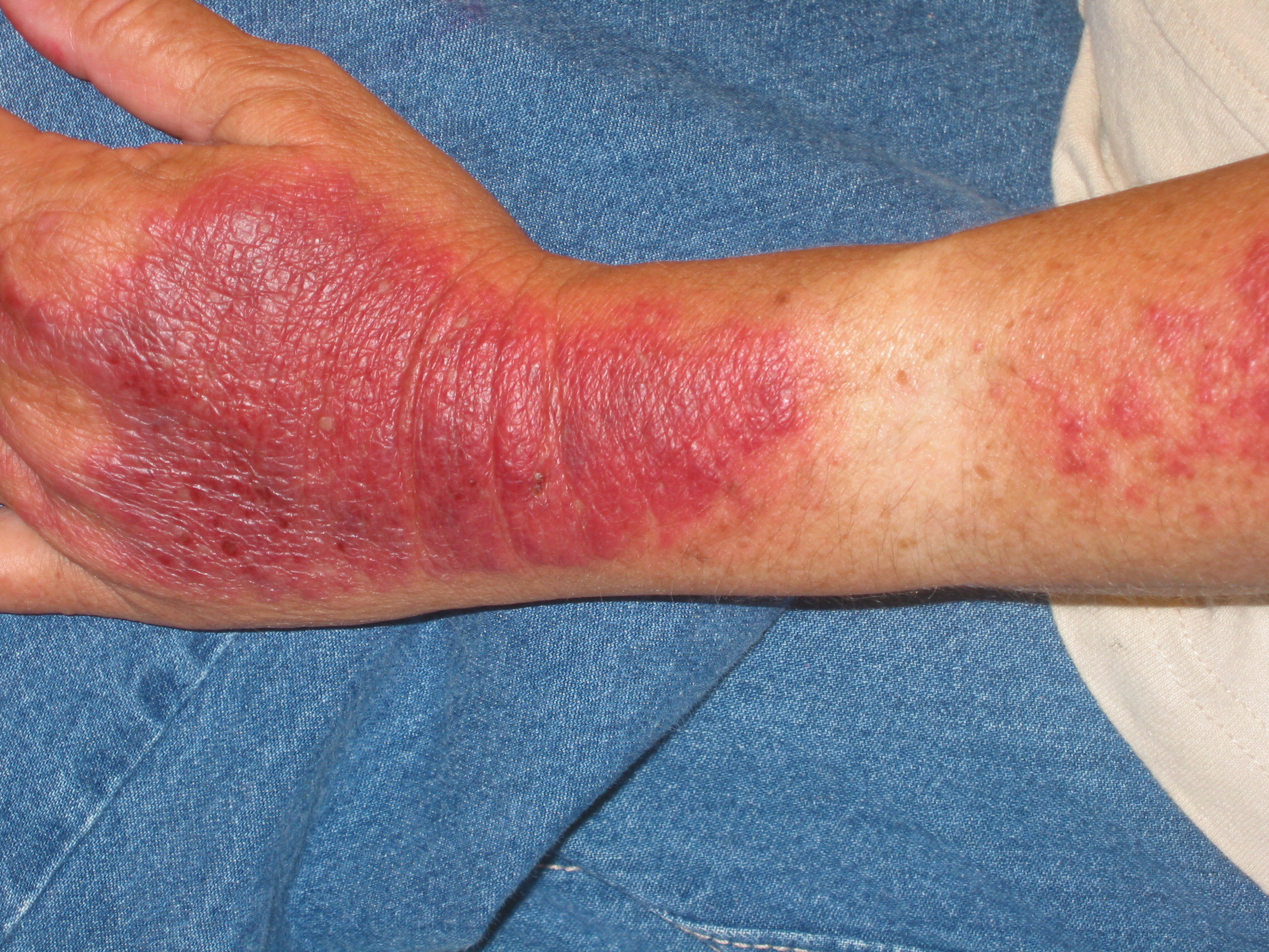
The family physician (FP) made the clinical diagnosis of polymorphous light eruption (PMLE). The sparing of the patient’s watch area made it clear that the plaques were photodistributed. PMLE may affect up to 10% of the population with a predilection for females. There are 3 common types of photodermatitis: PMLE, phototoxic eruption, and photoallergic eruption.
PMLE is an idiopathic, delayed type hypersensitivity reaction to ultraviolet A light and, to a lesser extent, ultraviolet B light. PMLE is the most common photo eruption encountered in clinical practice. The rash develops within hours to days of exposure to sunlight and lasts for several days to a week.
There is a broad range of photosensitivity with PMLE. Extremely sensitive individuals can tolerate only minutes of exposure, whereas others require prolonged exposure to sunlight before developing a reaction. PMLE is a recurrent condition that persists for many years in most patients.
The appearance of PMLE varies from person to person. Erythematous pruritic papules, sometimes with vesicles, are most common. Lesions may coalesce to form plaques. The rash typically involves the V of the neck and the arms, the legs, or both. The face tends to be spared. It tends to present in spring/summer, with the first significant UV exposure of the year.
The management of PMLE is aimed at prevention. Patients who have mild disease should avoid sun exposure, wear tightly woven clothing and hats, and use broad-spectrum sunscreens with a sun-protection factor of 50 or higher. Patients with severe PMLE can be desensitized in the spring with the use of phototherapy, and maintained in the nonreactive state with weekly one hour unprotected exposure to sunlight.
In this case, the FP told the patient to avoid sun exposure and started her on oral antihistamines and topical steroids to treat her symptoms.
Text for Photo Rounds Friday courtesy of Richard P. Usatine, MD. Photo courtesy of Chris Wenner, MD. This case was adapted from: Mayeaux EJ, Wenner C. Photodermatitis. In: Usatine R, Smith M, Mayeaux EJ, et al, eds. The Color Atlas of Family Medicine. New York, NY: McGraw-Hill; 2009:853-857.
To learn more about The Color Atlas of Family Medicine, see:
• http://www.amazon.com/Color-Atlas-Family-Medicine/dp/0071474641
You can now get The Color Atlas of Family Medicine as an app for mobile devices including the iPhone and iPad by clicking this link: