User login
| 
|
The family physician called his dermatology consultant and they agreed to start the patient on oral prednisone and to perform a punch biopsy, which revealed the diagnosis: pemphigus foliaceous.
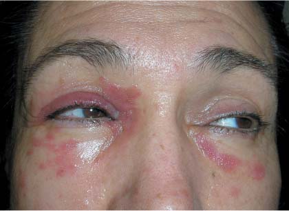
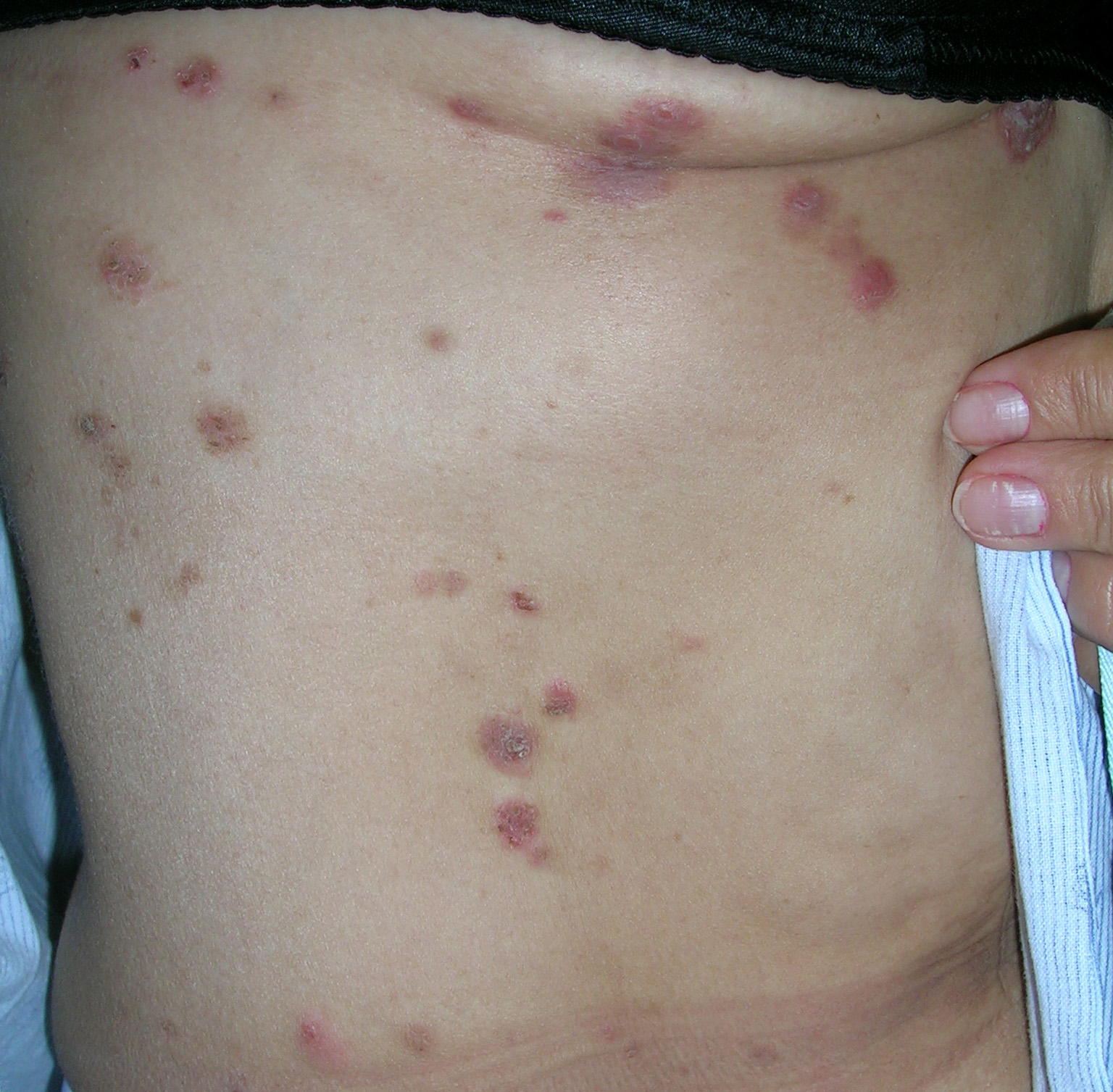
Pemphigus foliaceous causes multiple red, scaling, crusted, and pruritic lesions that look like “corn flakes.” Shallow erosions arise when crusts are removed; blisters are rare, as the disease is superficial.
Pemphigus foliaceous initially affects the face (FIGURE 1) and scalp and may progress to involve the chest (FIGURE 2) and back. Skin biopsy is essential for an accurate diagnosis. The depth of acantholysis and site of deposition of antibody complexes help differentiate pemphigus from other bullous diseases. Two biopsies are preferred but even a single biopsy can be used to make the diagnosis. The first biopsy should be sent in formalin for routine histopathology. This biopsy should be of the freshest lesion, with an intact bulla, if possible. The second biopsy should be taken from perilesional normal skin, and should be sent on a gauze pad soaked in normal saline or Michel’s solution for direct immunofluorescence (DIF). Routine histopathology demonstrates suprabasal acantholysis and DIF shows antibody deposition in the intercellular spaces of the epidermis.
Treatment of pemphigus should be undertaken in consultation with a dermatologist. In this case, the skin lesions were treated with topical high-potency steroids (clobetasol ointment). The facial lesions were treated with topical triamcinolone ointment.
Oral prednisone should be prescribed as soon as the diagnosis is suspected. The first priority of treatment should be disease control and remission, followed by disease suppression.
Photos and text for Photo Rounds Friday courtesy of Richard P. Usatine, MD. This case was adapted from: Mittal S. Pemphigus. In: Usatine R, Smith M, Mayeaux EJ, et al, eds. The Color Atlas of Family Medicine. New York, NY: McGraw-Hill; 2009:794-798.
To learn more about The Color Atlas of Family Medicine, see:
• http://www.amazon.com/Color-Atlas-Family-Medicine/dp/0071474641
The Color Atlas of Family Medicine is also available as an app for mobile devices. See
|
|
|
The family physician called his dermatology consultant and they agreed to start the patient on oral prednisone and to perform a punch biopsy, which revealed the diagnosis: pemphigus foliaceous.
Pemphigus foliaceous causes multiple red, scaling, crusted, and pruritic lesions that look like “corn flakes.” Shallow erosions arise when crusts are removed; blisters are rare, as the disease is superficial.
Pemphigus foliaceous initially affects the face (FIGURE 1) and scalp and may progress to involve the chest (FIGURE 2) and back. Skin biopsy is essential for an accurate diagnosis. The depth of acantholysis and site of deposition of antibody complexes help differentiate pemphigus from other bullous diseases. Two biopsies are preferred but even a single biopsy can be used to make the diagnosis. The first biopsy should be sent in formalin for routine histopathology. This biopsy should be of the freshest lesion, with an intact bulla, if possible. The second biopsy should be taken from perilesional normal skin, and should be sent on a gauze pad soaked in normal saline or Michel’s solution for direct immunofluorescence (DIF). Routine histopathology demonstrates suprabasal acantholysis and DIF shows antibody deposition in the intercellular spaces of the epidermis.
Treatment of pemphigus should be undertaken in consultation with a dermatologist. In this case, the skin lesions were treated with topical high-potency steroids (clobetasol ointment). The facial lesions were treated with topical triamcinolone ointment.
Oral prednisone should be prescribed as soon as the diagnosis is suspected. The first priority of treatment should be disease control and remission, followed by disease suppression.
Photos and text for Photo Rounds Friday courtesy of Richard P. Usatine, MD. This case was adapted from: Mittal S. Pemphigus. In: Usatine R, Smith M, Mayeaux EJ, et al, eds. The Color Atlas of Family Medicine. New York, NY: McGraw-Hill; 2009:794-798.
To learn more about The Color Atlas of Family Medicine, see:
• http://www.amazon.com/Color-Atlas-Family-Medicine/dp/0071474641
The Color Atlas of Family Medicine is also available as an app for mobile devices. See