User login
|
|
|
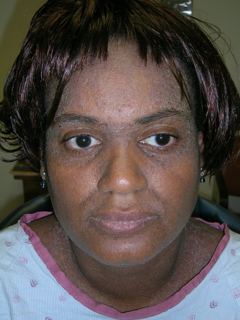
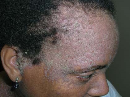
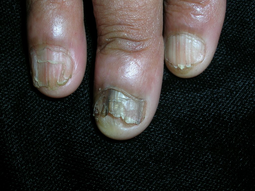
This patient was given a diagnosis of Darier’s disease, an autosomal dominant genodermatosis also known as keratosis follicularis. It starts with greasy, hyperkeratotic, yellowish-brown papules in a seborrheic distribution (FIGURE 1). (FIGURE 2 shows the papules on the patient’s scalp, which were visible after she removed her wig.) The feet may be covered with hyperkeratotic plaques, palms may have pits or keratotic papules, and the nails may have V-shaped nicking (FIGURE 3) and alternating longitudinal red and white bands. The keratotic papules can be intensely malodorous.
In early, mild, or partially treated disease, only the ears and postauricular areas may be affected. Skin biopsy reveals the characteristic histopathology. A test for the ATP2A2 gene mutation can be performed. The odor that accompanies the disease, as well as the facial involvement, often adversely affect the patient’s quality of life; thus, treatment is often warranted. Mild-to-moderate disease can be treated by avoiding exacerbating factors (sunlight, heat, and occlusion) and with topical medications; severe disease is best treated with oral retinoids.
Darier’s disease is so rare that there are no randomized controlled trials to guide treatment. Topical retinoids (adapalene, tretinoin, or tazarotene) are effective in some patients, but their main limitation is irritation. Topical corticosteroids may help. Lower-potency topical corticosteroids should be used on the face, groin, and axillae to minimize adverse effects in these areas. Systemic retinoids (acitretin or isotretinoin) are the most potent treatment. Patients on systemic retinoids require close monitoring and careful selection, as these agents are teratogenic (category X) and can cause hyperlipidemia, hypertriglyceridemia, mucous membrane dryness, alopecia, hepatotoxicity, and possibly mood disturbances. Cyclosporine can be used for acute flares. Laser, radiation, photodynamic therapy, and gene therapy are newer treatment modalities that are being investigated.
This patient was postmenopausal, so the physician was willing to treat her with acitretin. Acitretin is very expensive, and the patient’s health plan did not cover it. She was able to get it through a patient assistance program and benefitted from using it.
Photos and text for Photo Rounds Friday courtesy of Richard P. Usatine, MD. This case was adapted from: Babcock M. Genodermatosis. In: Usatine R, Smith M, Mayeaux EJ, et al, eds. The Color Atlas of Family Medicine. New York, NY: McGraw-Hill; 2009:881-885.
To learn more about The Color Atlas of Family Medicine, see:
• http://www.amazon.com/Color-Atlas-Family-Medicine/dp/0071474641
You can now get The Color Atlas of Family Medicine as an app for mobile devices including the iPhone and iPad by clicking this link:
|
|
|
|
|
|
This patient was given a diagnosis of Darier’s disease, an autosomal dominant genodermatosis also known as keratosis follicularis. It starts with greasy, hyperkeratotic, yellowish-brown papules in a seborrheic distribution (FIGURE 1). (FIGURE 2 shows the papules on the patient’s scalp, which were visible after she removed her wig.) The feet may be covered with hyperkeratotic plaques, palms may have pits or keratotic papules, and the nails may have V-shaped nicking (FIGURE 3) and alternating longitudinal red and white bands. The keratotic papules can be intensely malodorous.
In early, mild, or partially treated disease, only the ears and postauricular areas may be affected. Skin biopsy reveals the characteristic histopathology. A test for the ATP2A2 gene mutation can be performed. The odor that accompanies the disease, as well as the facial involvement, often adversely affect the patient’s quality of life; thus, treatment is often warranted. Mild-to-moderate disease can be treated by avoiding exacerbating factors (sunlight, heat, and occlusion) and with topical medications; severe disease is best treated with oral retinoids.
Darier’s disease is so rare that there are no randomized controlled trials to guide treatment. Topical retinoids (adapalene, tretinoin, or tazarotene) are effective in some patients, but their main limitation is irritation. Topical corticosteroids may help. Lower-potency topical corticosteroids should be used on the face, groin, and axillae to minimize adverse effects in these areas. Systemic retinoids (acitretin or isotretinoin) are the most potent treatment. Patients on systemic retinoids require close monitoring and careful selection, as these agents are teratogenic (category X) and can cause hyperlipidemia, hypertriglyceridemia, mucous membrane dryness, alopecia, hepatotoxicity, and possibly mood disturbances. Cyclosporine can be used for acute flares. Laser, radiation, photodynamic therapy, and gene therapy are newer treatment modalities that are being investigated.
This patient was postmenopausal, so the physician was willing to treat her with acitretin. Acitretin is very expensive, and the patient’s health plan did not cover it. She was able to get it through a patient assistance program and benefitted from using it.
Photos and text for Photo Rounds Friday courtesy of Richard P. Usatine, MD. This case was adapted from: Babcock M. Genodermatosis. In: Usatine R, Smith M, Mayeaux EJ, et al, eds. The Color Atlas of Family Medicine. New York, NY: McGraw-Hill; 2009:881-885.
To learn more about The Color Atlas of Family Medicine, see:
• http://www.amazon.com/Color-Atlas-Family-Medicine/dp/0071474641
You can now get The Color Atlas of Family Medicine as an app for mobile devices including the iPhone and iPad by clicking this link: