User login
SAN DIEGO – Researchers have taken a major step toward eliminating the need for routine pathology on every polyp removed during colonoscopy.
Use of a high-definition, dual-focus colonoscope allowed endoscopists to perform "significantly more accurate, high-confidence endoscopic diagnosis of diminutive polyps," Dr. Tonya Kaltenbach said at the annual Digestive Disease Week.
"In many hospitals, about 20%-25% of all pathology samples are small colon polyps, almost all of which are benign. This is a lot of money that can be saved," said Dr. Kaltenbach in a written statement. About 80% of all polyps found during screening colonoscopy are diminutive, 5 mm in diameter or less, and the current standard of care is to do a pathology analysis on each of these polyps.
As used by five gastroenterologists who participated in the study at three U.S. centers to assess 530 polyps in 277 people, the high-definition scope produced a 95.9% agreement in polyp assignment for follow-up surveillance, compared with pathology-laboratory analysis of the same polyps, and a 96.4% negative predictive value for adenomas, said Dr. Kaltenbach, a gastroenterologist at the VA Medical Center in Palo Alto, Calif.
These performance levels surpassed the greater than 90% targets for colonoscopic assessment of diminutive polyps set last year by a committee organized by the American Society of Gastrointestinal Endoscopy (Gastrointest. Endosc. 2011;73:419-22). Notably, the 90% threshold for assignment of postpolypectomy surveillance accuracy and negative predictive value was set by the ASGE committee before dual-focus colonoscopies like the one used in the current study were in widespread use, said Dr. Roy Soetikno, chief of endoscopy at the Palo Alto VA and senior investigator on the study.
Today, some of the scopes used in routine U.S. practice have the high-definition, dual focus features of the one used in the study, and within the next 5 years essentially all U.S. colonoscopies will have these performance characteristics, he said in an interview.
Although the new study results showed that endoscopists can surpass the thresholds set by the ASGE for visual assessment, widespread use of the method will require further validation, Dr. Soetikno said. "We need to develop programs to teach gastroenterologists and incorporate them into fellowship programs. We also need a way to store the images so that people can document their diagnoses. But [our study] is the beginning," he said.
Dr. Soetikno also noted that the idea of visual assessment of potentially cancerous or precancerous lesions is not new, as the same approach is routinely used in dermatology. With new technologies, gastroenterologists "may be able to do the same thing. We can save our resources, and not spend money on evaluating polyps of no consequence."
Dr. Soetikno, Dr. Kaltenbach, and their associates designed the Veterans Affairs Colorectal Lesion Interpretation and Diagnosis (VALID) study to test the hypothesis that colonoscopy using a high-definition, narrow-band imaging, dual focus colonoscope could increase the rate of accurate, high-confidence histology predictions of diminutive polyps. Five endoscopists working at three VA Medical Centers randomized 558 patients scheduled to undergo routine colonoscopy. The patients averaged 63 years old, 95% were men, and about 80% were white. Roughly 38% underwent colonoscopy for screening, 44% for surveillance following a history of polyps, and for 19% the examination was symptom driven (total is 101% due to rounding).
The protocol randomized 277 people to high-definition colonoscopy with an Olympus model CF-HQ190, and 281 people to examination with an older model without dual-focus capability, the Olympus CF-H180. Examination of the people in the dual-focus group found 710 polyps, including 530 diminutive lesions; the control group had 599 polyps, including 445 that were 5 mm or less.
The endoscopists made accurate, high-confidence endoscopic predictions on 75% of the diminutive polyps that they saw using the dual-focus device, compared with a 63% when they used the less advanced endoscope, a statistically significant difference. When compared with the pathologic assessments, 95.9% of the high-confidence, dual-focus diagnoses produced correct surveillance-interval recommendations, compared with a 95.2% rate using the standard-focus device. Negative predictive value was 96.4% using the dual-focus scope and 92.0% for the more conventional scope. In addition, the dual-focus scope produced no misdiagnoses of advanced histology as non-neoplasia, and use of this scope did not result in any meaningful increase in examination time, nor did it produce any bleeding or perforation.
The VALID study received partial funding from Olympus America, the company that markets the dual-focus, high-definition colonoscope tested. Dr. Soetikno said that he has been a consultant to Olympus. Dr. Kaltenbach and Dr. Kochman said they had no disclosures.
The Preservation and Incorporation of Valuable Endoscopic Innovations (PIVI) statement issued last year by the American Society of Gastrointestinal Endoscopy threw down a challenge to companies to come up with a colonoscope technology that could meet the threshold for clinical application of the "cut and discard" approach to managing diminutive colorectal polyp. The new report by Drs. Kaltenbach, Dr. Soetikno, and their associates is a major step in that direction. Their findings show that endoscopists really can see and accurately predict the histologic profile of colorectal polyps without producing any major adverse outcomes.

|
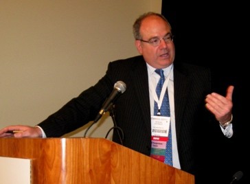
Mitchel L. Zoler/IMNG Medical Media
|
However, despite the success reported in this study, it is premature to extrapolate the efficacy of the colonoscopic methods they used to routine practice. Results are always better when physicians participate in a study and know they are being watched. We worry that there will be a fall in efficacy as the method diffuses out more widely. A better approach is what Dr. Soetikno suggested: waiting until a generation of endoscopists come into practice who have undergone thorough training with the new, high-definition scopes.
Dr. Michael L. Kochman is a gastroenterologist and professor of medicine at the University of Pennsylvania in Philadelphia. He said that he had no relevant disclosures. He made these remarks in a press conference during the DDW meeting.
The Preservation and Incorporation of Valuable Endoscopic Innovations (PIVI) statement issued last year by the American Society of Gastrointestinal Endoscopy threw down a challenge to companies to come up with a colonoscope technology that could meet the threshold for clinical application of the "cut and discard" approach to managing diminutive colorectal polyp. The new report by Drs. Kaltenbach, Dr. Soetikno, and their associates is a major step in that direction. Their findings show that endoscopists really can see and accurately predict the histologic profile of colorectal polyps without producing any major adverse outcomes.

|
Mitchel L. Zoler/IMNG Medical Media
|
However, despite the success reported in this study, it is premature to extrapolate the efficacy of the colonoscopic methods they used to routine practice. Results are always better when physicians participate in a study and know they are being watched. We worry that there will be a fall in efficacy as the method diffuses out more widely. A better approach is what Dr. Soetikno suggested: waiting until a generation of endoscopists come into practice who have undergone thorough training with the new, high-definition scopes.
Dr. Michael L. Kochman is a gastroenterologist and professor of medicine at the University of Pennsylvania in Philadelphia. He said that he had no relevant disclosures. He made these remarks in a press conference during the DDW meeting.
The Preservation and Incorporation of Valuable Endoscopic Innovations (PIVI) statement issued last year by the American Society of Gastrointestinal Endoscopy threw down a challenge to companies to come up with a colonoscope technology that could meet the threshold for clinical application of the "cut and discard" approach to managing diminutive colorectal polyp. The new report by Drs. Kaltenbach, Dr. Soetikno, and their associates is a major step in that direction. Their findings show that endoscopists really can see and accurately predict the histologic profile of colorectal polyps without producing any major adverse outcomes.

|
Mitchel L. Zoler/IMNG Medical Media
|
However, despite the success reported in this study, it is premature to extrapolate the efficacy of the colonoscopic methods they used to routine practice. Results are always better when physicians participate in a study and know they are being watched. We worry that there will be a fall in efficacy as the method diffuses out more widely. A better approach is what Dr. Soetikno suggested: waiting until a generation of endoscopists come into practice who have undergone thorough training with the new, high-definition scopes.
Dr. Michael L. Kochman is a gastroenterologist and professor of medicine at the University of Pennsylvania in Philadelphia. He said that he had no relevant disclosures. He made these remarks in a press conference during the DDW meeting.
SAN DIEGO – Researchers have taken a major step toward eliminating the need for routine pathology on every polyp removed during colonoscopy.
Use of a high-definition, dual-focus colonoscope allowed endoscopists to perform "significantly more accurate, high-confidence endoscopic diagnosis of diminutive polyps," Dr. Tonya Kaltenbach said at the annual Digestive Disease Week.
"In many hospitals, about 20%-25% of all pathology samples are small colon polyps, almost all of which are benign. This is a lot of money that can be saved," said Dr. Kaltenbach in a written statement. About 80% of all polyps found during screening colonoscopy are diminutive, 5 mm in diameter or less, and the current standard of care is to do a pathology analysis on each of these polyps.
As used by five gastroenterologists who participated in the study at three U.S. centers to assess 530 polyps in 277 people, the high-definition scope produced a 95.9% agreement in polyp assignment for follow-up surveillance, compared with pathology-laboratory analysis of the same polyps, and a 96.4% negative predictive value for adenomas, said Dr. Kaltenbach, a gastroenterologist at the VA Medical Center in Palo Alto, Calif.
These performance levels surpassed the greater than 90% targets for colonoscopic assessment of diminutive polyps set last year by a committee organized by the American Society of Gastrointestinal Endoscopy (Gastrointest. Endosc. 2011;73:419-22). Notably, the 90% threshold for assignment of postpolypectomy surveillance accuracy and negative predictive value was set by the ASGE committee before dual-focus colonoscopies like the one used in the current study were in widespread use, said Dr. Roy Soetikno, chief of endoscopy at the Palo Alto VA and senior investigator on the study.
Today, some of the scopes used in routine U.S. practice have the high-definition, dual focus features of the one used in the study, and within the next 5 years essentially all U.S. colonoscopies will have these performance characteristics, he said in an interview.
Although the new study results showed that endoscopists can surpass the thresholds set by the ASGE for visual assessment, widespread use of the method will require further validation, Dr. Soetikno said. "We need to develop programs to teach gastroenterologists and incorporate them into fellowship programs. We also need a way to store the images so that people can document their diagnoses. But [our study] is the beginning," he said.
Dr. Soetikno also noted that the idea of visual assessment of potentially cancerous or precancerous lesions is not new, as the same approach is routinely used in dermatology. With new technologies, gastroenterologists "may be able to do the same thing. We can save our resources, and not spend money on evaluating polyps of no consequence."
Dr. Soetikno, Dr. Kaltenbach, and their associates designed the Veterans Affairs Colorectal Lesion Interpretation and Diagnosis (VALID) study to test the hypothesis that colonoscopy using a high-definition, narrow-band imaging, dual focus colonoscope could increase the rate of accurate, high-confidence histology predictions of diminutive polyps. Five endoscopists working at three VA Medical Centers randomized 558 patients scheduled to undergo routine colonoscopy. The patients averaged 63 years old, 95% were men, and about 80% were white. Roughly 38% underwent colonoscopy for screening, 44% for surveillance following a history of polyps, and for 19% the examination was symptom driven (total is 101% due to rounding).
The protocol randomized 277 people to high-definition colonoscopy with an Olympus model CF-HQ190, and 281 people to examination with an older model without dual-focus capability, the Olympus CF-H180. Examination of the people in the dual-focus group found 710 polyps, including 530 diminutive lesions; the control group had 599 polyps, including 445 that were 5 mm or less.
The endoscopists made accurate, high-confidence endoscopic predictions on 75% of the diminutive polyps that they saw using the dual-focus device, compared with a 63% when they used the less advanced endoscope, a statistically significant difference. When compared with the pathologic assessments, 95.9% of the high-confidence, dual-focus diagnoses produced correct surveillance-interval recommendations, compared with a 95.2% rate using the standard-focus device. Negative predictive value was 96.4% using the dual-focus scope and 92.0% for the more conventional scope. In addition, the dual-focus scope produced no misdiagnoses of advanced histology as non-neoplasia, and use of this scope did not result in any meaningful increase in examination time, nor did it produce any bleeding or perforation.
The VALID study received partial funding from Olympus America, the company that markets the dual-focus, high-definition colonoscope tested. Dr. Soetikno said that he has been a consultant to Olympus. Dr. Kaltenbach and Dr. Kochman said they had no disclosures.
FROM THE ANNUAL DIGESTIVE DISEASE WEEK
Major Finding: High-definition, dual-focus colonoscopy produced 75% accurate, high-confidence diagnoses of diminutive polyps, vs. 63% using an older-model scope.
Data Source: Data came from a randomized, controlled trial with 558 people who underwent colonoscopy at three U.S. centers.
Disclosures: The VALID study received partial funding from Olympus America, the company that markets the dual-focus, high-definition colonoscope tested. Dr. Soetikno said that he has been a consultant to Olympus. Dr. Kaltenbach said that she had no disclosures.

