User login
| 
| 
|
| FIGURE 1 | FIGURE 2 | FIGURE 3 |
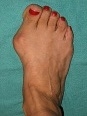
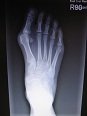
The family physician (FP) easily diagnosed a bunion deformity (FIGURE 1); an x-ray showed medial angulation of the first metatarsal and lateral deviation of the hallux (FIGURE 2).
Commonly associated signs that accompany a bunion include: hypermobility, flatfoot deformity, second MTP joint pain, pain under the second metatarsal head, overlapped second digit, decreased ankle dorsiflexion, concurrent gout, decreased first MTP joint range of motion, sesamoiditis, hyperkeratosis, and hammertoe deformity. A unilateral bunion deformity is often a result of limb length discrepancy.
Conservative treatment measures include:
- change in shoes
- placing a toe spacer in the first interdigital space to straighten the hallux and decreases the irritation caused by rubbing of the first and second digits
- padding to limit shearing force from shoes
- water-soluble cortisone injection into the first MTP joint for patients who complain of joint pain secondary to early-stage osteoarthritis
- custom-made orthotics to help slow the progression of the deformity caused by biomechanical factors
- resting, nonsteroidal anti-inflammatory drugs, and ice to help an inflamed joint and/or shoe irritation
- physical therapy to help improve joint range of motion, reduce edema, or decrease nerve pain.
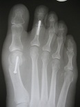
In this case, the patient required surgical correction. First metatarsal–cuneiform joint fusion was chosen to correct the medial angulation; dorsal elevation of the first metatarsal and lateral soft tissue release at the first MTP joint was done to correct the lateral deviation of the hallux. The medial aspect of the first metatarsal head was also resected (FIGURE 3).
The patient was placed in a short-leg cast for 6 weeks and slowly progressed to a regular shoe over the next month. The patient was encouraged to use the custom-made orthotics for her flatfoot to prevent a recurrence of the bunion.
Text for Photo Rounds Friday courtesy of Richard P. Usatine, MD. Images courtesy of Naohiro Shibuya, DPM. This case was adapted from: Shibuya N, Fontaine J. Bunion deformity. In: Usatine R, Smith M, Mayeaux EJ, et al, eds. The Color Atlas of Family Medicine. New York, NY: McGraw-Hill; 2009:896-899.
To learn more about The Color Atlas of Family Medicine, see:
• http://www.amazon.com/Color-Atlas-Family-Medicine/dp/0071474641
You can now get The Color Atlas of Family Medicine as an app for mobile devices including the iPhone and iPad by clicking this link:
|
|
|
|
| FIGURE 1 | FIGURE 2 | FIGURE 3 |
The family physician (FP) easily diagnosed a bunion deformity (FIGURE 1); an x-ray showed medial angulation of the first metatarsal and lateral deviation of the hallux (FIGURE 2).
Commonly associated signs that accompany a bunion include: hypermobility, flatfoot deformity, second MTP joint pain, pain under the second metatarsal head, overlapped second digit, decreased ankle dorsiflexion, concurrent gout, decreased first MTP joint range of motion, sesamoiditis, hyperkeratosis, and hammertoe deformity. A unilateral bunion deformity is often a result of limb length discrepancy.
Conservative treatment measures include:
- change in shoes
- placing a toe spacer in the first interdigital space to straighten the hallux and decreases the irritation caused by rubbing of the first and second digits
- padding to limit shearing force from shoes
- water-soluble cortisone injection into the first MTP joint for patients who complain of joint pain secondary to early-stage osteoarthritis
- custom-made orthotics to help slow the progression of the deformity caused by biomechanical factors
- resting, nonsteroidal anti-inflammatory drugs, and ice to help an inflamed joint and/or shoe irritation
- physical therapy to help improve joint range of motion, reduce edema, or decrease nerve pain.
In this case, the patient required surgical correction. First metatarsal–cuneiform joint fusion was chosen to correct the medial angulation; dorsal elevation of the first metatarsal and lateral soft tissue release at the first MTP joint was done to correct the lateral deviation of the hallux. The medial aspect of the first metatarsal head was also resected (FIGURE 3).
The patient was placed in a short-leg cast for 6 weeks and slowly progressed to a regular shoe over the next month. The patient was encouraged to use the custom-made orthotics for her flatfoot to prevent a recurrence of the bunion.
Text for Photo Rounds Friday courtesy of Richard P. Usatine, MD. Images courtesy of Naohiro Shibuya, DPM. This case was adapted from: Shibuya N, Fontaine J. Bunion deformity. In: Usatine R, Smith M, Mayeaux EJ, et al, eds. The Color Atlas of Family Medicine. New York, NY: McGraw-Hill; 2009:896-899.
To learn more about The Color Atlas of Family Medicine, see:
• http://www.amazon.com/Color-Atlas-Family-Medicine/dp/0071474641
You can now get The Color Atlas of Family Medicine as an app for mobile devices including the iPhone and iPad by clicking this link: