User login
| 
| 
|
| FIGURE 1 | FIGURE 2 | FIGURE 3 |
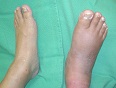
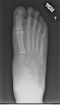
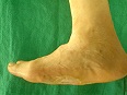
The appearance of the patient’s foot (FIGURE 1) prompted the physician to order a radiograph (FIGURE 2), which revealed midfoot osteopenia—an early sign of acute Charcot arthropathy. The incidence of Charcot arthropathy in diabetes ranges from 0.1% to 5%. Untreated Charcot foot may lead to a rockerbottom foot (FIGURE 3), which in turn leads to increased plantar pressure in the neuropathic foot. This cascade will result in ulceration and possible amputation. Charcot arthropathy is a gradual destruction of the joint in patients with neurosensory loss. Its most common presentation is in the diabetic neuropathic patient. Clinical features include a red, hot, swollen foot with neurosensory loss. The pathogenesis is unknown.
Radiographs are imperative for diagnosis. Usually, radiographs show arch collapse within the joints of the midfoot (tarsometatarsal joints). Radiographs can also show erosions and cystic degeneration of the tarsometatarsal joints. When infection is suspected, other imaging modalities such as a bone scan and magnetic resonance imaging may be ordered, but are often inconclusive because of the difficulty differentiating cellulitis, osteomyelitis, and Charcot arthropathy.
If osteomyelitis is suspected, bone cultures and bone biopsy are recommended. Cultures need to be taken during the bone biopsy so that the suspected infected bone can be visualized for accurate sampling.
Off-loading pressure from the foot is the standard of care. The total contact cast is most effective, and it covers the toes for protection. Other methods that are used include the removable cast boot, crutches, and a wheelchair.
If foot care is not optimized, a plantar ulcer can form under the Charcot’s joints. The bones will take approximately 4 to 5 months to heal in the presence of neuropathy. Oral antibiotics are not indicated unless infection is suspected. If deformity develops, custom-molded shoes and insoles must be ordered to prevent plantar ulcers that can lead to amputation.
Text for Photo Rounds Friday courtesy of Richard P. Usatine, MD. Photos courtesy of Javier La Fontaine, DPM. This case was adapted from: La Fontaine J, Shibuya, N. Charcot arthropathy. In: Usatine R, Smith M, Mayeaux EJ, et al, eds. The Color Atlas of Family Medicine. New York, NY: McGraw-Hill; 2009:909-911.
To learn more about The Color Atlas of Family Medicine, see:
• http://www.amazon.com/Color-Atlas-Family-Medicine/dp/0071474641
You can now get The Color Atlas of Family Medicine as an app for mobile devices including the iPhone and iPad by clicking this link:
|
|
|
|
| FIGURE 1 | FIGURE 2 | FIGURE 3 |
The appearance of the patient’s foot (FIGURE 1) prompted the physician to order a radiograph (FIGURE 2), which revealed midfoot osteopenia—an early sign of acute Charcot arthropathy. The incidence of Charcot arthropathy in diabetes ranges from 0.1% to 5%. Untreated Charcot foot may lead to a rockerbottom foot (FIGURE 3), which in turn leads to increased plantar pressure in the neuropathic foot. This cascade will result in ulceration and possible amputation. Charcot arthropathy is a gradual destruction of the joint in patients with neurosensory loss. Its most common presentation is in the diabetic neuropathic patient. Clinical features include a red, hot, swollen foot with neurosensory loss. The pathogenesis is unknown.
Radiographs are imperative for diagnosis. Usually, radiographs show arch collapse within the joints of the midfoot (tarsometatarsal joints). Radiographs can also show erosions and cystic degeneration of the tarsometatarsal joints. When infection is suspected, other imaging modalities such as a bone scan and magnetic resonance imaging may be ordered, but are often inconclusive because of the difficulty differentiating cellulitis, osteomyelitis, and Charcot arthropathy.
If osteomyelitis is suspected, bone cultures and bone biopsy are recommended. Cultures need to be taken during the bone biopsy so that the suspected infected bone can be visualized for accurate sampling.
Off-loading pressure from the foot is the standard of care. The total contact cast is most effective, and it covers the toes for protection. Other methods that are used include the removable cast boot, crutches, and a wheelchair.
If foot care is not optimized, a plantar ulcer can form under the Charcot’s joints. The bones will take approximately 4 to 5 months to heal in the presence of neuropathy. Oral antibiotics are not indicated unless infection is suspected. If deformity develops, custom-molded shoes and insoles must be ordered to prevent plantar ulcers that can lead to amputation.
Text for Photo Rounds Friday courtesy of Richard P. Usatine, MD. Photos courtesy of Javier La Fontaine, DPM. This case was adapted from: La Fontaine J, Shibuya, N. Charcot arthropathy. In: Usatine R, Smith M, Mayeaux EJ, et al, eds. The Color Atlas of Family Medicine. New York, NY: McGraw-Hill; 2009:909-911.
To learn more about The Color Atlas of Family Medicine, see:
• http://www.amazon.com/Color-Atlas-Family-Medicine/dp/0071474641
You can now get The Color Atlas of Family Medicine as an app for mobile devices including the iPhone and iPad by clicking this link: