User login
BALTIMORE – A new study hints that mapping the functional connectivity of the brain may allow surgeons to pinpoint previously unidentified epileptogenic networks more precisely, leading to more effective epilepsy surgery.
Functional connectivity mapping could not only identify epileptogenic networks, but also could predict successful and failed epilepsy surgeries, Dr. Hyang Woon Lee reported in a study of the resting state functional MRI (fMRI) scans of 29 patients who underwent surgery for intractable partial epilepsy.
"I was quite thrilled to find that intrinsic functional connectivity changes stand out in the epileptogenic zones," Dr. Lee said in an interview at the annual meeting of the American Epilepsy Society. She is in the departments of neurology at Yale University, New Haven, Conn., and at Ewha Womans University in Seoul, South Korea.
The research is based on a relatively new method of identifying brain regions that are structurally separate but functionally connected by previously unidentifiable neural networks. Dr. Lee’s colleague at Yale, R. Todd Constable, Ph.D., developed the technique of using a mathematical algorithm called intrinsic connectivity contrast (ICC) to identify interrelationships between the blood oxygenation level dependent signal of voxels measured with fMRI. A high degree of ICC for a particular voxel means that it has a strong connection to other brain areas. The resulting image shows the degree of these connections, and "provides a measure as to how well-connected any given tissue element is," Dr. Lee wrote in her poster.
The analysis can be done ipsilaterally, where connections to each voxel are compared with all others in the same hemisphere, and contralaterally, where connections to voxels in the other hemisphere are measured. The resulting maps identify functionally connected networks, Dr. Lee said. In the case of patients with focal epilepsy, they sometimes show that epileptogenic areas identified by intracranial EEG have additional connections, or are even part of a nodal network of abnormally functioning tissue.
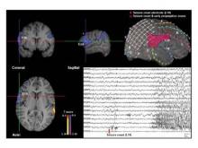
Each of the 29 patients underwent presurgical fMRI and intracranial EEG to localize the seizure foci. Dr. Lee, Dr. Constable, and their coauthors analyzed the fMRI images with the ICC algorithm, both ipsilaterally and contralaterally. Dr. Lee then compared the ICC results to the intracranial EEG data that guided the epilepsy surgery. The ICC maps were a good match for the seizure onset and propagation zones detected by intracranial EEG, she said.
"Intracranial EEG seizure onset zones might have decreased ICCs, since the epileptogenic foci are likely to be abnormal brain tissue, often due to neuronal damage," she said.
Overall, 15 patients had good surgical outcomes, defined as an Engels class I, meaning they were free of disabling seizures. The other 14 patients had poor surgical outcomes: Engels classes II (rare disabling seizures), III (worthwhile improvement), and IV (no worthwhile improvement).
The determination of the seizure onset zones with ICC was concordant with presurgical intracranial EEG testing in 13 of the 15 patients with good surgical outcomes and in 7 of the 14 patients with poor outcomes.
The concordance between ICC and intracranial EEG also varied by the type of epilepsy. The techniques were 100% concordant among patients with extratemporal lobe epilepsy who had good outcomes. These patients often have ictal onset zones that are diffuse and sometimes difficult to determine.
The measurement of functional connectivity with fMRI has the potential to become a noninvasive presurgical diagnostic tool in epilepsy surgery based on its strong ability to identify the nodes of an epileptogenic network, Dr. Lee said.
"It might also help in the diagnosis of other neuropsychiatric disorders, such as Alzheimer’s disease, stroke, and schizophrenia by elucidating altered functional connectivity in the neural network. Although it’s still in the early stages of development, I believe this approach can be widely used for both clinical and scientific purposes with further validation."
Dr. Lee said that she had no financial disclosures.
FROM THE ANNUAL MEETING OF THE AMERICAN EPILEPSY SOCIETY
Major Finding: The determination of seizure onset zones with ICC was concordant with presurgical intracranial EEG testing in 13 of 15 patients with good surgical outcomes and in 7 of 14 patients with poor outcomes.
Data Source: A retrospective case study of 29 patients with intractable epilepsy who underwent presurgical fMRI and intracranial EEG to localize their seizure foci.
Disclosures: Dr. Lee said that she had no financial disclosures.

