User login
Team reproduces HSPCs in artificial bone marrow

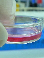
artificial bone marrow
Credit: C. Lee-Thedieck
Researchers say they have developed artificial bone marrow analogs that can be used to reproduce hematopoietic stem and progenitor cells (HSPCs).
The team created macroporous hydrogel scaffolds that mimic the stem cell niche of the bone marrow.
When they introduced mesenchymal stem cells (MSCs) from actual bone marrow into the analogs, the MSCs promoted HSPC proliferation.
In fact, the MSCs preserved HSPC stemness more effectively in the analogs than in standard 2-dimensional cell culture systems.
Annamarija Raic, of the Max Planck Institute for Intelligent Systems in Stuttgart, Germany, and her colleagues reported these results in Biomaterials.
The researchers noted that reproducing functional HSPCs in the lab has proven challenging. The cells cannot be cultured in vitro for a feasible period of time without differentiating.
So the team set out to create a culture system that mimics the important physical and biological parameters of the stem cell niche: the 3D architecture, the adhesive extracellular matrix, soluble factors, and the stromal cell compartment.
They used salt leaching technology to produce poly(ethylene glycol) diacrylate hydrogel scaffolds that could soak cells into their pores. To biofunctionalize the scaffolds, the investigators added an RGD peptide carrying an acrylate moiety.
They then introduced 3 different cell types into the scaffolds—the human osteosarcoma cell line CAL72, MSCs from bone marrow, and MSCs from umbilical cord blood—to see which best supported the proliferation of CD34+ HSPCs isolated from cord blood.
Each of the cell types supported HSPC proliferation, but bone marrow MSCs were the most effective. The researchers therefore decided to use bone marrow MSCs when they compared their 3D scaffolds to a 2D culture system.
The bone marrow MSCs had a beneficial effect on HSPC proliferation in the 2D cell cultures. Over 4 days, HSPCs divided 1 to 2 times more often when they were cultured with bone marrow MSCs than without the cells.
In the 3D scaffolds, HSPC proliferation was comparable or slightly lower than that observed in the 2D cultures. However, the scaffolds had a higher percentage of CD34+ HSPCs after 4 days.
The investigators therefore concluded that their hydrogel scaffolds meet the basic requirements for creating artificial stem cell niches. ![]()
Controlling cells after transplant
Credit: Umberto Salvagnin
Scientists say they have devised a method for engineering cells that are more easily controlled after transplantation.
The team loaded cells with microparticles that release phenotype-altering agents for days to weeks after transplantation.
With this method, the researchers were able to control cells’ secretome, viability, proliferation, and differentiation. The approach was also successful in delivering drugs and other factors to the cell’s microenvironment.
The scientists described this method in Nature Protocols.
They provided step-by-step instructions for generating micrometer-sized agent-doped poly(lactic-co-glycolic) acid (PLGA) particles using a single-emulsion evaporation technique, engineering cultured cells, and confirming particle internalization.
“Once those particles are internalized into the cells, which can take on the order of 6 to 24 hours, we can deliver the transplant immediately or even cryopreserve the cells,” said study author Jeffrey Karp, PhD, of the Harvard Stem Cell Institute in Cambridge, Massachusetts.
“When the cells are thawed at the patient’s bedside, they can be administered, and the agents will start to be released inside the cells to control differentiation, immune modulation, or matrix production, for example.”
Of course, it could take more than a decade for this type of cell therapy to be a common medical practice. But Dr Karp and his colleagues detailed this research in Nature Protocols to encourage others in the scientific community to use the technique and potentially speed up the pace of this research.
The team’s paper shows the range of different cell types that can be particle-engineered, including stem cells, immune cells, and pancreatic cells.
“With this versatile platform . . . , we’ve demonstrated the ability to track cells in the body, control stem cell differentiation, and even change the way cells interact with immune cells,” said study author James Ankrum, PhD, who was a graduate student in Dr Karp’s lab when this research was conducted but is now at the University of Minnesota in Minneapolis.
“We’re excited to see what applications other researchers will imagine using this platform.” ![]()
Drug can prevent CMV in HSCT recipients
A new drug can prevent cytomegalovirus (CMV) in patients undergoing hematopoietic stem cell transplant (HSCT), according to a study published in The New England Journal of Medicine.
The drug, called CMX001, is an oral nucleotide analog lipid-conjugate that blocks replication of double-stranded DNA viruses.
HSCT recipients who took CMX001 after engraftment were less likely to develop CMV than HSCT patients who received placebo, researchers found.
“With current agents, between 3% and 5% of allogeneic transplant patients develop CMV disease within 6 months of transplantation, and a small number of them may die of it,” said investigator Francisco Marty, MD, of the Dana-Farber Cancer Institute in Boston.
“There clearly is a need for better treatments with fewer adverse effects. This clinical trial examined whether the disease can be prevented, rather than waiting for blood tests to show that treatment is needed.”
The phase 2 trial involved 230 HSCT recipients treated at 27 centers across the US. The patients were randomized to receive placebo or CMX001 at doses ranging from 40 mg a week to 200 mg twice a week.
Treatment began after engraftment, at about 2 to 3 weeks post-transplant, and continued for 9 to 11 weeks.
The study’s primary efficacy outcome was a “CMV event,” which was defined as CMV that affects the lung, digestive tract, or other organs, or a detectable amount of CMV in the blood at the end of treatment.
CMV events occurred in 25% (43/171) of patients who received CMX001 and 37% (22/59) of patients who received placebo. However, when CMX001 was given at the optimal dose—100 mg twice a week—only 10% of patients had a CMV event.
“The results show the effectiveness of CMX001 in preventing CMV infections in this group of patients,” Dr Marty said. “Because CMX001 is known to be active against other herpes viruses and against adenoviruses that sometimes affect transplant patients, it may be useful as a preventive or treatment agent for those infections as well.”
Most of the side effects associated with CMX001 were gastrointestinal in nature. Diarrhea was common and often serious in patients who received the drug at 200 mg twice a week.
Patients in this dose group were more likely to experience elevated alanine aminotransferase levels as well. But this was not associated with increases in levels of bilirubin or aspartate aminotransferase.
This research was funded by Chimerix, the company developing CMX001.![]()
Overcoming limitations of haploidentical HSCT
Researchers say they have found a strategy to overcome the limitations of haploidentical hematopoietic stem cell transplantation (HSCT).
To prevent the early, severe graft-versus-host disease (GVHD) associated with haploidentical HSCT, donor T cells reacting with recipient antigens are eliminated from the graft prior to transplant.
However, the depletion of T cells can lead to delayed immune reconstitution in the transplant recipient, which increases the risk of infection and death.
Results of a new study may help clinicians decrease those risks. The study showed that the infusion of specially engineered haploidentical donor T cells induced early reconstitution of post-HSCT immunity. These cells were also able to control GVHD and preserve a graft-versus-leukemia effect.
This study appeared in the May issue of The Lancet Oncology and was funded by the biotech company MolMed SpA.
Claudio Bordignon, MD, from the Raffaele Scientific Institute, Milan, Italy, and colleagues conducted this phase 1/2, multicenter, nonrandomized trial of haploidentical T-cell depleted HSCT in 50 high-risk leukemia patients in remission.
Of the 50 patients, 28 patients received T cells engineered to carry the herpes simplex thymidine kinase suicide gene (TK cells).
To prepare the TK cells, the researchers used the haploidentical donor T lymphocytes that were collected prior to mobilization with G-CSF or marrow harvesting of stem cells. The T lymphocytes were expanded in vitro and then transduced with the herpes simplex thymidine kinase suicide gene. This rendered the cells sensitive to the antiviral agent ganciclovir, which enabled the researchers to selectively eliminate the cells upon the development of GVHD.
Twenty-eight patients received a first dose of TK cells. If patients did not achieve immune reconstitution 30 days later, they received up to 3 additional monthly infusions of TK cells. Transplant recipients did not receive GVHD prophylaxis following TK cell infusion.
Twenty-two patients achieved immune reconstitution at a median time of 75 days after HSCT and 23 days following TK cell infusion. Immune reconstitution was dependent on the dose of TK cells.
A progressive decline in the number and severity of infectious complications occurred in patients with immune reconstitution. Patients without immune reconstitution continued to have more frequent and more severe infectious complications.
Nonrelapse mortality at 100 days posttransplant was lower in patients who achieved immune reconstitution than in those who did not, at 14% and 60%, respectively. The researchers said this was possibly due to protection from late infectious mortality.
Effective immune reconstitution did not increase the incidence of GVHD, the researchers said. Rates of GVHD were similar to rates reported in other studies. Ten patients developed grades 1 to 4 acute GVHD, and 1 patient developed chronic GVHD.
Dr Bordignon and colleagues said acute GVHD was directly associated with infiltration of the TK cells at affected lesions. The team was able to control acute GVHD by administering ganciclovir, thereby activating the suicide gene and eliminating the TK cells. ![]()