User login
Venetoclax can produce short-term responses in AML

Results of a phase 2 trial suggest the BCL2 inhibitor venetoclax can produce responses in patients with acute myelogenous leukemia (AML) who do not respond to or cannot tolerate chemotherapy.
However, the overall response rate in this trial was low, and responses were not durable.
All of the patients studied discontinued venetoclax, most due to disease progression.
The study was published in Cancer Discovery. It was funded by AbbVie in collaboration with Genentech/Roche.
The trial included 32 patients with AML and a median age of 71 (range, 19–84). Thirteen patients had an antecedent hematologic disorder or myeloproliferative neoplasm, and 4 had therapy-related AML with complex cytogenetics.
Twelve patients had mutations in IDH genes, and 6 had a high BCL2-sensitive protein index.
Thirty patients had received at least 1 prior therapy, and 13 had received at least 3 prior treatment regimens. Two patients were considered unfit for intensive chemotherapy and were treatment-naive at study entry.
The patients received venetoclax at 800 mg daily. All 32 patients received at least 1 dose, and 26 patients received at least 4 weeks of therapy.
Efficacy
The overall response rate was 19%. Two patients had a complete response (CR), and 4 had a CR with incomplete blood count recovery. Three of the 6 responders had an antecedent hematologic disorder.
“[E]ven among pretreated patients whose AML was refractory to intensive chemotherapy, there was evidence of exceptional sensitivity to selective BCL2 inhibition, even to the point of complete remissions,” said study author Anthony Letai, MD, PhD, of the Dana-Farber Cancer Institute in Boston, Massachusetts.
The median duration of therapy in responders was 144.5 days, and the median duration of CR was 48 days.
The 4 patients who had CRs with incomplete count recovery had IDH mutations. Response to the drug correlated with biomarker results, including indices of BCL2 protein expression and BH3 profiling.
“This is significant as it supports the mechanism of action of venetoclax as an on-target inhibitor of BCL2,” Dr Letai said. “Moreover, it offers the possibility of using BH3 profiling as a potential predictive biomarker for clinical use of BH3 mimetics.”
Safety and discontinuation
All of the patients discontinued therapy—29 due to progressive disease and 1 due to an adverse event (terminal ileitis). One patient withdrew consent, and 1 proceeded to allogeneic transplant after achieving stable disease.
All of the patients experienced treatment-emergent adverse events. The most common were nausea (59%), diarrhea (56%), hypokalemia (41%), vomiting (41%), fatigue (34%), headache (34%), hypomagnesemia (34%), febrile neutropenia (31%), and hypophosphatemia (31%).
Serious adverse events occurred in 84% of patients. These included febrile neutropenia (28%), pneumonia (16%), abdominal pain (6%), acute renal failure (6%), failure to thrive (6%), hypotension (6%), sepsis (6%), and urinary tract infection (6%).
Based on the results of this trial, the researchers concluded that venetoclax may be a viable treatment option for AML patients when used in combination with other therapies.
“We believe that venetoclax will soon become an equal partner to standard-of-care chemotherapy in elderly patients with AML when used in combinations with hypomethylating agents and other approaches,” said study author Marina Konopleva, MD, PhD, of MD Anderson Cancer Center in Houston, Texas.
“Planned studies will test the hypothesis that venetoclax may likewise improve outcomes in younger AML patients when combined with high-dose chemotherapy.” ![]()
Protein promotes hematopoietic regeneration
![]()
Photo by Chad McNeeley
The protein angiogenin (ANG) plays a significant role in the regulation of hematopoiesis, according to a group of researchers.
The team discovered that ANG suppresses the proliferation of hematopoietic stem and progenitor cells (HSPCs) while promoting the proliferation of myeloid progenitor cells.
They also showed that treatment with recombinant ANG protein improved survival in irradiated mice and enhanced the regenerative capabilities of HSPCs.
The researchers believe these findings have significant implications for hematopoietic stem cell transplant (HSCT) and bone marrow injury.
The team reported the findings in Cell.
“We knew that ANG was involved in promoting cell growth, so it was not unexpected to find that ANG stimulates proliferation of myeloid progenitor cells,” said study author Guo-fu Hu, PhD, of Tufts Medical Center in Boston, Massachusetts.
“But it was surprising to find that ANG also suppresses growth of stem cells and that it accomplishes these divergent promotion or suppression functions through RNA processing events specific to individual cell types.”
The researchers discovered that, in HSPCs, ANG induces processing of tiRNA, which suppresses global protein synthesis. And in myeloid progenitor cells, ANG induces processing of rRNA, which enhances protein synthesis.
The team also tested ANG’s ability to prevent and mitigate radiation-induced bone marrow failure. They found that treating mice with recombinant ANG protein, either before or after lethal irradiation, increased survival, improved bone marrow cellularity, and enhanced peripheral blood content.
Finally, the researchers assessed the effects of ANG in the context of HSCT in mice. They found that treating mouse long-term HSCs with ANG ex vivo resulted in a “dramatic” increase in multi-lineage reconstitution over 24 weeks after HSCT.
Upon secondary transplant, enhanced regeneration occurred over 16 weeks, and mice had elevated peripheral blood counts at 1 year post-HSCT, without any signs of leukemia.
The researchers observed similar results in experiments with human cells. They transplanted CD34+ cord blood cells—cultured in the presence or absence of ANG—into mice. Treatment with ANG resulted in enhanced multi-lineage regeneration and enhanced reconstitution upon secondary transplant.
“Proper blood cell production is dependent on functioning hematopoietic stem and progenitor cells that are destroyed during conditioning procedures for transplantation or following bone marrow injury,” said study author Kevin A. Goncalves, of Tufts Medical Center.
“Our study demonstrates that ANG regulates critical functions of both clinically relevant cell types.” ![]()
Chemo during pregnancy may impact baby’s fertility

Photo by Nina Matthews
Preclinical research suggests the chemotherapy drug etoposide may have adverse effects on the developing ovaries of female fetuses.
Researchers found that etoposide can damage the development of mouse ovary tissue in vitro.
However, if the drug is given after ovarian follicles have developed, the damage is not significant.
The researchers said further study is needed to assess whether etoposide has similar effects on human tissue.
“In a study involving mouse tissue, we have shown that etoposide can damage the development of the ovaries while a fetus is in the womb,” said study author Norah Spears, DPhil, of the University of Edinburgh in the UK.
“The drug affects the germ cells in the ovaries, which are the cells that give rise to eggs. This is important because it could mean that the fertility of the offspring could be affected in later life.”
Dr Spears and her colleagues reported these findings in BMC Cancer.
The researchers noted that etoposide is considered safe for use in the second and third trimester of pregnancy because it has a low risk of miscarriage and birth defects. However, little is known about the effects of the drug on the unborn baby in later life.
A woman’s reproductive lifespan is determined before birth, while the ovaries are developing in the womb. The second and third trimesters are particularly important, as that’s when female germ cells form ovarian follicles, which determine how many eggs a woman will be able to release in her lifetime.
Ovarian follicles each contain an oocyte. The process by which oocytes become enclosed in follicles starts about 17 weeks into fetal development and is only completed in late pregnancy.
Dr Spears and her colleagues found that exposing mouse ovaries to etoposide before follicles had formed caused the death of most germ cells. The few remaining germ cells went on to form unhealthy follicles. Once oocytes were enclosed in follicles, however, etoposide had no significant adverse effects.
The researchers collected fetal and neonatal ovaries from mice and cultured them in the lab. The team then exposed groups of 6 ovaries each to different doses of etoposide.
When fetal ovaries were treated with etoposide prior to follicle formation, this resulted in dose-dependent damage. Total follicle numbers declined by 72% to 90% in response to medium and high doses of etoposide, respectively.
In neonatal ovaries after follicle formation, etoposide only had minor effects, even at doses higher than those used to treat fetal ovaries.
“Our work indicates that female mouse germ cells are particularly susceptible to damage by etoposide at a specific early developmental stage, immediately prior to follicle formation, and that there could be possible consequences to the fertility of females born to women who were treated with etoposide during the second trimester of their pregnancy,” Dr Spears said.
She and her colleagues noted that additional research is needed to determine if these effects might occur in humans. And research is needed to determine whether the adverse effects of etoposide in germ cells are caused by damage to the DNA or because etoposide affects other processes such as transcription. ![]()
CAR T-cell therapy granted orphan designation

Photo courtesy of NIAID
The US Food and Drug Administration (FDA) has granted orphan drug designation for a CD4-directed chimeric antigen receptor (CD4CAR) T-cell therapy to treat peripheral T-cell lymphoma (PTCL).
The CD4CAR therapy, also known as ICG122, consists of properly matched allogeneic T cells engineered to express an anti-CD4 single-chain variable fragment antibody domain.
ICG122 is being developed by iCell Gene Therapeutics.
The company is planning a phase 1 trial of ICG122 in cooperation with the National Institutes of Health, Indiana Clinical and Translational Sciences Institute, Stony Brook Hospital, and the James Graham Brown Cancer Center at University of Louisville.
“CD4CAR could significantly enhance currently available treatment options for [PTCL] patients,” said Yupo Ma, MD, PhD, a professor at Stony Brook University and chairman and chief scientific officer at iCell Gene Therapeutics.
“The orphan drug designation is an important achievement as we advance our development plans for this promising treatment in T-cell hematologic cancers.”
The FDA grants orphan designation to drugs and biologics intended to treat, diagnose, or prevent diseases/disorders that affect fewer than 200,000 people in the US.
The designation provides incentives for sponsors to develop products for rare diseases. This may include tax credits toward the cost of clinical trials, prescription drug user fee waivers, and 7 years of market exclusivity if the drug is approved. ![]()
Immunotherapy conditioning proves successful in mice

Photo by Aaron Logan
Research in mice suggests it’s feasible to use an immunotherapy conditioning regimen rather than radiation or chemotherapy prior to hematopoietic stem cell transplant (HSCT).
Investigators found that combining an antibody against the HSC receptor c-Kit with a CD47-blocking therapy could eliminate host HSCs and allow for successful engraftment of donor HSCs in immunocompetent recipient mice.
Adding T-cell-depleting antibodies to the mix allowed for robust HSC engraftment in a clinically relevant model of allogeneic HSCT.
Irving Weissman, MD, of Stanford University School of Medicine in California, and his colleagues conducted this research and reported the results in Science Translational Medicine.
The researchers first found that ACK2, an antibody against c-Kit, successfully depleted HSCs in immune-deficient mice.
“However, this antibody alone would not be effective in immune-competent recipients, who represent a majority of potential bone marrow transplant recipients,” said study author Akanksha Chhabra, PhD, of Stanford University School of Medicine.
So the researchers sought to enhance the effectiveness of ACK2 by combining it with antibodies or biologic agents that block CD47. They found that blocking CD47—particularly with an antagonist known as CV1mb—liberated macrophages to engulf target cells.
In this way, the immune system effectively depleted host HSCs in the immunocompetent mice, clearing the way for donor HSCs to take up residence in the bone marrow.
Finally, the researchers set out to determine whether conditioning with an anti-c-Kit antibody and CD47-blocking therapy could be extended to a clinically relevant model of allogeneic HSCT, in which the donor and recipient are matched through human leukocyte antigen alleles but mismatched at minor histocompatibility complex (mHC) antigens.
So the team conditioned mice with either ACK2 and CV1mb or ACK2 and the anti-CD47 antibody MIAP410. And they achieved immune ablation with T-cell-depleting antibodies—GK1.5 (anti-CD4) and YTS169.4 (anti-CD8). The mice then received mHC-mismatched HSCs.
The researchers found that either conditioning regimen, when combined with a T-cell-depleting regimen, resulted in substantial granulocyte, B-cell, T-cell, and NK-cell chimerism, as well as HSC engraftment in the bone marrow.
The success of these techniques in mice raises the researchers’ hopes that similar techniques will succeed in humans.
“If it works in humans like it did in mice, we would expect that the risk of death from blood stem cell transplant would drop from 20% to effectively 0,” said study author Judith Shizuru, MD, PhD, of Stanford University School of Medicine.
“If and when this is accomplished, it will be a whole new era in disease treatment and regenerative medicine,” Dr Weissman said. ![]()
FDA authorizes use of Zika assay

Photo by Juan D. Alfonso
The US Food and Drug Administration (FDA) has granted emergency use authorization (EUA) for the xMAP® MultiFLEX™ Zika RNA Assay.
This multiplex nucleic acid test is designed to detect Zika virus RNA in blood serum, plasma, or urine (collected alongside a patient-matched serum or plasma specimen).
The xMAP® MultiFLEX™ Zika RNA Assay is available for purchase by laboratories that are certified under the Clinical Laboratory Improvement Amendments of 1988 (CLIA) to perform high complexity tests.
The assay uses the Luminex® 100/200™ analyzer, MAGPIX® system, or other authorized instruments to simultaneously test for 6 genetic targets of the Zika virus.
The xMAP® MultiFLEX™ Zika RNA Assay was designed by GenArraytion, Inc. and is marketed by Luminex Corporation.
For more information on the test, see the fact sheet for healthcare providers on the Luminex website.
About the EUA
The EUA does not mean the xMAP® MultiFLEX™ Zika RNA Assay is FDA cleared or approved.
An EUA allows for the use of unapproved medical products or unapproved uses of approved medical products in an emergency.
The products must be used to diagnose, treat, or prevent serious or life-threatening conditions caused by chemical, biological, radiological, or nuclear threat agents, when there are no adequate alternatives.
This means the xMAP® MultiFLEX™ Zika RNA Assay is only authorized as long as circumstances exist to justify the authorization of the emergency use of in vitro diagnostics for the detection of Zika virus, unless the authorization is terminated or revoked sooner. ![]()
FDA approves drug for prevention of CINV

Photo by Rhoda Baer
The US Food and Drug Administration (FDA) has approved granisetron extended-release injection (Sustol®) for the prevention of chemotherapy-induced nausea and vomiting (CINV) in adults.
Extended-release granisetron is a serotonin-3 (5-HT3) receptor antagonist that utilizes Biochronomer® polymer-based drug delivery technology to maintain therapeutic levels of granisetron for at least 5 days, covering both the acute and delayed phases of CINV.
The product is intended for use in combination with other anti-emetics to prevent acute and delayed nausea and vomiting associated with initial and repeat courses of moderately emetogenic chemotherapy (MEC) or anthracycline and cyclophosphamide (AC) combination chemotherapy regimens.
“Despite advances in the management of CINV, up to half of patients receiving chemotherapy can still experience CINV, with delayed CINV being particularly challenging to control,” said Ralph V. Boccia, MD, of the Center for Cancer and Blood Disorders in Bethesda, Maryland.
“In our experience, other 5-HT3 receptor antagonists, including palonosetron, are generally effective for 48 hours or less. Sustol, due to its extended-release profile, represents a novel option that can protect patients from CINV for a full 5 days.”
Extended-release granisetron (formerly known as APF530) is a product of Heron Therapeutics, Inc. The US commercial launch of the drug is planned for the fourth quarter of 2016.
Phase 3 trials
The global phase 3 development program of extended-release granisetron consisted of 2 large, guideline-based clinical trials of more than 2000 cancer patients.
In one trial, researchers compared extended-release granisetron to palonosetron for the prevention of acute and delayed CINV after MEC or highly emetogenic chemotherapy (HEC).
Results suggested extended-release granisetron was non-inferior to palonosetron. The most common adverse events observed in patients receiving granisetron were injection-site reactions and constipation.
In another trial, researchers compared extended-release granisetron to ondansetron for control of delayed CINV after HEC. Patients received extended-release granisetron, dexamethasone, and fosaprepitant or ondansetron, dexamethasone, and fosaprepitant.
A higher percentage of patients in the granisetron arm had delayed-phase complete response. The incidence of treatment-emergent adverse events was similar between the treatment arms.
“The Sustol clinical trial populations and results are highly representative of cancer patients in our real-world clinical practice,” said Jeffrey Vacirca, MD, of North Shore Hematology Oncology Associates in East Setauket, New York.
“Use of MEC regimens is widespread, and AC-based regimens are among the most commonly prescribed highly emetogenic chemotherapy regimens. The most significant challenge for my breast cancer patients receiving AC is chemotherapy-induced nausea and vomiting. Sustol represents a better option to manage this devastating side effect of therapy.”
For more details on the drug, access the full prescribing information at www.SUSTOL.com. ![]()
Photo by Rhoda Baer
The US Food and Drug Administration (FDA) has approved granisetron extended-release injection (Sustol®) for the prevention of chemotherapy-induced nausea and vomiting (CINV) in adults.
Extended-release granisetron is a serotonin-3 (5-HT3) receptor antagonist that utilizes Biochronomer® polymer-based drug delivery technology to maintain therapeutic levels of granisetron for at least 5 days, covering both the acute and delayed phases of CINV.
The product is intended for use in combination with other anti-emetics to prevent acute and delayed nausea and vomiting associated with initial and repeat courses of moderately emetogenic chemotherapy (MEC) or anthracycline and cyclophosphamide (AC) combination chemotherapy regimens.
“Despite advances in the management of CINV, up to half of patients receiving chemotherapy can still experience CINV, with delayed CINV being particularly challenging to control,” said Ralph V. Boccia, MD, of the Center for Cancer and Blood Disorders in Bethesda, Maryland.
“In our experience, other 5-HT3 receptor antagonists, including palonosetron, are generally effective for 48 hours or less. Sustol, due to its extended-release profile, represents a novel option that can protect patients from CINV for a full 5 days.”
Extended-release granisetron (formerly known as APF530) is a product of Heron Therapeutics, Inc. The US commercial launch of the drug is planned for the fourth quarter of 2016.
Phase 3 trials
The global phase 3 development program of extended-release granisetron consisted of 2 large, guideline-based clinical trials of more than 2000 cancer patients.
In one trial, researchers compared extended-release granisetron to palonosetron for the prevention of acute and delayed CINV after MEC or highly emetogenic chemotherapy (HEC).
Results suggested extended-release granisetron was non-inferior to palonosetron. The most common adverse events observed in patients receiving granisetron were injection-site reactions and constipation.
In another trial, researchers compared extended-release granisetron to ondansetron for control of delayed CINV after HEC. Patients received extended-release granisetron, dexamethasone, and fosaprepitant or ondansetron, dexamethasone, and fosaprepitant.
A higher percentage of patients in the granisetron arm had delayed-phase complete response. The incidence of treatment-emergent adverse events was similar between the treatment arms.
“The Sustol clinical trial populations and results are highly representative of cancer patients in our real-world clinical practice,” said Jeffrey Vacirca, MD, of North Shore Hematology Oncology Associates in East Setauket, New York.
“Use of MEC regimens is widespread, and AC-based regimens are among the most commonly prescribed highly emetogenic chemotherapy regimens. The most significant challenge for my breast cancer patients receiving AC is chemotherapy-induced nausea and vomiting. Sustol represents a better option to manage this devastating side effect of therapy.”
For more details on the drug, access the full prescribing information at www.SUSTOL.com.
How procoagulant platelets develop
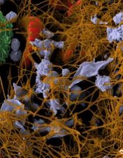
Image by Andre E.X. Brown
Researchers say they have determined how procoagulant platelets develop.
One of the mysteries in the field of thrombosis and hemostasis is how platelets are divided into two kinds when activated—“ordinary” platelets capable of aggregation and “super-activated,” procoagulant platelets.
The new study suggests that, to become super-activated, platelets must die. And the platelets need mitochondria to commit suicide.
Researchers were able to show how this programmed death—mitochondrial necrosis—follows a chain of events that lead to the platelets’ transition to a super-activated state.
“It was not clear before how a platelet makes the decision of what type to become,” said study author Mikhail Panteleev, of Lomonosov Moscow State University in Russia.
“We have deciphered the sequence of events: how the signal goes within the platelet and how the cell decides to die.”
Panteleev and his colleagues described these events in the Journal of Thrombosis and Haemostasis.
The team noted that platelets have many activators, but the chief among them are collagen, ADP, and thrombin.
Platelets detect different concentrations of an activator and respond with a varying frequency of calcium impulses in the cytoplasm.
The platelets’ mitochondria absorb and store the calcium, and when its concentration exceeds the critical level, the process of mitochondrial necrosis starts.
Calcium and reactive oxygen species are released from mitochondria, ATPases begin to destroy ATP instead of synthesizing it, the cell cytoskeleton collapses, and the platelets greatly increase in size.
As a result, at the outer membrane of the enlarged platelets, a lipid called phosphatidylserine appears, which is responsible for rapid blood clotting. And all this happens in seconds.
Stable INRs uncommon with long-term warfarin, study suggests

Results of a large study suggest most patients taking warfarin long-term do not maintain stable international normalized ratio (INR) values.
Researchers analyzed data from more than 3700 patients and found that 74% did not have stable INRs during the first 6 months of analysis.
Of the patients who did have stable INRs during this period, only 34% maintained stable INRs over the subsequent year.
Sean D. Pokorney, MD, of Duke University Medical Center in Durham, North Carolina, and his colleagues reported these findings in JAMA.
The researchers analyzed data from a prospective registry of patients with atrial fibrillation treated at 176 clinics. The patients were enrolled from June 2010 through August 2011 and followed for 3 years through November 2014.
Patients receiving warfarin at study entry with 3 or more INR values in the first 6 months and 6 or more in the subsequent year were included.
Of 10,132 registry patients, 6383 were not taking warfarin or had insufficient INR values and were excluded.
So there were 3749 eligible patients taking warfarin. Their average age was 75. Forty-three percent of patients were female, and 91% self-identified as white.
The patients’ median time from their first warfarin prescription to baseline was 3.9 years (range, 1.5-7.5 years). Thirty-seven percent of patients were also taking aspirin, and 5% were taking clopidogrel.
Results
INR stability was defined as 80% or more INRs in the therapeutic range (2.0-3.0).
Twenty-six percent of the patients met this definition during the first 6 months, and 34% of these patients maintained stable INRs over the subsequent year.
Ten percent of all patients had 100% of their INR values in the therapeutic range during the first 6 months. Of these patients, 37% met the definition of stability over the subsequent year.
Of the patients who had 80% or more of their INR values in the therapeutic range at baseline, 36% had 1 or more well-out-of-range INRs in the following year.
Of the patients with 100% of their baseline INR values in the therapeutic range, 33% had 1 or more well-out-of-range INRs in the subsequent year.
The researchers said these results suggest warfarin stability is difficult to predict, and this study challenges the notion that patients who have done well taking warfarin should continue taking warfarin.
Device could be used to monitor antiplatelet therapy
Photo courtesy of the
Wyss Institute at Harvard
A novel microfluidic device can be used to monitor thrombus formation and platelet function in patients receiving antiplatelet therapy, according to research published in Biomedical Microdevices.
The device is designed to mimic cellular and vascular flow conditions inside the human body.
Investigators say it demonstrates how endothelial cells contribute to hemostasis, even though it does not contain living endothelial cells.
The device contains microfluidic channels lined with chemically fixed human endothelial cells.
“It’s a bioinspired device that contains the endothelial function of a diseased patient without having actual living cells, and this greatly increases the robustness of the device,” explained study author Abhishek Jain, PhD, of Texas A&M University in College Station, Texas.
D Jain and his colleagues said their device retains the ability to modulate hemostasis under continuous flow in vitro, even after a few days of storage.
And the team successfully used the device to measure thrombus formation and platelet function in small amounts of whole blood from patients receiving antiplatelet therapy.
“Abnormal blood coagulation and platelet activation are major medical problems, and the ways we study them now are overly simplified,” said study author Donald Ingber, MD, PhD, of the Wyss Institute for Biologically Inspired Engineering at Harvard University in Boston, Massachusetts.
“Clinicians currently do not have tools to monitor hemostasis that take into account physiologically important interactions between endothelial cells and flowing blood.”
In a previous study, Dr Ingber and his colleagues showed that recreating the physicality and blood flow of vasculature within microfluidic channels allowed them to predict precise times that blood might clot, with potential applications in real-time monitoring of patients receiving anticoagulants.
The group’s new device adds another layer of complexity by embedding the functionality of the vascular endothelium within a tool that might be manufactured, stored, and shipped for clinical use.
“This is one of the first examples of how a microfluidic cell culture system could have added value in clinical diagnostics,” said study author Andries van der Meer, PhD, of University of Twente in Enschede, Netherlands.
“Using chemically fixed tissue that is no longer alive offers a clear, low-risk path toward further testing and product development.”