User login
Pre-labor cesarean delivery linked to ALL

Photo by Nina Matthews
Researchers have found a potential correlation between pre-labor cesarean delivery (PLCD) and acute lymphoblastic leukemia (ALL).
The team analyzed data from 13 studies and found a 23% increase in the risk of ALL in children born by PLCD.
However, there was no link between PLCD and acute myeloid leukemia (AML), and there was no correlation between emergency cesareans and ALL or AML.
Erin Marcotte, PhD, of the University of Minnesota, and her colleagues reported these results in The Lancet Haematology.
The researchers analyzed data from the Childhood Leukemia International Consortium. They looked at 33,571 subjects overall, including 23,351 control subjects, 8780 cases of ALL, and 1332 cases of AML.
The analyses were controlled for a number of outside factors, including breastfeeding, parental education levels, and ethnicity.
“Our goal was to determine if there was an association between cesarean deliveries and ALL [and] to identify potential new targets for research into cancer prevention if there is a correlation,” Dr Marcotte said.
“While the link between overall cesarean delivery and childhood leukemia was not statistically significant, it was notable to find an association between pre-labor cesarean delivery and ALL.”
The data suggested AML was not associated with cesarean delivery. The odds ratios (ORs) were 0.99 for all cesarean deliveries, 0.83 for PLCD, and 1.05 for emergency cesarean delivery.
The ORs for ALL were 1.06 overall, 1.02 for emergency cesarean delivery, and 1.23 (P=0.018) for PLCD.
The reason for the increased risk of ALL with PLCD is not known. The researchers said several mechanisms may be at play, including the stress response in the fetus caused by labor and the colonization of microbiota a newborn experiences during a vaginal delivery that is missed during a cesarean birth.
“The most plausible explanation for the association between ALL and pre-labor cesarean delivery is in the cortisol, or stress-related, mechanism,” Dr Marcotte said. “Because ALL is not associated with all cesarean deliveries, it seems less likely the microbiota colonization is a significant factor in this phenomenon. We believe further investigation into this cortisol-mechanism link is warranted due to these findings.”
The researchers said the strength of association in these findings is comparable to other studies looking at cesarean delivery rates and other childhood outcomes, including Type I diabetes and asthma. They believe further investigation into this study’s findings is needed, utilizing more detailed and reliable delivery information.
“This association deserves a closer look to better determine what’s behind the link,” said Logan Spector, PhD, of the University of Minnesota.
“Cortisol exposure is plausible since similar compounds are used to treat ALL. We also know that some are born with cells that are on the path to becoming leukemia. Thus, our working hypothesis is that cortisol exposure at birth may eliminate these pre-leukemic cells.” ![]()
FDA authorizes CDC’s test for Zika virus
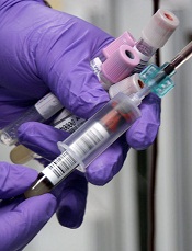
Photo by Jeremy L. Grisham
The US Food and Drug Administration (FDA) has issued an Emergency Use Authorization (EUA) for a laboratory test used to detect the Zika virus.
The test, which was developed by the Centers for Disease Control and Prevention (CDC), is called the Zika IgM Antibody Capture Enzyme-Linked Immunosorbent Assay (Zika MAC-ELISA).
It will be distributed to certain laboratories in the US and abroad over the next 2 weeks.
The test will not be available in US hospitals or other primary care settings.
The Zika MAC-ELISA test can detect human immunoglobulin M (IgM) antibodies to the Zika virus. These antibodies appear in the blood of an infected person 4 to 5 days after the start of the illness, and they remain in the blood for about 12 weeks.
The Zika MAC-ELISA test is intended for use in sera or cerebrospinal fluid when submitted with a patient-matched serum sample from individuals meeting CDC Zika clinical and epidemiological criteria for testing. This includes people with a history of symptoms associated with Zika and/or people who have recently traveled to an area that has active Zika transmission.
Results of Zika MAC-ELISA tests require careful interpretation. The test can give false-positive results when someone has been infected with a virus closely related to Zika (such as dengue virus).
When positive or inconclusive results occur, additional testing (plaque reduction neutralization test) to confirm the presence of antibodies to Zika virus will be performed by the CDC or a CDC-authorized laboratory.
Moreover, a negative test result does not necessarily mean a person has not been infected with Zika virus. If a sample is collected just after a person becomes ill, there may not be enough IgM antibodies for the test to measure, resulting in a false-negative.
Similarly, if the sample was collected more than 12 weeks after illness, it is possible that the body has successfully fought the virus and IgM antibody levels have dropped below the detectable limit.
The CDC will begin distributing the Zika MAC-ELISA test over the next 2 weeks to qualified laboratories in the Laboratory Response Network, an integrated network of domestic and international laboratories that can respond to public health emergencies.
About the EUA
An EUA allows the use of unapproved medical products or unapproved uses of approved medical products in an emergency. The products must be used to diagnose, treat, or prevent serious or life-threatening conditions caused by chemical, biological, radiological, or nuclear threat agents, when there are no adequate alternatives.
As there are no commercially available diagnostic tests approved by the FDA for the detection of Zika virus infection, the agency decided an EUA was crucial to ensure timely access to a diagnostic tool.
The FDA issued the EUA for the Zika MAC-ELISA test based on data submitted by the CDC and on the US Secretary of Health and Human Services’ declaration that circumstances exist to justify the emergency use of in vitro diagnostic tests for the detection of Zika virus and Zika virus infection. This EUA will end when the Secretary’s declaration ends, unless the FDA revokes it sooner. ![]()
FDA approves obinutuzumab for FL

The US Food and Drug Administration (FDA) has approved obinutuzumab (Gazyva) for certain patients with previously treated follicular lymphoma (FL).
Obinutuzumab is a glycoengineered, humanized, monoclonal antibody that selectively binds to the extracellular domain of the CD20 antigen on B cells.
The drug was previously approved by the FDA for use in combination with chlorambucil to treat patients with previously untreated chronic lymphocytic leukemia.
Now, obinutuzumab is approved for use in combination with bendamustine, followed by obinutuzumab alone, to treat patients with FL who did not respond to a rituximab-containing regimen or whose FL returned after such treatment.
The recommended dose and schedule for the regimen is:
- Obinutuzumab at 1000 mg by intravenous infusion on days 1, 8, and 15 of cycle 1; on day 1 of cycles 2-6 (28-day cycles); then every 2 months for 2 years.
- Bendamustine at 90 mg/m2 by intravenous infusion on days 1 and 2 of cycles 1-6.
Full prescribing information for obinutuzumab is available on the FDA website or at www.Gazyva.com.
Phase 3 study
The approval for obinutuzumab in FL is based on results from the phase 3 GADOLIN study. The trial included 413 patients with rituximab-refractory non-Hodgkin lymphoma, including 321 patients with FL, 46 with marginal zone lymphoma, and 28 with small lymphocytic lymphoma.
The patients were randomized to receive bendamustine alone (control arm) or a combination of bendamustine and obinutuzumab followed by obinutuzumab maintenance (every 2 months for 2 years or until progression).
The primary endpoint of the study was progression-free survival (PFS), as assessed by an independent review committee (IRC). The secondary endpoints were PFS as assessed by investigator review, best overall response, complete response (CR), partial response (PR), duration of response, overall survival, and safety profile.
Among patients with FL, the obinutuzumab regimen improved PFS compared to bendamustine alone, as assessed by IRC (hazard ratio [HR]=0.48, P<0.0001). The median PFS was not reached in patients receiving the obinutuzumab regimen but was 13.8 months in those receiving bendamustine alone.
Investigator-assessed PFS was consistent with IRC-assessed PFS. Investigators said the median PFS with the obinutuzumab regimen was more than double that with bendamustine alone—29.2 months vs 13.7 months (HR=0.48, P<0.0001).
Best overall response for patients receiving the obinutuzumab regimen was 78.7% (15.5% CR, 63.2% PR), compared to 74.7% for those receiving bendamustine alone (18.7% CR, 56% PR), as assessed by the IRC.
The median duration of response was not reached for patients receiving the obinutuzumab regimen and was 11.6 months for those receiving bendamustine alone.
The median overall survival has not yet been reached in either study arm.
The most common grade 3/4 adverse events observed in patients receiving the obinutuzumab regimen were neutropenia (33%), infusion reactions (11%), and thrombocytopenia (10%).
The most common adverse events of any grade were infusion reactions (69%), neutropenia (35%), nausea (54%), fatigue (39%), cough (26%), diarrhea (27%), constipation (19%), fever (18%), thrombocytopenia (15%), vomiting (22%), upper respiratory tract infection (13%), decreased appetite (18%), joint or muscle pain (12%), sinusitis (12%), anemia (12%), general weakness (11%), and urinary tract infection (10%).
About obinutuzumab
Obinutuzumab is being studied in a large clinical program, including the phase 3 GOYA and GALLIUM studies.
In GOYA, researchers are comparing obinutuzumab head-to-head with rituximab plus CHOP chemotherapy in first-line diffuse large B-cell lymphoma. In GALLIUM, researchers are comparing obinutuzumab plus chemotherapy head-to-head with rituximab plus chemotherapy in first-line indolent non-Hodgkin lymphoma.
Additional combination studies investigating obinutuzumab with other approved or investigational medicines, including cancer immunotherapies and small-molecule inhibitors, are planned or underway across a range of blood cancers.
Obinutuzumab was discovered by Roche Glycart AG, a wholly owned, independent research unit of Roche. In the US, obinutuzumab is part of a collaboration between Genentech and Biogen.
Genentech has a patient assistance program, Genentech Access Solutions, that can help qualifying patients access obinutuzumab and other Genentech medications.
The program is designed to help people navigate the access and reimbursement process and provide assistance to eligible patients in the US who are uninsured or cannot afford the out-of-pocket costs for their medicine. For more information, visit www.Genentech-Access.com. ![]()
DNA delivery vehicles may circumvent drug resistance in AML

Image courtesy of PNAS
DNA origami nanostructures may be used to overcome drug resistance in acute myeloid leukemia (AML), according to preclinical research published in the journal Small.
Researchers found they could create these nanostructures in 10 minutes and load them with the anthracycline daunorubicin.
When the team introduced the structures to daunorubicin-resistant AML cells, the drug delivery vehicles entered the cells via endocytosis.
This allowed the drug to bypass defenses in the cell membrane that are effective against the free drug.
Once the nanostructures broke down, daunorubicin flooded the cells and killed them off.
Other research groups have used this delivery technique to overcome drug resistance in solid tumors, but this is the first time researchers have shown the same technique works on drug-resistant leukemia cells.
To create the DNA origami nanostructures, the researchers used the genome of a common bacteriophage and synthetic strands that were designed to fold up the bacteriophage DNA.
Although the folded-up shape performs a function, the DNA itself does not, explained Patrick Halley, a graduate student at The Ohio State University in Columbus.
“[T]he DNA capsule doesn’t do anything except hold a shape,” Halley said. “It’s just a static, rigid structure that carries things. It doesn’t encode any proteins or do anything else that we normally think of DNA as doing.”
The researchers tested the DNA origami nanostructures in AML cell lines that had developed resistance to daunorubicin. When molecules of daunorubicin enter these cells, the cells recognize the drug molecules and eject them through openings in the cell wall.
“Cancer cells have novel ways of resisting drugs, like these ‘pumps,’ and the exciting part of packaging the drug this way is that we can circumvent those defenses so that the drug accumulates in the cancer cell and causes it to die,” said John Byrd, MD, of The Ohio State University.
“Potentially, we can also tailor these structures to make them deliver drugs selectively to cancer cells and not to other parts of the body where they can cause side effects.”
In tests, the resistant AML cells effectively absorbed molecules of daunorubicin when they were hidden inside the rod-shaped nanostructures.
The researchers tracked the nanostructures inside the cells using fluorescent tags. Each structure measures about 15 nanometers wide and 100 nanometers long, and each has 4 hollow, open-ended interior compartments.
Study author Christopher Lucas, PhD, of The Ohio State University, said the design of the nanostructures maximizes the surface area available to carry the drug.
“The way daunorubicin works is it tucks into the cancer cell’s DNA and prevents it from replicating,” Dr Lucas said. “So we designed a capsule structure that would have lots of accessible DNA base-pairs for it to tuck into. When the capsule breaks down, the drug molecules are freed to flood the cell.”
The researchers said they designed the nanostructures to be strong and stable so they wouldn’t fully disintegrate and release the bulk of the drug until it was too late for the cells to eject them.
And that’s what the team observed with a fluorescence microscope. The cells drew the nanostructures into the organelles that would normally digest them (if they were food).
When the nanostructures broke down, the drug flooded the cells and caused them to disintegrate. Most cells died within the first 15 hours after consuming the nanostructures.
“DNA origami nanostructures have a lot of potential for drug delivery, not just for making effective drug delivery vehicles, but enabling new ways to study drug delivery,” said Carlos Castro, PhD, of The Ohio State University.
“For instance, we can vary the shape or mechanical stiffness of a structure very precisely and see how that affects entry into cells.”
Dr Castro said he hopes to create a streamlined and economically viable process for building DNA origami nanostructures as part of a modular drug delivery system.
Dr Byrd said the technique should work on most any form of drug-resistant cancer if further research shows it can be translated to animal models. ![]()
AAs have lower rate of most blood cancers than NHWs

receiving treatment
Photo by Rhoda Baer
A new report suggests African Americans (AAs) have significantly lower rates of most hematologic malignancies than non-Hispanic white (NHW) individuals in the US.
AAs of both sexes had significantly lower rates of leukemia, Hodgkin lymphoma (HL), and non-Hodgkin lymphoma (NHL) than NHWs, but the rate of myeloma was significantly higher among AAs.
The death rates for these malignancies followed the same patterns, with the exception of HL. There was no significant difference in HL mortality between AAs and NHWs of either sex.
These findings can be found in the report, “Cancer Statistics for African Americans, 2016,” appearing in CA: A Cancer Journal for Clinicians.
To compile this report, the researchers used data from the Surveillance, Epidemiology, and End Results program and the Centers for Disease Control and Prevention’s National Program of Cancer Registries.
Incidence
For part of the report, the researchers compared the incidence of cancers between AAs and NHWs (divided by gender) for the period from 2008 to 2012.
Among females, the incidence of leukemia was 8.6 per 100,000 in AAs and 10.7 per 100,000 in NHWs (P<0.05). Among males, the incidence was 13.2 per 100,000 in AAs and 17.7 per 100,000 in NHWs (P<0.05).
The incidence of HL in females was 2.4 per 100,000 in AAs and 2.7 per 100,000 in NHWs (P<0.05). The incidence of HL in males was 3.2 per 100,000 in AAs and 3.4 per 100,000 in NHWs (P<0.05).
The incidence of NHL in females was 12.0 per 100,000 in AAs and 16.6 per 100,000 in NHWs (P<0.05). The incidence of NHL in males was 17.2 per 100,000 in AAs and 24.1 per 100,000 in NHWs (P<0.05).
The incidence of myeloma in females was 11.1 per 100,000 in AAs and 4.3 per 100,000 in NHWs (P<0.05). The incidence of myeloma in males was 14.8 per 100,000 in AAs and 7.0 per 100,000 in NHWs (P<0.05).
Mortality
The researchers also compared cancer mortality between AAs and NHWs (divided by gender) for the period from 2008 to 2012.
The death rate for female leukemia patients was 4.8 per 100,000 in AAs and 5.4 per 100,000 in NHWs (P<0.05). The death rate for male leukemia patients was 8.1 per 100,000 in AAs and 9.9 per 100,000 in NHWs (P<0.05).
The death rate for female HL patients was 0.3 per 100,000 for both AAs and NHWs. The death rate for male HL patients was 0.4 per 100,000 for AAs and 0.5 per 100,000 in NHWs (not significant).
The death rate for female NHL patients was 3.6 per 100,000 in AAs and 5.0 per 100,000 in NHWs (P<0.05). The death rate for male NHL patients was 5.9 per 100,000 in AAs and 8.3 per 100,000 in NHWs (P<0.05).
The death rate for female myeloma patients was 5.4 per 100,000 in AAs and 2.4 per 100,000 in NHWs (P<0.05). The death rate for male myeloma patients was 7.8 per 100,000 in AAs and 4.0 per 100,000 in NHWs (P<0.05).
The researchers noted that the reasons for the higher rates of myeloma and myeloma death among AAs are, at present, unknown. ![]()
Team identifies potential target for T-ALL therapy

New research suggests T-cell acute lymphoblastic leukemia (T-ALL) cells use the tricarboxylic acid (TCA) cycle to support their growth and survival.
Investigators say the findings could aid the development of therapeutics that can kill T-ALL cells by targeting an enzyme that exists in the TCA cycle—dihydrolipoamide S-succinyltransferase (DLST).
The team described this research in the journal Leukemia.
“Researchers have wrongly assumed that cancer cells do not use the TCA cycle to support their growth,” said study author Hui Feng, MD, PhD, of Boston University Medical Center in Massachusetts.
“Our new findings provide solid evidence that these cancer cells depend on the TCA cycle for their survival. Additionally, we demonstrated the importance of DLST in T-cell leukemia development and have identified a targetable enzyme for T-cell leukemia treatment.”
For this study, the investigators set out to examine the mechanisms underlying MYC-mediated tumorigenesis in T-ALL.
They used a zebrafish model of MYC-induced T-ALL to screen for genes that contribute to disease onset. The results suggested the TCA-cycle enzyme DLST is an important contributor to T-ALL development.
And experiments showed that heterozygous inactivation of DLST significantly delayed disease onset in the zebrafish, apparently without affecting the development of the fish.
Further analysis revealed that inhibiting the activity of DLST could effectively kill human T-ALL cells. Specifically, RNAi knockdown of DLST decreased cell viability and induced apoptosis in human T-ALL cell
lines.
The investigators found that knockdown of DLST disrupted the TCA cycle in the human T-ALL cells. But adding succinate, the downstream TCA-cycle intermediate, to the cells rescued defects in cell viability caused by DLST knockdown.
The investigators said the therapeutic benefit of DLST inhibition may extend to cancers other than T-ALL as well. ![]()
Orphan designation recommended for BTK inhibitor

The European Medicines Agency’s Committee for Orphan Medicinal Products (COMP) is recommending orphan designation for the second-generation BTK inhibitor acalabrutinib (ACP-196) for 3 indications.
The COMP is recommending the drug receive orphan designation as a treatment for chronic lymphocytic leukemia (CLL)/small lymphocytic lymphoma (SLL), mantle cell lymphoma, and Waldenström’s macroglobulinemia.
The COMP adopts an opinion on orphan drug designation, and that opinion is submitted to the European Commission (EC) for endorsement.
To be granted orphan designation by the EC, a medicine must be intended for the treatment, prevention, or diagnosis of a disease that is life-threatening and has a prevalence of up to 5 in 10,000 in the European Union. Additionally, the medicine must aim to provide significant benefit to those affected by the condition.
Orphan designation provides companies with development and market exclusivity incentives for designated compounds and medicines.
About acalabrutinib
Acalabrutinib is under development by AstraZeneca and Acerta Pharma BV. The drug is currently being evaluated in trials of patients with CLL/SLL, mantle cell lymphoma, Waldentröm’s macroglobulinemia, and a range of other hematologic malignancies and solid tumor cancers.
Data from a phase 1/2 trial of acalabrutinib in CLL were presented at the 2015 ASH Annual Meeting and simultaneously published in NEJM.
The researchers reported on 61 patients with relapsed CLL who had a median age of 62 (range, 44-84) and a median of 3 prior therapies (range, 1-13).
Patients enrolled in the phase 1 portion of the study received escalating doses of acalabrutinib, with a maximum dose of 400 mg once daily. Patients involved in the phase 2 portion of the study were treated with a 100 mg dose twice daily.
At a median follow-up of 14.3 months (range, 0.5 to 20), 53 patients were still receiving treatment.
The most common adverse events of all grades (occurring in at least 20% of patients) were headache (43%), diarrhea (39%), increased weight (26%), pyrexia (23%), upper respiratory tract infection (23%), fatigue (21%), peripheral edema (21%), hypertension (20%), and nausea (20%).
Grade 3/4 adverse events included diarrhea (2%), increased weight (2%), pyrexia (3%), fatigue (3%), hypertension (7%), and arthralgia (2%).
The overall response rate among the 60 evaluable patients was 95%. This included partial responses in 85% of patients and partial responses with lymphocytosis in 10%. The rate of stable disease was 5%.
The researchers noted that responses occurred in all dosing cohorts, and the response rate increased over time. ![]()
The European Medicines Agency’s Committee for Orphan Medicinal Products (COMP) is recommending orphan designation for the second-generation BTK inhibitor acalabrutinib (ACP-196) for 3 indications.
The COMP is recommending the drug receive orphan designation as a treatment for chronic lymphocytic leukemia (CLL)/small lymphocytic lymphoma (SLL), mantle cell lymphoma, and Waldenström’s macroglobulinemia.
The COMP adopts an opinion on orphan drug designation, and that opinion is submitted to the European Commission (EC) for endorsement.
To be granted orphan designation by the EC, a medicine must be intended for the treatment, prevention, or diagnosis of a disease that is life-threatening and has a prevalence of up to 5 in 10,000 in the European Union. Additionally, the medicine must aim to provide significant benefit to those affected by the condition.
Orphan designation provides companies with development and market exclusivity incentives for designated compounds and medicines.
About acalabrutinib
Acalabrutinib is under development by AstraZeneca and Acerta Pharma BV. The drug is currently being evaluated in trials of patients with CLL/SLL, mantle cell lymphoma, Waldentröm’s macroglobulinemia, and a range of other hematologic malignancies and solid tumor cancers.
Data from a phase 1/2 trial of acalabrutinib in CLL were presented at the 2015 ASH Annual Meeting and simultaneously published in NEJM.
The researchers reported on 61 patients with relapsed CLL who had a median age of 62 (range, 44-84) and a median of 3 prior therapies (range, 1-13).
Patients enrolled in the phase 1 portion of the study received escalating doses of acalabrutinib, with a maximum dose of 400 mg once daily. Patients involved in the phase 2 portion of the study were treated with a 100 mg dose twice daily.
At a median follow-up of 14.3 months (range, 0.5 to 20), 53 patients were still receiving treatment.
The most common adverse events of all grades (occurring in at least 20% of patients) were headache (43%), diarrhea (39%), increased weight (26%), pyrexia (23%), upper respiratory tract infection (23%), fatigue (21%), peripheral edema (21%), hypertension (20%), and nausea (20%).
Grade 3/4 adverse events included diarrhea (2%), increased weight (2%), pyrexia (3%), fatigue (3%), hypertension (7%), and arthralgia (2%).
The overall response rate among the 60 evaluable patients was 95%. This included partial responses in 85% of patients and partial responses with lymphocytosis in 10%. The rate of stable disease was 5%.
The researchers noted that responses occurred in all dosing cohorts, and the response rate increased over time.
FDA investigates issues with rivaroxaban trial

The US Food and Drug Administration (FDA) is investigating issues surrounding a defect in the device that was used to test international normalized ratios (INRs) in the ROCKET AF trial.
ROCKET AF was used to support approval for the direct oral anticoagulant rivaroxaban (Xarelto) in the US and European Union.
According to a legal brief filed in federal court earlier this week, the FDA has asked rivaroxaban’s manufacturer, Johnson & Johnson, if there was evidence of the defect in the INR-measuring device while ROCKET AF was underway.
The trial was a comparison of rivaroxaban and warfarin in patients with nonvalvular atrial fibrillation.
Results suggested rivaroxaban was noninferior to warfarin for preventing stroke or systemic embolism. And there was no significant difference between the treatment arms with regard to major or nonmajor clinically relevant bleeding.
However, an article recently published in The BMJ questioned these results because the Alere INRatio Monitor System (INRatio Monitor or INRatio2 Monitor and INRatio Test Strips), which was used to measure patients’ INRs during the trial, was recalled in December 2014 after giving falsely low test results.
The article suggested the system could have provided falsely low INR values in some patients in the warfarin arm. The lower values could have led investigators to give incorrect doses of warfarin, increasing the risk of bleeding for those patients and therefore giving a false impression of the comparative safety of rivaroxaban.
Two days after The BMJ article was published, the European Medicines Agency released a statement saying the defect with the INRatio system does not change the overall conclusions of ROCKET AF.
Researchers at the Duke Clinical Research Institute, which oversaw ROCKET AF, also assessed the impact of the INRatio defect. Their findings, published in NEJM, suggested the defect did not affect the trial’s outcome.
However, it appears the FDA still has some concerns. The agency has asked Johnson & Johnson whether there was evidence of the INRatio defect during ROCKET AF, according to a legal brief filed by lawyers representing individuals claiming injuries resulting from rivaroxaban.
The brief also cited internal emails that, according to the lawyers, showed some ROCKET AF investigators questioned the accuracy of the INRatio system during the trial. In fact, the lawyers said a special program was set up to investigate the malfunctions, but Johnson & Johnson never disclosed this information to the FDA.
Gene therapy could treat aplastic anemia

with telomeres in green
Image by Claus Azzalin
Researchers say they have found a new way to fight aplastic anemia—using a therapy designed to delay aging.
Four years ago, the group created telomerase gene therapy, an antiaging treatment based on repairing telomeres.
Now, they have found evidence to suggest this therapy can be effective against both acquired and inherited aplastic anemia.
The team reported preclinical results with the treatment in Blood.
In 2012, Maria A. Blasco, PhD, of Centro Nacional de Investigaciones Oncologicas in Madrid, Spain, and her colleagues described a strategy to repair telomeres.
They used adeno-associated virus (AAV9) vectors to deliver telomerase (Tert) gene therapy, which attenuated or reverted aging-associated telomere erosion in peripheral blood mononuclear cells.
For the current study, the researchers tested the therapy in a mouse model of acquired aplastic anemia and one of inherited aplastic anemia.
Acquired aplastic anemia
For the model of acquired aplastic anemia, the researchers depleted the TRF1 shelterin protein in the bone marrow. The team said this causes severe telomere uncapping and provokes a persistent DNA damage response at telomeres, which leads to fast clearance of hematopoietic stem and progenitor cells (HSPCs) deficient for Trf1.
The remaining HSPCs then undergo additional rounds of compensatory proliferation to regenerate the bone marrow, which leads to rapid telomere attrition. So this model recapitulates the compensatory hyperproliferation and short-telomere phenotype observed in acquired aplastic anemia.
The researchers induced Trf1 deletion with polyinosinic-polycytidylic acid injections given 3 times a week for 5 weeks. At that point, the mice began to show signs of aplastic anemia. A week after the last injection, the mice received either AAV9-Tert or AAV9-empty vectors.
Eighty-seven percent of the AAV9-Tert mice were still alive at 100 days, compared to 55% of mice in the empty vector group (P=0.0025).
In addition, 13% (4/31) of the mice treated with AAV9-Tert actually developed aplastic anemia, while 44% (16/36) of the control mice died showing “clear signs” of aplastic anemia (P=0.0006).
Finally, the researchers found that AAV9-Tert reversed telomere shortening in peripheral blood and bone marrow cells.
Inherited aplastic anemia
For the model of inherited aplastic anemia, the researchers transplanted irradiated wild-type mice with bone marrow from third-generation telomerase-deficient Tert knockout mice. These mice have short telomeres resulting from telomerase deficiency over 3 generations.
As with the previous model, these mice received AAV9-Tert or AAV9-empty vectors. The AAV9-Tert mice had a superior survival rate that nearly reached statistical significance (P=0.058).
The researchers also found that, compared to controls, AAV9-Tert-treated mice had significant increases in hemoglobin levels (P=0.003), erythrocyte counts (P=0.006), and platelet counts (P=0.035), as well as a trend toward an increase in leukocyte counts (P=0.09).
In addition, AAV9-Tert treatment led to a net increase in average telomere length of 5.18Kb, while control mice had a slight telomere shortening of 1.76Kb.
The researchers noted that there are types of aplastic anemia not associated with short telomeres. However, they believe these results provide proof of concept that gene therapy is a valid strategy for treating aplastic anemia.
Agent can mobilize HSCs for transplant, study suggests

in the bone marrow
HONOLULU—Early results from a pilot study suggest an investigational agent can effectively mobilize hematopoietic stem cells (HSCs) in healthy transplant donors.
The agent, CDX-301 (recombinant human Flt3 ligand), is a hematopoietic cytokine designed to expand dendritic cells and HSCs.
For the study, researchers tested CDX-301 in sibling-matched donors for hematopoietic stem cell transplant (HSCT) recipients with hematologic malignancies.
The team reported early results with 4 donor/recipient pairs at the 2016 BMT Tandem Meetings (abstract 479). The research was sponsored by Celldex Therapeutics, Inc., the company developing CDX-301.
Donor results
The donors received CDX-301 at 75 μg/kg/day for 5 days. Leukapheresis began on day 6 if the peripheral blood CD34+ count was 7/μL or greater. The goal CD34+ yield was at least 2x10⁶ per kilogram of recipient weight collected in 2 days of leukapheresis or less.
The donors could receive rescue plerixafor if the peripheral blood CD34+ count was less than 7/μL by day 8 or if the total CD34+ yield was less than 1x10⁶/kg after the second day of leukapheresis.
The researchers analyzed CDX-301-mobilized cells by flow cytometry and compared them to historical data for peripheral blood grafts mobilized by granulocyte colony-stimulating factor (G-CSF).
Compared to the G-CSF-mobilized grafts, CDX-301-mobilized grafts had an increase in CD127+ (IL-7R) naïve T cells, γδ T cells, natural killer cells, and B cells.
The CDX-301-mobilized grafts were more enriched with plasmacytoid dendritic cells. And dendritic cells from the CDX-301 grafts had a more mature phenotype, expressing CD80 and CD86.
“From these data and preclinical studies, CDX-301 appears to be an effective, targeted approach to mobilization comparable to G-CSF,” said Steven Devine, MD, of The Ohio State Comprehensive Cancer Center in Columbus.
“With a relatively short course of treatment, we are observing specificity for mobilized stem cells and a lack of toxicity, instead of broad cellular mobilization and side effects.”
CDX-301 related adverse events included dizziness (grade 2), back pain (grade 1), arthralgia (grade 1), injection site reaction (2 grade 1), dyspepsia (1 grade 1, 1 grade 2), and decreased platelet count (2 grade 1). There were no grade 3/4 adverse events.
Recipient results
Thus far, 4 recipients have undergone HSCT with CDX-301-mobilized grafts. Recipient 1 had acute myeloid leukemia, recipients 2 and 3 had mantle cell lymphoma, and recipient 4 had chronic myeloid leukemia.
Recipients 1 through 3 received myeloablative conditioning, while recipient 4 received reduced-intensity conditioning. All patients received standard graft-vs-host disease (GVHD) prophylaxis (tacrolimus and methotrexate).
Recipient 4 has not yet engrafted. The other 3 had both neutrophil engraftment (day 17, 18, and 19) and platelet engraftment (day 14, 25, and 40).
There have been no infectious complications, and recipients 1 and 3 have not developed GVHD.
Recipient 2 developed steroid-responsive, grade 2, upper gastrointestinal GVHD on day 18, followed by grade 1 skin GVHD on day 50 that progressed to stage 3. Overall, the patient had grade 2 GVHD on day 64. The patient’s lymphoma has also progressed.
The researchers said these results suggest CDX-301 is well tolerated and can mobilize CD34+ cells when given as a single agent. They said recipients experienced successful engraftment in an expected time frame.
And the additional naïve T cells and plasmacytoid dendritic cells in CDX-301-mobilized grafts (compared to G-CSF-mobilized grafts) may provide better outcomes in HSCT recipients.
Now, the researchers plan to explore CDX-301 in combination with plerixafor. Additional donor/patient pairs are being accrued to a second study cohort.