User login
Adding mAb to treatment extends PFS in MM

Photo by Linda Bartlett
The monoclonal antibody (mAb) elotuzumab may be a useful addition to the multiple myeloma (MM) arsenal, according to investigators involved in the phase 3 ELOQUENT-2 trial.
The trial showed that adding elotuzumab to treatment with lenalidomide and dexamethasone extended progression-free survival (PFS) in relapsed MM patients by about 5 months, on average, when compared to treatment with just lenalidomide and dexamethasone.
“It appears that, for patients with relapsed multiple myeloma who would otherwise be offered lenalidomide and dexamethasone, addition of this new targeted drug makes the outcomes even better,” said study investigator Sagar Lonial, MD, of Emory University in Atlanta, Georgia.
Dr Lonial presented data from ELOQUENT-2 at a presscast in advance of the 2015 ASCO Annual Meeting. Full data from the study will be presented at the meeting on June 2 as abstract 8508.
The study was funded by Bristol-Myers Squibb and AbbVie, the companies developing elotuzumab.
Dr Lonial explained that elotuzumab attaches to the cell surface protein SLAMF7, which is found on MM cells and natural killer (NK) cells. Scientists believe that elotuzumab mounts a 2-pronged attack on MM by targeting myeloma cells directly and by enhancing NK cells’ ability to kill myeloma cells.
In ELOQUENT-2, 646 patients with recurrent MM were randomized to receive elotuzumab plus lenalidomide and dexamethasone or only lenalidomide and dexamethasone (control).
The patients’ median age was 66. They had failed 1 to 3 prior treatments, and 35% of them were refractory to their last therapy. Thirty-two percent of patients had del(17p), and 9% had t[4;14].
At a median follow-up of 24 months, elotuzumab had reduced the risk of MM progression and death by 30%. Patients in the elotuzumab arm had significantly longer PFS than patients in the control arm—a median of 19.4 months and 14.9 months, respectively (P=0.0004).
The 1-year PFS was 68% in the elotuzumab arm and 57% in the control arm. The 2-year PFS was 41% and 27%, respectively. Dr Lonial pointed out that, unlike some other therapies, elotuzumab continued to improve PFS over time.
“[T]he idea of the maintenance of benefit over time really speaks to the power of an immune-based approach when we treat cancer,” he said.
“Patients who received elotuzumab had a longer duration of remission [and] a higher overall response rate, and this improvement in clinical parameters occurred without a significant increase in adverse events or toxicity. In fact, there was no reduction in quality of life for [patients in the] 3-drug arm.”
Mild infusion reactions occurred after the first few doses in 10% of patients who received elotuzumab. Most of these reactions were grade 1 or 2.
Common grade 3-4 adverse events (occurring in ≥ 15% of patients) in both the elotuzumab and control arms were neutropenia (25% and 33%, respectively) and anemia (15% and 16%, respectively).
In all, 210 patients died, 94 in the elotuzumab arm and 116 in the control arm.
“Based on this randomized, phase 3 trial, we hope that we will soon have a new treatment option for patients with relapsed or refractory myeloma . . . ,” Dr Lonial said.
The US Food and Drug Administration has already granted elotuzumab breakthrough therapy designation to treat MM patients who have received at least 1 prior therapy. ![]()
Modified T cells may treat GVHD
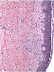
Image from PLOS ONE
A T-cell therapy designed to mitigate graft-vs-host disease (GVHD) is both feasible and safe, according to results of a pilot study published in Molecular Therapy.
To create this therapy, researchers transduced donor T cells with γ-retroviruses carrying a CD34-TK75 fusion gene.
The team said this allows them to track the cells via PET/CT and induce apoptosis by administering the antiviral drug ganciclovir if patients begin showing signs of GVHD.
“If donor T cells expand and cause severe graft-vs-host disease, we can give the patient ganciclovir, and it should kill the T cells and stop the process,” said study author John F. DiPersio, MD, PhD, of the Washington University School of Medicine in St Louis, Missouri.
Dr DiPersio and his colleagues tested the CD34-TK75-enriched T cells in 8 patients—4 with acute myeloid leukemia and 4 with myelodysplastic syndromes—who relapsed after allogeneic transplant. Six patients underwent [18F]FHBG PET/CT to track the T cells at several time points after infusion.
Patients received T-cell infusions ranging from 0.1 × 10⁶ cells/kg to 1.3 × 10⁶ cells/kg. Seven of the 8 patients received chemotherapy before T-cell infusion.
Four patients achieved a complete remission, 1 had progressive disease, and 3 patients died before the researchers could evaluate them for response.
Two patients developed GVHD—1 before T-cell infusion and 1 after. The patient who developed GVHD before infusion had grade 2 liver GVHD that resolved after an increased dose of steroids. The patient ultimately died of relapsed leukemia.
The patient who developed GVHD after T-cell infusion did not respond to chemotherapy or T-cell infusion. At 64 days after infusion, he exhibited symptoms consistent with grade 4 liver GVHD. He was treated with high-dose steroids and ganciclovir but did not respond. His primary cause of death was relapsed/progressive disease.
The mean overall survival after T-cell infusion was 165 days, 4 patients were still alive at 6 months, and 1 patient lived 408 days. All of the patients ultimately died.
The researchers said they did not detect any replication competent retrovirus or any antibodies against CD34-TK in any of the patients. The team also said there were no toxicities related to the CD34-TK75-enriched T cells.
Among patients who underwent imaging, there was no clear distinction between the [18F]FHBG biodistribution at baseline and later time points. And there was no difference between images in the patient who developed GVHD after infusion and patients who did not.
Past work from Dr DiPersio’s lab showed that leukemic mice that receive donor T cells and go on to develop GVHD show a characteristic migration pattern of the T cells through the body. When the cells gather early in the thymus, the mice develop GVHD. When T cells don’t migrate to the thymus, GVHD doesn’t occur.
In the current study, the patients received a relatively small number of CD34-TK75-enriched T cells, so the researchers were not able to show whether the migration patterns in mice were similar in humans.
However, based on the results of this study, Dr DiPersio and his colleagues are participating in a larger trial in partnership with a company based in Italy. The researchers are planning to include patients from multiple medical centers, and each participant should receive about 50 times more of the CD34-TK75-enriched T cells than were administered in this trial.
Washington University is the only center in the new trial that will be able to perform the imaging studies examining migration patterns of the T cells. ![]()
Company stops phase 3 PTCL trial

for use in a clinical trial
Photo by Esther Dyson
Takeda Pharmaceutical Company Limited has announced its decision to discontinue its phase 3 trial of the aurora A kinase inhibitor alisertib (MLN8237) in patients with relapsed or refractory peripheral T-cell lymphoma (PTCL).
Results of a pre-specified interim analysis indicated that alisertib was unlikely to meet the study’s primary endpoint: providing superior progression-free survival over the standard of care for PTCL.
Takeda said patients enrolled in the trial may continue to receive alisertib if they are thought to be benefitting from it and no safety concerns are present.
The company is encouraging patients to consult their study investigators to address any questions and before making any changes to their medication.
Takeda is working with trial investigators and local regulatory authorities to ensure that PTCL patients who participated in the study receive appropriate care.
The company is still investigating alisertib for use in small-cell lung cancer.
“While we are disappointed that alisertib will not be further investigated for relapsed or refractory peripheral T-cell lymphoma, we are optimistic about alisertib’s clinical development program in small-cell lung cancer,” said Michael Vasconcelles, MD, global head of the Takeda Oncology Therapeutic Unit.
“The randomized, phase 2 study of alisertib in small-cell lung cancer will continue as planned and is currently underway. Takeda also continues to support investigator-initiated research with alisertib and will evaluate its potential use in other oncology indications going forward.” ![]()
Equation provides new insight into blood flow

Engineers have devised an equation that yields simple predictions as to how quickly blood cells will migrate away from blood-vessel walls, how they will behave when they collide with each other, and how they will segregate during flow.
In the long run, these insights could help practitioners manipulate the mechanics of blood to design better blood transfusions, new techniques for drug delivery, and new processes for isolating blood-borne tumor cells.
Mike Graham, PhD, of the University of Wisconsin-Madison, and his colleagues described this work in Physical Review Letters.
“I’m really excited about this paper because it’s the first analytical theory for this phenomenon,” Dr Graham said. “It’s not very common that theory is ahead of experiments, but we’re in that position now.”
Dr Graham and his colleagues created complex computer simulations that showed how relatively stiff white blood cells and platelets interact with more flexible red blood cells.
As the different cells collide during blood flow, white cells tend to be pushed toward the walls of a blood vessel. This segregation process, called margination, creates some advantages; for example, letting white blood cells quickly exit the blood vessel to head to the site of an injury or infection.
However, the mechanical details of blood could spell both good news and bad in areas ranging from drug delivery to blood disorders to the spread of disease.
“I view my role as providing a fundamental basis of understanding for practitioners and for other engineers who are more directly connected with applications,” Dr Graham said.
Now, he is aiming to draw a firmer connection between mechanical insights and the biological functions they might impact. His group is working to refine the new equation to suit more complex flow situations and pursuing an experimental collaboration with Wilbur Lam, MD, PhD, a hematologist at Georgia Tech and Emory University in Atlanta.
Building on Dr Graham’s theoretical and simulation work, Dr Lam’s research group is creating microfluidic devices to study the behavior of blood cells. Dr Lam has developed a way to grow endothelial cells inside the artificial channels of the microfluidic devices.
“I think, together, our labs have really stumbled on how fluid mechanics may be able to explain a lot of the biological phenomena we see in blood,” Dr Lam said. “This can be related to a new way of understanding inflammation, infections, even transfusion medicine. It really pervades many different problems we see in hematology.”
Dr Graham said that capturing the physical nuances of blood vessels’ shape, size, and relative stiffness has tremendous value, even given the myriad other forces at work in the human body.
“We’d like to be able to convince practitioners that you don’t have to worry about all the details to capture the fundamental understanding of what’s going on,” Dr Graham said. “It’s extremely challenging to incorporate all the phenomena that might be important into a simulation. You have to make your case convincingly—if you want somebody to apply this research—that you’ve kept the important parts.”
Both researchers pointed out that sickle cell anemia has long been understood as both a mechanical and a biological problem. The defective red blood cells the disease causes are not only misshapen, but also stiffer than healthy red blood cells, meaning they block blood flow.
Yet, on a more detailed mechanical level, Drs Graham and Lam believe that sickle cells may literally poke and irritate the inner walls of blood vessels. If so, that would make sickle cell anemia not just a blood disorder but a disorder of the entire circulatory system. Their combined research strengths now create an opportunity to test that hypothesis.
“Biologists and hematologists have known for decades that these cells can get stuck, but what is less understood is that the blood vessel walls in the entire patient are really inflamed, and we don’t really know why,” Dr Lam said.
The researchers noted that a better understanding of blood-flow mechanics could help to make blood transfusions safer as well. Transfusions can sometimes set off heart attacks or lung damage, and the medical community isn’t entirely sure why. Dr Lam wants to find out if certain cells in stored, donated blood have mechanical properties that put patients at greater risk.
Though the collaboration between Drs Graham and Lam is still in an early stage, both researchers see the possibility of opening a new frontier in blood research.
“This would be a whole new category of things we could be looking at, and that’s why it’s so exciting,” Dr Lam said. “Suddenly, we have applications where the mechanics can be just as important.” ![]()
Familial factors linked to child’s risk of blood cancer

A new study has linked a father’s age at his child’s birth to the risk that the child will develop a hematologic malignancy as an adult, but this risk only proved significant among children without siblings.
Only-children whose fathers were 35 or older at the child’s birth were significantly more likely to develop hematologic malignancies than only-children with fathers who were younger than 25 at the child’s birth.
There was no association between these cancers and a mother’s age, either among only-children or those with siblings.
A previous study of more than 100,000 women also showed an association between paternal—but not maternal—age at a child’s birth and the risk of hematologic malignancy.
To further investigate the association, Lauren Teras, PhD, of the American Cancer Society in Atlanta, Georgia, and her colleagues analyzed data from women and men enrolled in the American Cancer Society Cancer Prevention Study-II Nutrition Cohort.
The team reported their findings in the American Journal of Epidemiology.
Among the 138,003 participants, there were 2532 cases of hematologic malignancies diagnosed between 1992 and 2009.
Subjects’ mothers tended to be younger at their birth than fathers, with median ages of 27 and 31, respectively. Almost a third of the fathers were 35 or older when a subject was born, compared with 17% of the mothers.
In the categorical analysis, the researchers found a positive association between older paternal age at a subject’s birth and the risk of hematologic malignancies in male, but not female, subjects. The hazard ratio (HR) was 1.35 for male subjects with fathers aged 35 and older compared to those whose fathers were younger than 25.
On the other hand, when paternal age was modeled as a continuous variable, there was no association with the risk of hematologic malignancy for males or females. Likewise, there was no association between maternal age at a subject’s birth and the risk of hematologic malignancy in male or female subjects.
However, among subjects without siblings, there was a significant, positive association with paternal age and the risk of hematologic malignancy (P=0.002).
When the researchers separated only-children by sex, they found a suggestive positive association between paternal age and hematologic malignancy for females (HR=1.40) and a significant association for males (HR=1.84). However, the linear spline was significant for males (P=0.01) and females (P=0.04).
There was no association between paternal age at a subject’s birth and the risk of hematologic malignancy among subjects with at least 1 sibling (HR=1.06).
The researchers said the fact that the association between paternal age and malignancy was significant in subjects with no siblings suggests it may be related to the “hygiene hypothesis”—the idea that exposure to mild infections in childhood, which might be more numerous with more siblings, are important to immune system development and may reduce the risk of immune-related diseases.
It is possible that the combination of having an older father and no siblings may promote cell proliferation in those individuals with an underdeveloped immune system and, as such, favors the development of cancers related to the immune system, the team said.
They added that this study suggests a need for further research to better understand the association between paternal age at a child’s birth and hematologic malignancies.
“The lifetime risk of these cancers is fairly low—about 1 in 20 men and women will be diagnosed with lymphoma, leukemia, or myeloma at some point during their lifetime—so people born to older fathers should not be alarmed,” Dr Teras said.
“Still, the study does highlight the need for more research to confirm these findings and to clarify the biologic underpinning for this association, given the growing number of children born to older fathers in the United States and worldwide.” ![]()
New method to assess cancer risk from pollutants

Photo by Tiffany Dawn Nicholson
Scientists say they have developed a faster, more accurate method to assess cancer risk from certain common environmental pollutants.
The group found they could analyze the immediate genetic responses of the skin cells of exposed mice and apply statistical approaches to determine whether or not those cells would eventually become cancerous.
The study focused on a class of pollutants known as polycyclic aromatic hydrocarbons (PAHs) that commonly occur in the environment as mixtures such as diesel exhaust and cigarette smoke.
“After only 12 hours, we could predict the ability of certain PAH mixtures to cause cancer, rather than waiting 25 weeks for tumors to develop,” said study author Susan Tilton, PhD, of Oregon State University in Corvallis.
For at least some PAH mixtures, the new method is not only quicker but produces more accurate cancer-risk assessments than are currently possible, she added.
Dr Tilton and her colleagues described the method in Toxicological Sciences.
“Our work was intended as a proof of concept,” Dr Tilton noted. “The method needs to be tested with a larger group of chemicals and mixtures. But we now have a model that we can use to develop larger-scale screening tests with human cells in a laboratory dish.”
The researchers believe the model will be particularly useful for screening PAHs, a large class of pollutants that result from combustion of organic matter and fossil fuels. PAHs are widespread contaminants of air, water, and soil. There are hundreds of different kinds, and some are known carcinogens, but many have not been tested.
Humans are primarily exposed to PAHs in the environment as mixtures, which makes it harder to assess their cancer risk. The standard calculation, Dr Tilton said, is to identify the risk of each element in the mix—if it’s known—and add them together.
But this method doesn’t work with most PAH mixes. It assumes the risk for each component is known, as well as which components are in a given mix. Often, that information is not available.
For this study, Dr Tilton and her colleagues examined 3 PAH mixtures that are common in the environment—coal tar, diesel exhaust, and cigarette smoke—and various mixtures of them.
The group found that each substance touched off a rapid and distinctive cascade of biological and metabolic changes in the skin cells of a mouse. The response amounted to a unique “fingerprint” of the genetic changes that occur as cells reacted to exposure to each chemical.
By matching patterns of genetic changes known to occur as cells become cancerous, the researchers found that some of the cellular responses were early indicators of developing cancers.
They also found that the standard method to calculate carcinogenic material underestimated the cancer risk of some mixtures and overestimated the combined risk of others.
“Our study is a first step in moving away from risk assessments based on individual components of these PAH mixtures and developing more accurate methods that look at the mixture as a whole,” Dr Tilton said. “We’re hoping to bring the methodology to the point where we no longer need to use tumors as our endpoint.” ![]()
Microbubbles can treat, track thrombosis

Image by Andre E.X. Brown
SAN FRANCISCO—Microbubbles can be used to simultaneously treat and monitor thrombosis, according to preclinical research presented at the ATVB/PVD 2015 Scientific Sessions.
The microbubbles double as agents for contrast-enhanced ultrasound imaging and targeted drug-delivery vehicles.
Experiments in mice showed that the microbubbles could deliver treatment directly to a blood clot, noticeably reducing its size without causing abnormal bleeding.
Karlheinz Peter, MD, PhD, of Baker IDI Heart and Diabetes Institute in Melbourne, Victoria, Australia, presented these results at the meeting as abstract 36.
He and his colleagues combined the thrombolytic agent urokinase with an activated-platelet-specific, single-chain antibody and placed the duo on the surface of microbubbles.
The researchers then tracked the microbubbles’ progress in a mouse model of carotid artery thrombosis induced by ferric-chloride.
The team monitored clot size in these mice with ultrasound imaging and measured bleeding time after experimental injury, while comparing 4 treatment groups:
- Microbubbles coated with both the targeting antibody and urokinase
- Targeted microbubbles and a high dose of urokinase administered separately
- Targeted microbubbles and a low dose of urokinase administered separately
- A control group with targeted microbubbles and no urokinase.
Microbubbles with urokinase on their surface significantly reduced clot size after 45 minutes, but microbubbles without urokinase did not. The mean percentage change in thrombus size from baseline was 97.16 ± 4.3 and 37.09 ± 5.6, respectively (P<0.001).
Urokinase administered alone could only match the efficacy of the treatment-loaded microbubbles if the drug was administered at a high dose. However, that significantly prolonged bleeding time from baseline—1079.25 ± 260.7 seconds vs 79.25 ± 6.5 seconds (P<0.001)—whereas, treatment with microbubbles did not.
While this research is in the early stages, the researchers believe the technology could help patients who develop venous thromboembolism and related conditions, such as heart attack and stroke. The team also believes the technology could have a major impact on treatment of patients in the emergency department. ![]()
Image by Andre E.X. Brown
SAN FRANCISCO—Microbubbles can be used to simultaneously treat and monitor thrombosis, according to preclinical research presented at the ATVB/PVD 2015 Scientific Sessions.
The microbubbles double as agents for contrast-enhanced ultrasound imaging and targeted drug-delivery vehicles.
Experiments in mice showed that the microbubbles could deliver treatment directly to a blood clot, noticeably reducing its size without causing abnormal bleeding.
Karlheinz Peter, MD, PhD, of Baker IDI Heart and Diabetes Institute in Melbourne, Victoria, Australia, presented these results at the meeting as abstract 36.
He and his colleagues combined the thrombolytic agent urokinase with an activated-platelet-specific, single-chain antibody and placed the duo on the surface of microbubbles.
The researchers then tracked the microbubbles’ progress in a mouse model of carotid artery thrombosis induced by ferric-chloride.
The team monitored clot size in these mice with ultrasound imaging and measured bleeding time after experimental injury, while comparing 4 treatment groups:
- Microbubbles coated with both the targeting antibody and urokinase
- Targeted microbubbles and a high dose of urokinase administered separately
- Targeted microbubbles and a low dose of urokinase administered separately
- A control group with targeted microbubbles and no urokinase.
Microbubbles with urokinase on their surface significantly reduced clot size after 45 minutes, but microbubbles without urokinase did not. The mean percentage change in thrombus size from baseline was 97.16 ± 4.3 and 37.09 ± 5.6, respectively (P<0.001).
Urokinase administered alone could only match the efficacy of the treatment-loaded microbubbles if the drug was administered at a high dose. However, that significantly prolonged bleeding time from baseline—1079.25 ± 260.7 seconds vs 79.25 ± 6.5 seconds (P<0.001)—whereas, treatment with microbubbles did not.
While this research is in the early stages, the researchers believe the technology could help patients who develop venous thromboembolism and related conditions, such as heart attack and stroke. The team also believes the technology could have a major impact on treatment of patients in the emergency department.
FDA issues draft guidance on blood donation
The US Food and Drug Administration (FDA) has released a draft guidance recommending changes to current policies aimed at reducing the risk of HIV transmission via blood products.
Among the recommended changes is a proposal to alter the policy that prevents men who have sex with men (MSM) from donating blood.
The FDA’s draft guidance is recommending that MSM be allowed to donate blood if they have abstained from sexual contact for 1 year.
If this draft guidance is implemented, the US would follow other countries that have lifted the lifetime ban on MSM blood donors in recent years, such as the UK, Canada, and South Africa.
Human rights groups—such as the Human Rights Campaign, the US’s largest lesbian, gay, bisexual, and transgender civil rights organization—have said the FDA’s proposed policy change is still discriminatory.
“While the new policy is a step in the right direction toward an ideal policy that reflects the best scientific research, it still falls far short of a fully acceptable solution because it continues to stigmatize gay and bisexual men,” said Human Rights Campaign Government Affairs Director David Stacy.
“This policy prevents men from donating life-saving blood based solely on their sexual orientation rather than actual risk to the blood supply. It simply cannot be justified in light of current scientific research and updated blood screening technology.”
On the other side of the debate, blood banking groups—including the American Red Cross, America’s Blood Centers, and the American Association of Blood Banks—have voiced their support of a 1-year deferral period for MSM, as data have suggested this group has an increased risk of contracting HIV.
According to the Centers for Disease Control and Prevention, MSM are more severely affected by HIV than any other group in the US.
“This change in policy would align the donor deferral period for MSM with criteria for other activities that may pose a similar risk of transfusion-transmissible infections,” the blood banking groups said in a joint statement.
“We believe the current FDA indefinite blood donation deferral for a man who has [had] sex with another man since 1977 is medically and scientifically unwarranted. The blood banking community strongly supports the use of rational, scientifically based deferral periods that are applied fairly and consistently among blood donors who engage in similar-risk activities.”
The FDA’s draft guidance seems to reflect that idea, as the 1-year deferral period does not only pertain to MSM. It also pertains to individuals who have a history of receiving a transfusion of whole blood or blood components, individuals with a history of syphilis or gonorrhea, and individuals who have had a tattoo or piercing in the last year, among others.
The draft guidance also includes recommendations pertaining to donor education material and donor history questionnaires, donor requalification, product retrieval and quarantine, testing requirements, and other issues.
The full guidance, available here, is open for comment.
Team links telomere degeneration and MDS

telomeres in green
Image by Claus Azzalin
New research has revealed a direct link between telomere degeneration and myelodysplastic syndromes (MDS).
“MDS risk correlates with advancing age, therapy-induced DNA damage, and/or shorter telomeres, but whether telomere erosion directly causes MDS is unknown,” said Simona Colla, PhD, of the MD Anderson Cancer Center in Houston, Texas.
“Our study provided genetic evidence that DNA damage caused by telomere loss is linked to this disorder.”
Dr Colla and her colleagues described this study in Cancer Cell.
The team’s in vitro and in vivo work showed that DNA damage caused by dysfunctional telomeres resulted in repressed expression of the gene SRSF2.
SRSF2 is an RNA splicing gene that plays a role in cellular processes. This change impacted common myeloid progenitors (CMPs), affecting their ability to differentiate or fully mature.
“This study established an intimate link across telomere biology, aberrant RNA splicing, and CMP differentiation,” said Ron DiPinho, MD, also of the MD Anderson Cancer Center.
“This may suggest that strategies to mitigate this DNA damage may be useful for preventing and/or treating MDS.”
Dr Colla added that the researchers’ findings “were consistent with long-standing observations that poor prognosis in MDS correlates strongly with short telomeres and elevated DNA damage in CMP cells.”
“This improved understanding should provide highly specific risk biomarkers for preventing and treating this incurable disease,” she said.
BMI may affect bleeding risk with warfarin

SAN FRANCISCO—Obese patients have a higher risk of bleeding while taking warfarin than patients of normal weight, according to research presented at the ATVB/PVD 2015 Scientific Sessions.
In a study of more than 800 patients taking the anticoagulant, obese individuals were almost twice as likely as normal-weight individuals to experience a major bleed.
And patients in the highest obesity class had the greatest risk of bleeding.
Adedotun A. Ogunsua, MD, of the University of Massachusetts Medical School in Worcester, presented these findings at the meeting as abstract 388.
He and his colleagues analyzed 863 patients taking warfarin. The researchers divided patients according to their body mass index (BMI) and looked at the rate of abnormal bleeding, including major gastrointestinal and brain bleeds and minor bleeds.
According to BMI, 21% of the patients were of normal weight, 38% were overweight, and 41.3% were obese, including class I (21%), class II (9%), and class III (11.3%).
Overall, 8.2% of patients (n=71) had abnormal bleeding during the study. Among the reported bleeds, about a third (35.2%) were major and two-thirds (64.8%) were minor.
Multivariable analyses showed that obesity (having a BMI higher than 30) significantly increased the risk of having a major bleed. The hazard ratio was 1.84 (P<0.001).
In univariate analyses, the risk of major bleeding increased with higher obesity categories. The hazard ratios were 1.30 for class I, 1.85 for class II, and 1.93 for class III.
The researchers said these results do not suggest patients should stop taking warfarin because of their weight. However, future studies are needed to understand why obesity increases the bleeding risk for patients receiving warfarin and whether similar risks exist for other anticoagulants.