User login
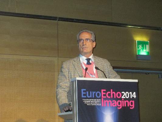
VIENNA – Three-dimensional transesophageal echocardiography “may be considered the gatekeeper for assessing the feasibility of and planning the strategy for percutaneous mitral valve repair,” Dr. Giovanni La Canna said at the annual meeting of the European Association of Association of Cardiovascular Imaging.
“Real-time, three-dimensional transesophageal echo is a new way of looking at the mitral valve,” and it supplies a highly accurate and reproducible anatomic view. It adds important additional information when selecting patients for percutaneous mitral valve repair by giving a comprehensive picture of annular dimensions and shape, intercommissural extension of target-leaflet lesions, and the extent of calcification on the annulus and leaflets, said Dr. La Canna, an echocardiographer at San Raffaele Hospital in Milan.
Three-dimensional echo aids both forms of percutaneous mitral valve repair: annuloplasty and clipping with the MitraClip system.
When performing mitral-leaflet clipping, use of 3D echo adds incremental information beyond 2D echo that aids in the selection of the site for transseptal puncture; facilitates device alignment; optimizes grasping of the leaflet with the clip; and helps in assessment of mitral-valve area and residual regurgitation, the need for additional clips, and any residual defect in the interatrial septum. For especially challenging cases, data from 2D echo can be integrated with the 3D data for the most complete imaging guidance, Dr. La Canna said.
Despite its value, 3D echo still has limitations. Resolution is currently low, parts of the imaging can “drop out” or contain artifacts, the catheter can produce an image shadow, and the image can decay during the course of the procedure, he said.
Dr. La Canna has been a consultant to Abbott Vascular, which markets the MitraClip.
On Twitter @mitchelzoler
EXPERT ANALYSIS FROM EUROECHO-IMAGING 2014

