User login
Case
An 82-year-old woman presented to the ED for evaluation of left eye pain. She stated the pain began earlier in the day as a mild discomfort but progressed and acutely worsened 2 hours prior to presentation. She rated the pain as a “9” out of “10” on a pain scale; she described the pain as constant, with throbbing behind her left eye. There was no pain associated with extraocular movements. Photosensitivity and increased lacrimation of the left eye were present, along with associated nausea. The patient denied any ocular trauma or previous surgery.
On examination, the patient’s left pupil measured 4 mm, was oval in shape, and was nonreactive with surrounding scleral edema. Visual acuity on the right eye was 20/50, but on the left eye, she had only finger-counting at 2 feet. Since tonometry was unavailable, bedside ultrasound images of the affected eye (Figure 1) and a comparison image of the patient’s normal, unaffected eye (Figure 2) were taken, revealing acute angle closure glaucoma (AACG) in the patient’s left eye.
Ocular Ultrasound
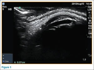
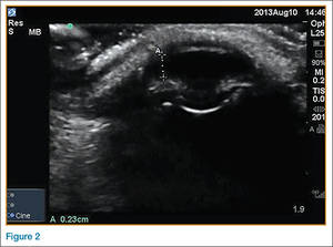
Diagnosis of AACG in the ED is generally made through clinical examination and tonometry. Tonometry, however, may be either unavailable or malfunctioning. In such cases, bedside ultrasound can serve as an alternative diagnostic tool. Ocular ultrasound is also beneficial in diagnosing AACG in patients who do not present with classic signs and symptoms of the condition. The abnormal bedside ultrasound can prompt earlier specialist consultation, which may decrease negative long-term sequelae.
Dr Rose is ultrasound fellow and clinical instructor in the department of emergency medicine, University of Kentucky, Lexington. Dr Cuevas is a resident in the department of emergency medicine, University of Kentucky, Lexington. Dr Dawson is an associate professor, director of ultrasound fellowship, and director of point-of-care ultrasound in the department of emergency medicine, University of Kentucky, Lexington.
- Rippey, J. Ultrasound of Acute angle closure glaucoma. The SonoCave Web site. Available at: http://thesonocave.com/2013/04/ultrasound-of-acute-angle-closure-glaucoma. Accessed February 23, 2016.
- Feng MT, Belin MW, Ambrósio R Jr, et al. Anterior chamber depth in normal subjects by rotating scheimpflug imaging. Saudi J Ophthalmol. 2011;25(3):255-259. doi:10.1016/j.sjopt.2011.04.005.
Case
An 82-year-old woman presented to the ED for evaluation of left eye pain. She stated the pain began earlier in the day as a mild discomfort but progressed and acutely worsened 2 hours prior to presentation. She rated the pain as a “9” out of “10” on a pain scale; she described the pain as constant, with throbbing behind her left eye. There was no pain associated with extraocular movements. Photosensitivity and increased lacrimation of the left eye were present, along with associated nausea. The patient denied any ocular trauma or previous surgery.
On examination, the patient’s left pupil measured 4 mm, was oval in shape, and was nonreactive with surrounding scleral edema. Visual acuity on the right eye was 20/50, but on the left eye, she had only finger-counting at 2 feet. Since tonometry was unavailable, bedside ultrasound images of the affected eye (Figure 1) and a comparison image of the patient’s normal, unaffected eye (Figure 2) were taken, revealing acute angle closure glaucoma (AACG) in the patient’s left eye.
Ocular Ultrasound
Diagnosis of AACG in the ED is generally made through clinical examination and tonometry. Tonometry, however, may be either unavailable or malfunctioning. In such cases, bedside ultrasound can serve as an alternative diagnostic tool. Ocular ultrasound is also beneficial in diagnosing AACG in patients who do not present with classic signs and symptoms of the condition. The abnormal bedside ultrasound can prompt earlier specialist consultation, which may decrease negative long-term sequelae.
Dr Rose is ultrasound fellow and clinical instructor in the department of emergency medicine, University of Kentucky, Lexington. Dr Cuevas is a resident in the department of emergency medicine, University of Kentucky, Lexington. Dr Dawson is an associate professor, director of ultrasound fellowship, and director of point-of-care ultrasound in the department of emergency medicine, University of Kentucky, Lexington.
- Rippey, J. Ultrasound of Acute angle closure glaucoma. The SonoCave Web site. Available at: http://thesonocave.com/2013/04/ultrasound-of-acute-angle-closure-glaucoma. Accessed February 23, 2016.
- Feng MT, Belin MW, Ambrósio R Jr, et al. Anterior chamber depth in normal subjects by rotating scheimpflug imaging. Saudi J Ophthalmol. 2011;25(3):255-259. doi:10.1016/j.sjopt.2011.04.005.