User login

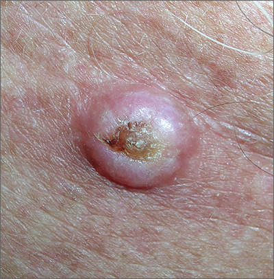
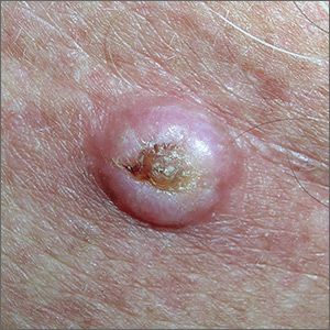
The FP noted that the lesion had a central keratin core (resembling a volcano) surrounded by a somewhat pearly raised border. He suspected that this was a squamous cell carcinoma (SCC) of the keratoacanthoma type and recommended a biopsy.
A shave biopsy was performed, and the result confirmed the diagnosis of SCC of the keratoacanthoma type. (See the Watch & Learn video on “Shave biopsy.”) The pathologist added that the lesion was well-differentiated (which was to be expected with a keratoacanthoma). While some keratoacanthomas actually resolve spontaneously, the standard of care is to treat them with either excision, electrodesiccation and curettage, or cryotherapy. On the follow-up visit, these options were presented to the patient and he decided to have cryotherapy. He accepted the risk of hypopigmentation in the treated area, but said that he preferred to avoid surgery.
The area was anesthetized with an injection of 1% lidocaine and epinephrine, and cryotherapy was performed with a 5 mm margin. The area was frozen with liquid nitrogen spray for 30 seconds and allowed to thaw. A second freeze for 30 seconds was then applied. Sun protection and sun avoidance were encouraged. At a 1-year follow-up, there was no recurrence and there was hypopigmentation at the treatment site.
Photos and text for Photo Rounds Friday courtesy of Richard P. Usatine, MD. This case was adapted from: Guzman A, Usatine R. Keratoacanthoma. In: Usatine R, Smith M, Mayeaux EJ, et al. Color Atlas of Family Medicine. 2nd ed. New York, NY: McGraw-Hill; 2013:977-980.
To learn more about the Color Atlas of Family Medicine, see: www.amazon.com/Color-Family-Medicine-Richard-Usatine/dp/0071769641/.
You can now get the second edition of the Color Atlas of Family Medicine as an app by clicking on this link: usatinemedia.com.
