User login
Birds are among the most popular pets in the United States, ranking fourth behind dogs, cats, and fish. According to a commercial survey,1 6.4 million US households own at least one pet bird—and are therefore at risk of a number of bacterial, protozoal, fungal, viral, or parasitic zoonoses (infectious diseases of animals that are communicable to humans).2
This review focuses on the most common pet bird-associated diseases, with implications for bird-keepers’ health. Unless specifically stated otherwise, these diseases are not routinely transmissible from human to human.
NO NEED TO AVOID OWNING A BIRD
Although one can indeed acquire an infection from a pet bird, this possibility need not discourage people from owning birds.3,4
The ability of a microorganism to make a person sick varies with the virulence of the organism, the dose to which a person is exposed, and the route of infection. For a bird owner, prevention often involves simple hygiene, handwashing, sanitation, and regular veterinary care for the bird.3 Many of these diseases are transmitted by ingestion of food contaminated by avian fecal matter or inhalation of contaminated dust. Therefore, bird owners should take steps to minimize dander in the environment. Furthermore, wearing a mask during cage cleaning is suggested.5 Obtaining pet birds from reputable domestic sources decreases the risk of acquiring a bird with an infectious disease.4,6

These hygienic measures are especially important for people who are more susceptible to infection, eg, the very young or very old, those in poor health to begin with, and those with compromised immunity.
Most of the illnesses one can acquire from birds are asymptomatic or self-limited in humans, but they should be considered if a bird-keeper has persistent or unusual symptoms of infection or if the pet bird was recently acquired or has been ill or died.
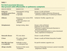
DISEASES PRESENTING WITH FLU-LIKE OR PULMONARY SYMPTOMS
Psittacosis (ornithosis, parrot fever, chlamydiosis)
Psittacosis is caused by Chlamydophila (formerly Chlamydia) psittaci. The organism has been reported to be present in 40% of birds.4,7 Humans are considered incidental hosts.
Transmission of C psittaci is usually via inhalation of aerosolized particles contaminated with infected bird excreta, nasal secretions, tissue, or feathers. Beak-to-mouth transmission and transmission via bird bites have been reported.8 Unlike Chlamydophila pneumoniae, C psittaci has not conclusively been reported to be transmitted from human to human.
Many infected pet birds show no signs of the disease, but others may have conjunctivitis, liver disease, or generalized signs of severe infection such as ruffled feathers, loss of appetite, diarrhea, or lime-green urates (R. Stevenson, personal communication, 2007).2,5,9,10
Recognizing that the patient has been exposed to birds is clinically useful, as in one study 90% of patients with psittacosis had been exposed to birds as pets or in their occupations.11
Atypical pneumonia is the most common presentation of psittacosis in humans. The symptoms typically begin 7 to 14 days after exposure, with fever, chills, prominent headache, photophobia, and cough. Hepatosplenomegaly is clinically detectable in 10% to 70% of patients. Serious but uncommon presentations include pericarditis, myocarditis, bacterial culture-negative endocarditis, mental status changes, and thrombophlebitis. The combination of pneumonitis and hepatosplenomegaly should prompt consideration of psittacosis.11
Influenza
All known subtypes of influenza A virus can infect birds (influenza B virus cannot). However, there are substantial genetic differences between the subtypes of influenza A that typically infect both people and birds.12
Avian influenza viruses affecting birds and humans (H5N1, H7N7, H9N2, and others), commonly called “bird flu,” emerged in 1997 in association with poultry.13,14 Highly pathogenic H5N1 viral infections have attracted particular attention, as they have killed millions of birds and infected several hundred humans, half of whom have died.13 Various avian influenza viruses that can affect humans have been isolated from captive birds such as parrots, canaries, and poultry and from wild waterfowl and migrating birds.15 To our knowledge, pet birds have not been implicated in transmission, but they are a potential source of infection if they have been in contact with infected birds.
Transmission from bird to human has usually been via inhaled droplets and, less likely, from contaminated environmental sources. According to the US Centers for Disease Control and Prevention, family clusters of H5N1 infection have been observed,16 but person-to-person transmission has been very rare, limited, and unsustained.13
Signs of influenza in birds can range from none to respiratory tract infection, decreased egg production, systemic illness, and death.15
Avian influenza should be suspected in people with flu symptoms who have been exposed to sick poultry or wild birds, have travelled to an endemic area, or have had direct contact with a person known or suspected to be infected with avian influenza.17 Information about areas of the world in which avian influenza is endemic or is breaking out is available at the World Health Organization Epidemic and Pandemic Alert and Response Web page (www.who.int/csr/disease/avian_influenza/updates/en/index.html).
Signs and symptoms of avian influenza in humans include fever, upper respiratory tract infection, cough, and gastrointestinal symptoms. With highly pathogenic viruses such as H5N1, the disease can progress very rapidly from onset to death. Less-pathogenic viruses such as H9N2 cause milder symptoms.16
Histoplasmosis
Histoplasma capsulatum is a fungus that colonizes the gastrointestinal tract of birds and contaminates the soil via bird and bat droppings. The most highly endemic regions of the world are the Ohio and Mississippi River valleys. Typical pet birds such as canaries and parrots are not susceptible to symptomatic infection, but doves and pigeons (often treated as pets by bird-lovers) may become colonized if they contact contaminated birds or excreta (R. Stevenson, personal communication, 2007).5,18
Humans commonly acquire the organisms when they inhale disrupted soil contaminated with the organism. Human-to-human transmission has not been reported.
The degree of human illness depends on the inoculum size and the immunity of the person infected. In more than 90% of cases, the primary infection is minimally symptomatic or goes unnoticed. The typical incubation time is 7 to 21 days. In patients who become ill, symptoms include fever, chills, headache, nonproductive cough, and adenopathy-mediated chest pain. Disseminated disease generally occurs in immunocompromised patients and presents with fever, weight loss, hepatosplenomegaly, and pancytopenia.18
Newcastle disease (avian pneumoencephalitis)
The virus that causes Newcastle disease, avian paramyxovirus 1, can affect animals, reptiles, birds, and people. It is most common in wild birds, but parrots are also highly susceptible and can be reservoirs that continue to shed the virus for up to 12 months after the acute illness has subsided. Illegally imported Amazon parrots are the most likely source of infection for US households. The virus is spread through an infected bird’s feces and secretions from the nose, mouth, and eyes and can be carried on a person’s clothing, footwear, and equipment.
Some infected birds show no signs of it; others have respiratory signs, green diarrhea, muscle tremors, circling, paralysis, or swelling of tissues around the eyes and neck. The mortality rate in infected birds can be up to 100%.19
Human infection most often results in conjunctivitis. Chills, fever, and lethargy are exceptionally rare.15 Because the virus is prevalent in poultry, poultry workers are at greatest risk of infection. Rapid recovery in humans is common. People with conjunctivitis due to Newcastle disease virus should avoid contact with birds.2,20
Q fever
Q fever is caused by Coxiella burnetii, a gram-negative pleomorphic bacillus. Ticks and vertebrates (goats, sheep, and, less commonly, birds) are natural reservoirs for the organism. Human infection results from contact with infected animals or inhalation of dust contaminated with infected excreta or placental tissue.2 Birds may harbor the infection in experimental and natural settings.21,22
Symptoms in humans typically include fever, pneumonitis, severe headache, and photophobia. Meningitis, hepatitis, and thromboses are seen in more-severe disease. Infection acquired in pregnancy may lead to prematurity, abortion, or stillbirth.18,21
West Nile fever and West Nile encephalitis
Wild birds such as corvids and raptors harbor the West Nile virus; pet songbirds (passerines) can harbor it as well.23,24 The principal means of transmission of West Nile infection from birds to humans is via a mosquito biting an infected bird and then biting a human. Direct bird-to-human transmission has not been described.18 Infected birds may be asymptomatic or appear ill or reluctant to fly and die of disseminated viral infection.23
The incubation period in humans is generally 3 to 14 days, followed by the sudden onset of fever, malaise, nausea, vomiting, rash, lymphadenopathy, and retro-orbital pain. Neurologic presentations—ataxia, extrapyramidal signs, cranial nerve abnormalities, myelitis, optical neuritis, and seizures—are quite rare and generally occur in the elderly or immunocompromised. Fewer than 1% of affected people develop more-severe disease, such as acute encephalitis, aseptic meningitis, or Guillain-Barré syndrome.25
Allergic alveolitis
Allergic alveolitis (hypersensitivity pneumonitis, parakeet dander pneumoconiosis, pigeon lung disease, bird-breeder’s lung, bird-fancier’s disease) is not a zoonosis; the term describes diffuse parenchymal lung disease caused by repeated exposure to an inhaled allergen.26 However, it should be considered in patients with pulmonary symptoms and bird exposure. Avian proteins are a known trigger.27
Acute allergic alveolitis is clinically indistinguishable from a respiratory infection. It is characterized by the abrupt onset of an intense nonproductive cough, chest tightness, dyspnea, chills, fever, myalgia, and malaise. Symptoms gradually improve over 24 to 48 hours without antigen exposure but recur with repeated exposure.
Subacute illness presents with similar symptoms, which gradually worsen over weeks to months, and it can be indistinguishable from interstitial lung disease. Many patients with chronic allergic alveolitis are suspected of having tuberculosis or fungal pneumonia or receive a misdiagnosis of idiopathic pulmonary fibrosis.
Patients usually have a favorable outcome if the allergen is removed. If exposure continues, irreversible pulmonary fibrosis may develop.28
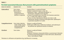
DISEASES PRESENTING WITH GASTROINTESTINAL SYMPTOMS
Salmonellosis
Nontyphoidal Salmonella species colonize the gastrointestinal tract of many animals, including birds. Up to 80% of chicken eggs are contaminated with this gram-negative bacterium.
Spread of nontyphoidal Salmonella to humans is much more common from poultry, poultry products, and pet reptiles than from pet birds, although ducks and baby chicks have transmitted infection to humans. Hand-to-mouth spread occurs after contact with pets or pet excreta.29,30 Infected birds may be healthy carriers, may develop enteritis or hemorrhagic hepatosplenic disease, or may even die.2,9
Gastroenteritis due to nontyphoidal Salmonella in humans begins with nausea, vomiting, fever, and loose, nonbloody diarrhea about 48 hours after ingestion. Most gastroenteritis infections are self-limited, with resolution of fever within 48 to 72 hours and resolution of diarrhea within 4 to 10 days.31
Systemic or severe infection warranting “preemptive” therapy is more likely in immunosuppressed patients, in patients with reduced gastric acid or impaired gastrointestinal mucosal integrity, in infants less than 3 months of age, and in patients with chronic gastrointestinal tract disease, malignant neoplasms, hemoglobinopathies, or infection with the human immunodeficiency virus.31 Systemic nontyphoidal Salmonella infection may settle in structurally abnormal sites such as in existing fractures, severe degenerative joint disease, organs affected by stones, or abnormal lung tissue. Large-vessel arteritis due to non-typhoidal Salmonella should be suspected in a person at risk (particularly if the person is elderly) who presents with back, chest, or abdominal pain preceded by gastroenteritis.31,32
Campylobacteriosis
The main reservoirs for Campylobacter jejuni are wild birds and poultry, although this bacterium can also affect other animals and pet birds.2 The most commonly affected pet birds are psittaciforms (parrots) and passeriforms (finches and canaries).
The organism colonizes the small intestine and colon of birds and can be spread to humans through contact with feces or carcasses of infected animals.33–35 Birds with campylobacteriosis develop hepatitis, lethargy, loss of appetite, weight loss, and yellow diarrhea and often die of the illness (R. Stevenson, personal communication, 2007).
The most important mode of transmission to humans is through handling or consuming chicken, milk, or other products contaminated with feces of carrier animals. However, in up to 24% of cases, the source of infection is unknown.33,34
Human infection with C jejuni most commonly leads to an acute, self-limited gastrointestinal illness characterized by fever, diarrhea, and abdominal cramps. The diarrhea is typically watery or bloody and occurs 8 to 10 times a day at peak illness. Fever can persist for up to a week. Most cases resolve within 7 days, but some patients may have a relapsing diarrheal illness lasting several weeks. Between 20% and 40% of cases of Guillain-Barré syndrome are preceded by infection with C jejuni.35
Giardiasis
Giardiasis is an intestinal protozoal infection caused by Giardia species (primarily G lamblia) that affect humans and other mammals. The parasite is found in bird droppings, but the role of birds in transmission to humans is unclear. Most infections are transmitted via contaminated surface water supplies, although person-to-person transmission has been documented.36 Infected pet birds have signs of gastroenteritis and can be treated, but reinfection often occurs.37
Giardia infections in humans are often asymptomatic, but about 50% of patients have diarrhea, abdominal pain, bloating, belching, nausea, and vomiting 3 days to 3 weeks after ingesting the parasite. A clinical clue may be new-onset lactose intolerance. Symptoms usually resolve after a week. Prolonged infection occurs in up to 20% of patients. People with hypochlorhydria or hypogammaglobulinemia, children, and travelers to endemic areas are at higher risk of infection.37
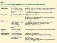
DISEASES PRESENTING WITH SKIN SYMPTOMS
Pasteurellosis
Pasteurellosis is caused by Pasteurella multocida, an inhabitant of the healthy nasopharynx of some birds and also the causative agent of avian cholera.38 Many pet birds that acquire systemic Pasteurella infection from a cat bite die of avian cholera (Stevenson R, personal communication, 2007).
Pasteurella organisms are transmissible to humans via bites or scratches from pet birds. Infected wounds in humans are usually red and painful, but the physical findings may lead one to underestimate the severity of infection. Transmission via respiratory droplets is rare but may cause acute or subacute bronchitis, pneumonia, or septicemia.38
Erysipeloid
Erysipeloid, caused by the bacterium Erysipelothrix rhusiopathiae, is transmissible to humans via contact with domestic or wild fowl. Infection in pet birds can cause sepsis but is rarely seen in veterinary practice (R. Stevenson, personal communication, 2007).38,39
Human infection typically affects broken skin, causing a dramatic, localized skin infection that is painful and pruritic; at first it is livid-red, then blue-red. The infection can spread to nearby joints. Septicemia and endocarditis in humans are rare complications.40
Cryptococcosis
Cryptococcosis, caused by the encapsulated yeast Cryptococcus neoformans, can be harbored and transmitted by asymptomatic pet birds such as cockatoos via colonization of the gastrointestinal tract.38,41 The organism is found in soil contaminated by feces of colonized birds.
Pulmonary symptoms and meningitis are more typical of cryptococcal disease in general, although when contracted from a pet bird via a break in the skin, cutaneous cryptococcosis usually presents with skin lesions resembling cellulitis, molluscum, herpes, and Kaposi sarcomalike papulonodules.42 Infection beyond the skin in immunocompromised patients may involve the lungs and the central nervous system.41 Prostate and eye infections have also been reported.38
Avian mite dermatitis
Birds carry several kinds of mites: feather or “red” mites (which do not affect humans) and mites that can affect humans such as Ornithonyssus sylviarum (the northern fowl mite) and Dermanyssus gallinae (the poultry mite or chicken mite) (R. Stevenson, personal communication, 2007). O sylviarum and D gallinae are found in the commercial poultry industry, but uncommonly, pet birds can harbor them (R. Stevenson, personal communication, 2007).42–44
In humans, mites can cause an intensely pruritic, papular-papulovesicular eruption.42
Nontuberculous mycobacteriosis
Nontuberculous mycobacteria (Mycobacterium species chelonae, abscessus, fortuitum, avium, kansasii, ulcerans, and marinum) are ubiquitous in the environment and can colonize animals. M avium subsp avium causes avian tuberculosis.38 Birds may carry mycobacterial organisms on beaks, claws, and talons, facilitating passage to humans (J.M. Gaskin, personal communication, 2006). In one reported case, M chelonae skin infection was probably transmitted in this manner to a bird-keeper via a bird bite.45
In symptomatic nontuberculous mycobacterial infections, the site of inoculation usually determines the presenting signs. Mycobacteriosis should be suspected when skin or lung infections fail to improve with empiric treatment. Patients with structurally abnormal lungs or immunosuppression may be at higher risk of pulmonary or disseminated disease from infected birds.46,47
Acknowledgment: The authors extend special thanks to Jack M. Gaskin, DVM, PhD, Department of Infectious Disease and Pathology, University of Florida, Gainesville, FL; Rhoda Stevenson, DVM, Exotic Bird Hospital, Jacksonville, FL; and Stephanie L. Hines, MD, Section of General Internal Medicine, Mayo Clinic, Jacksonville, FL, for their review of this paper. Editing, proofreading, and reference verification were provided by the Mayo Clinic Section of Scientific Publications.
- American Pet Product Manufacturer’s Association. 2005/2006 National Pet Owners Survey. In: Greenwich CT, American Pet Products Manufacturers Association 2005. www.appma.org.
- Krauss H, Weber A, Appel M, et al. Zoonoses: Infectious Diseases Transmissible From Animals to Humans, 3rd ed. Washington, DC: American Society for Microbiology Press, 2003.
- Hemsworth S, Pizer B. Pet ownership in immunocompromised children—a review of the literature and survey of existing guidelines. Eur J Oncol Nurs 2006; 10:117–127.
- Smith KA, Bradley KK, Stobierski MG, Tengelsen LA; National Association of State Public Health Veterinarians Psittacosis Compendium Committee. Compendium of measures to control Chlamydophila psittaci (formerly Chlamydia psittaci) infection among humans (psittacosis) and pet birds, 2005. J Am Vet Med Assoc 2005; 226:532–539.
- Jacob JP, Gaskin JM, Wilson HR, Mather FB. Avian diseases transmissible to humans. University of Florida Institute of Food and Agricultural Sciences Extension. http://edis.ifas.ufl.edu/PS019. Accessed 1/31/2009.
- PAWS (Pets Are Wonderful Support). Safe Pet Guidelines: A Comprehensive Guide for Immunocompromised Animal Guardians. www.pawssf.org/SafePetGuide/SPG8.pdf(pp10–13). Accessed 1/31/2009.
- Moroney JF, Guevara R, Iverson C, et al. Detection of chlamydiosis in a shipment of pet birds, leading to recognition of an outbreak of clinically mild psittacosis in humans. Clin Infect Dis 1998; 26:1425–1429.
- Glaser C, Lewis P, Wong S. Pet-, animal-, and vector-borne infections. Pediatr Rev 2000; 21:219–232.
- Grimes JE. Zoonoses acquired from pet birds. Vet Clin North Am Small Anim Pract 1987; 17:209–218.
- Spenser EL. Common infectious diseases of psittacine birds seen in practice. Vet Clin North Am Small Anim Pract 1991; 21:1213–1230.
- Richards MJ. Psittacosis. In: Rose B, editor: UpToDate. Waltham, MA: UpToDate, 2008.
- US Centers for Disease Control and Prevention. Avian influenza A viruses. www.cdc.gov/flu/avian/gen-info/avian-influenza.htm. Accessed 1/31/2009.
- US Centers for Disease Control and Prevention. Key facts about avian influenza (bird flu) and avian influenza A (H5N1) virus. www.cdc.gov/flu/avian/gen-info/facts.htm. Accessed 1/31/2009.
- Swayne DE, King DJ. Avian influenza and Newcastle disease. J Am Vet Med Assoc 2003; 222:1534–1540.
- Capua I, Alexander DJ. Human health implications of avian influenza viruses and paramyxoviruses. Eur J Clin Microbiol Infect Dis 2004; 23:1–6.
- US Centers for Disease Control and Prevention. Avian influenza A virus infections of humans. www.cdc.gov/flu/avian/gen-info/avian-flu-humans.htm. Accessed 1/31/2009.
- US Centers for Disease Control and Prevention. Updated Interim Guidance for Laboratory Testing of Persons with Suspected Infection with Avian Influenza A (H5N1) Virus in the United States. www2a.cdc.gov/han/ArchiveSys/ViewMsgV.asp?AlertNum=00246. Accessed 11/2008.
- Deepe GS. Histoplasma capsulatum. In: Mandell GL, Douglas RG, Bennett JE, Dolin R, editors. Principles and Practice of Infectious Diseases, 6th ed. New York: Churchill Livingstone, 2005:3012–3025.
- Cross GM. Newcastle disease. Vet Clin North Am Small Anim Pract 1991; 21:1231–1239.
- Beard C. Velogenic Newcastle disease. In: Committee on Foreign Animal Diseases of the United States. Foreign Animal Diseases. Richmond, VA: Animal Health Association, 1998:370–376.
- Behymer D, Riemann HP. Coxiella burnetii infection (Q fever). J Am Vet Med Assoc 1989; 194:764–767.
- To H, Sakai R, Shirota K, et al. Coxiellosis in domestic and wild birds from Japan. J Wild Dis 1998; 34:310–316.
- Weingartl HM, Neufeld JL, Copps J, Marszal P. Experimental West Nile virus infection in blue jays (Cyanocitta cristata) and crows (Corvus brachyrhynchos). Vet Pathol 2004; 41:362–370.
- Komar N, Panella NA, Langevin SA, et al. Avian hosts for West Nile virus in St. Tammany Parish, Louisiana, 2002. Am J Trop Med Hyg 2005; 73:1031–1037.
- Petersen LR. Clinical manifestations, diagnosis, and treatment of West Nile virus infection. In: Rose B, editor: UpToDate. Waltham, MA: UpToDate, 2008.
- King TE. Classification and clinical manifestations of hypersensitivity pneumonitis (extrinsic allergic alveolitis). In: Rose B, editor: UpToDate. Waltham, MA: UpToDate, 2008.
- du Marchie Sarvaas GJ, Merkus PJ, de Jongste JC. A family with extrinsic allergic alveolitis caused by wild city pigeons: a case report. Pediatrics 2000; 105:E62.
- King TE. Treatment and prognosis of hypersensitivity pneumonitis (extrinsic allergic alveolitis). In :Rose B, editor: UpToDate. Waltham, MA: UpToDate, 2008.
- Woodward DL, Khakhria R, Johnson WM. Human salmonellosis associated with exotic pets. J Clin Microbiol 1997; 35:2786–2790.
- Hohmann EL. Microbiology and epidemiology of Salmonellosis. In: Rose B, editor: UpToDate. Waltham, MA: UpToDate, 2008.
- Hohmann EL. Approach to the patient with nontyphoidal Salmonella in a stool culture. In: Rose B, editor: UpToDate. Waltham, MA: UpToDate, 2008.
- Pegues DA, Miller SI. Salmonellosis. In: Kasper DL, Fauci AS, Longo DL, et al, editors. Harrison's Principles of Internal Medicine, 17th ed. New York: McGraw-Hill Medical, 2008:956–957.
- Allos BM. Microbiology, pathogenesis, and epidemiology of Campylobacter infection. In: Rose B, editor: UpToDate. Waltham, MA: UpToDate, 2008.
- Padungton P, Kaneene JB. Campylobacter spp in human, chickens, pigs and their antimicrobial resistance. J Vet Med Sci 2003; 65:161–170.
- Allos BM. Clinical features and treatment of Campylobacter infection. In: Rose B, editor: UpToDate. Waltham, MA: UpToDate, 2008.
- Harris JM. Zoonotic diseases of birds. Vet Clin North Am Small Anim Pract 1991; 21:1289–1298.
- Leder K, Weller PF. Giardiasis in adults. In: Rose B, editor: UpToDate. Waltham, MA, 2008.
- Hubálek Z. An annotated checklist of pathogenic microorganisms associated with migratory birds. J Wild Dis 2004; 40:639–659.
- Gartrell BD, Alley MR, Mack H, Donald J, McInnes K, Jansen P. Erysipelas in the critically endangered kakapo (Strigops habroptilus). Avian Pathol 2005; 34:383–387.
- Hand WL, Ho H. Erysipelothrix infection. In: Rose B, editor: UpToDate. Waltham, MA: UpToDate, 2008.
- Nosanchuk JD, Shoham S, Fries BC, Shapiro DS, Levitz SM, Casadevall A. Evidence of zoonotic transmission of Cryptococcus neoformans from a pet cockatoo to an immunocompromised patient. Ann Intern Med 2000; 132:205–208.
- Rosen T, Jablon J. Infectious threats from exotic pets: dermatological implications. Dermatol Clin 2003; 21:229–236.
- Gupta AK, Billings JK, Ellis CN. Chronic pruritis: an uncommon cause. Avian mite dermatitis caused by Ornithonyssus sylviarum. Arch Dermatol 1988; 124:1102–1103,1105–1106.
- Orton D, Warren L, Wilkinson J. Avian mite dermatitis. Clin Exp Dermatol 2000; 25:129–131.
- Larson J, Gerlach S, Thompson K, et al. Mycobacterium chelonae/abcessus infection caused by a bird bite. Infect Dis Clin Pract 2008; 16:60–61.
- Griffith DE, Wallace JR. Clinical manifestations of non-tuberculous mycobacterial pulmonary infections in HIV-negative patients. In: Rose B, editor: UpToDate. Waltham, MA: UpToDate, 2008.
- Griffith DE, Wallace JR. Epidemiology of nontuberculous mycobacterial infections. In: Rose B, editor: UpToDate. Waltham, MA: UpToDate, 2006.
Birds are among the most popular pets in the United States, ranking fourth behind dogs, cats, and fish. According to a commercial survey,1 6.4 million US households own at least one pet bird—and are therefore at risk of a number of bacterial, protozoal, fungal, viral, or parasitic zoonoses (infectious diseases of animals that are communicable to humans).2
This review focuses on the most common pet bird-associated diseases, with implications for bird-keepers’ health. Unless specifically stated otherwise, these diseases are not routinely transmissible from human to human.
NO NEED TO AVOID OWNING A BIRD
Although one can indeed acquire an infection from a pet bird, this possibility need not discourage people from owning birds.3,4
The ability of a microorganism to make a person sick varies with the virulence of the organism, the dose to which a person is exposed, and the route of infection. For a bird owner, prevention often involves simple hygiene, handwashing, sanitation, and regular veterinary care for the bird.3 Many of these diseases are transmitted by ingestion of food contaminated by avian fecal matter or inhalation of contaminated dust. Therefore, bird owners should take steps to minimize dander in the environment. Furthermore, wearing a mask during cage cleaning is suggested.5 Obtaining pet birds from reputable domestic sources decreases the risk of acquiring a bird with an infectious disease.4,6

These hygienic measures are especially important for people who are more susceptible to infection, eg, the very young or very old, those in poor health to begin with, and those with compromised immunity.
Most of the illnesses one can acquire from birds are asymptomatic or self-limited in humans, but they should be considered if a bird-keeper has persistent or unusual symptoms of infection or if the pet bird was recently acquired or has been ill or died.
DISEASES PRESENTING WITH FLU-LIKE OR PULMONARY SYMPTOMS
Psittacosis (ornithosis, parrot fever, chlamydiosis)
Psittacosis is caused by Chlamydophila (formerly Chlamydia) psittaci. The organism has been reported to be present in 40% of birds.4,7 Humans are considered incidental hosts.
Transmission of C psittaci is usually via inhalation of aerosolized particles contaminated with infected bird excreta, nasal secretions, tissue, or feathers. Beak-to-mouth transmission and transmission via bird bites have been reported.8 Unlike Chlamydophila pneumoniae, C psittaci has not conclusively been reported to be transmitted from human to human.
Many infected pet birds show no signs of the disease, but others may have conjunctivitis, liver disease, or generalized signs of severe infection such as ruffled feathers, loss of appetite, diarrhea, or lime-green urates (R. Stevenson, personal communication, 2007).2,5,9,10
Recognizing that the patient has been exposed to birds is clinically useful, as in one study 90% of patients with psittacosis had been exposed to birds as pets or in their occupations.11
Atypical pneumonia is the most common presentation of psittacosis in humans. The symptoms typically begin 7 to 14 days after exposure, with fever, chills, prominent headache, photophobia, and cough. Hepatosplenomegaly is clinically detectable in 10% to 70% of patients. Serious but uncommon presentations include pericarditis, myocarditis, bacterial culture-negative endocarditis, mental status changes, and thrombophlebitis. The combination of pneumonitis and hepatosplenomegaly should prompt consideration of psittacosis.11
Influenza
All known subtypes of influenza A virus can infect birds (influenza B virus cannot). However, there are substantial genetic differences between the subtypes of influenza A that typically infect both people and birds.12
Avian influenza viruses affecting birds and humans (H5N1, H7N7, H9N2, and others), commonly called “bird flu,” emerged in 1997 in association with poultry.13,14 Highly pathogenic H5N1 viral infections have attracted particular attention, as they have killed millions of birds and infected several hundred humans, half of whom have died.13 Various avian influenza viruses that can affect humans have been isolated from captive birds such as parrots, canaries, and poultry and from wild waterfowl and migrating birds.15 To our knowledge, pet birds have not been implicated in transmission, but they are a potential source of infection if they have been in contact with infected birds.
Transmission from bird to human has usually been via inhaled droplets and, less likely, from contaminated environmental sources. According to the US Centers for Disease Control and Prevention, family clusters of H5N1 infection have been observed,16 but person-to-person transmission has been very rare, limited, and unsustained.13
Signs of influenza in birds can range from none to respiratory tract infection, decreased egg production, systemic illness, and death.15
Avian influenza should be suspected in people with flu symptoms who have been exposed to sick poultry or wild birds, have travelled to an endemic area, or have had direct contact with a person known or suspected to be infected with avian influenza.17 Information about areas of the world in which avian influenza is endemic or is breaking out is available at the World Health Organization Epidemic and Pandemic Alert and Response Web page (www.who.int/csr/disease/avian_influenza/updates/en/index.html).
Signs and symptoms of avian influenza in humans include fever, upper respiratory tract infection, cough, and gastrointestinal symptoms. With highly pathogenic viruses such as H5N1, the disease can progress very rapidly from onset to death. Less-pathogenic viruses such as H9N2 cause milder symptoms.16
Histoplasmosis
Histoplasma capsulatum is a fungus that colonizes the gastrointestinal tract of birds and contaminates the soil via bird and bat droppings. The most highly endemic regions of the world are the Ohio and Mississippi River valleys. Typical pet birds such as canaries and parrots are not susceptible to symptomatic infection, but doves and pigeons (often treated as pets by bird-lovers) may become colonized if they contact contaminated birds or excreta (R. Stevenson, personal communication, 2007).5,18
Humans commonly acquire the organisms when they inhale disrupted soil contaminated with the organism. Human-to-human transmission has not been reported.
The degree of human illness depends on the inoculum size and the immunity of the person infected. In more than 90% of cases, the primary infection is minimally symptomatic or goes unnoticed. The typical incubation time is 7 to 21 days. In patients who become ill, symptoms include fever, chills, headache, nonproductive cough, and adenopathy-mediated chest pain. Disseminated disease generally occurs in immunocompromised patients and presents with fever, weight loss, hepatosplenomegaly, and pancytopenia.18
Newcastle disease (avian pneumoencephalitis)
The virus that causes Newcastle disease, avian paramyxovirus 1, can affect animals, reptiles, birds, and people. It is most common in wild birds, but parrots are also highly susceptible and can be reservoirs that continue to shed the virus for up to 12 months after the acute illness has subsided. Illegally imported Amazon parrots are the most likely source of infection for US households. The virus is spread through an infected bird’s feces and secretions from the nose, mouth, and eyes and can be carried on a person’s clothing, footwear, and equipment.
Some infected birds show no signs of it; others have respiratory signs, green diarrhea, muscle tremors, circling, paralysis, or swelling of tissues around the eyes and neck. The mortality rate in infected birds can be up to 100%.19
Human infection most often results in conjunctivitis. Chills, fever, and lethargy are exceptionally rare.15 Because the virus is prevalent in poultry, poultry workers are at greatest risk of infection. Rapid recovery in humans is common. People with conjunctivitis due to Newcastle disease virus should avoid contact with birds.2,20
Q fever
Q fever is caused by Coxiella burnetii, a gram-negative pleomorphic bacillus. Ticks and vertebrates (goats, sheep, and, less commonly, birds) are natural reservoirs for the organism. Human infection results from contact with infected animals or inhalation of dust contaminated with infected excreta or placental tissue.2 Birds may harbor the infection in experimental and natural settings.21,22
Symptoms in humans typically include fever, pneumonitis, severe headache, and photophobia. Meningitis, hepatitis, and thromboses are seen in more-severe disease. Infection acquired in pregnancy may lead to prematurity, abortion, or stillbirth.18,21
West Nile fever and West Nile encephalitis
Wild birds such as corvids and raptors harbor the West Nile virus; pet songbirds (passerines) can harbor it as well.23,24 The principal means of transmission of West Nile infection from birds to humans is via a mosquito biting an infected bird and then biting a human. Direct bird-to-human transmission has not been described.18 Infected birds may be asymptomatic or appear ill or reluctant to fly and die of disseminated viral infection.23
The incubation period in humans is generally 3 to 14 days, followed by the sudden onset of fever, malaise, nausea, vomiting, rash, lymphadenopathy, and retro-orbital pain. Neurologic presentations—ataxia, extrapyramidal signs, cranial nerve abnormalities, myelitis, optical neuritis, and seizures—are quite rare and generally occur in the elderly or immunocompromised. Fewer than 1% of affected people develop more-severe disease, such as acute encephalitis, aseptic meningitis, or Guillain-Barré syndrome.25
Allergic alveolitis
Allergic alveolitis (hypersensitivity pneumonitis, parakeet dander pneumoconiosis, pigeon lung disease, bird-breeder’s lung, bird-fancier’s disease) is not a zoonosis; the term describes diffuse parenchymal lung disease caused by repeated exposure to an inhaled allergen.26 However, it should be considered in patients with pulmonary symptoms and bird exposure. Avian proteins are a known trigger.27
Acute allergic alveolitis is clinically indistinguishable from a respiratory infection. It is characterized by the abrupt onset of an intense nonproductive cough, chest tightness, dyspnea, chills, fever, myalgia, and malaise. Symptoms gradually improve over 24 to 48 hours without antigen exposure but recur with repeated exposure.
Subacute illness presents with similar symptoms, which gradually worsen over weeks to months, and it can be indistinguishable from interstitial lung disease. Many patients with chronic allergic alveolitis are suspected of having tuberculosis or fungal pneumonia or receive a misdiagnosis of idiopathic pulmonary fibrosis.
Patients usually have a favorable outcome if the allergen is removed. If exposure continues, irreversible pulmonary fibrosis may develop.28
DISEASES PRESENTING WITH GASTROINTESTINAL SYMPTOMS
Salmonellosis
Nontyphoidal Salmonella species colonize the gastrointestinal tract of many animals, including birds. Up to 80% of chicken eggs are contaminated with this gram-negative bacterium.
Spread of nontyphoidal Salmonella to humans is much more common from poultry, poultry products, and pet reptiles than from pet birds, although ducks and baby chicks have transmitted infection to humans. Hand-to-mouth spread occurs after contact with pets or pet excreta.29,30 Infected birds may be healthy carriers, may develop enteritis or hemorrhagic hepatosplenic disease, or may even die.2,9
Gastroenteritis due to nontyphoidal Salmonella in humans begins with nausea, vomiting, fever, and loose, nonbloody diarrhea about 48 hours after ingestion. Most gastroenteritis infections are self-limited, with resolution of fever within 48 to 72 hours and resolution of diarrhea within 4 to 10 days.31
Systemic or severe infection warranting “preemptive” therapy is more likely in immunosuppressed patients, in patients with reduced gastric acid or impaired gastrointestinal mucosal integrity, in infants less than 3 months of age, and in patients with chronic gastrointestinal tract disease, malignant neoplasms, hemoglobinopathies, or infection with the human immunodeficiency virus.31 Systemic nontyphoidal Salmonella infection may settle in structurally abnormal sites such as in existing fractures, severe degenerative joint disease, organs affected by stones, or abnormal lung tissue. Large-vessel arteritis due to non-typhoidal Salmonella should be suspected in a person at risk (particularly if the person is elderly) who presents with back, chest, or abdominal pain preceded by gastroenteritis.31,32
Campylobacteriosis
The main reservoirs for Campylobacter jejuni are wild birds and poultry, although this bacterium can also affect other animals and pet birds.2 The most commonly affected pet birds are psittaciforms (parrots) and passeriforms (finches and canaries).
The organism colonizes the small intestine and colon of birds and can be spread to humans through contact with feces or carcasses of infected animals.33–35 Birds with campylobacteriosis develop hepatitis, lethargy, loss of appetite, weight loss, and yellow diarrhea and often die of the illness (R. Stevenson, personal communication, 2007).
The most important mode of transmission to humans is through handling or consuming chicken, milk, or other products contaminated with feces of carrier animals. However, in up to 24% of cases, the source of infection is unknown.33,34
Human infection with C jejuni most commonly leads to an acute, self-limited gastrointestinal illness characterized by fever, diarrhea, and abdominal cramps. The diarrhea is typically watery or bloody and occurs 8 to 10 times a day at peak illness. Fever can persist for up to a week. Most cases resolve within 7 days, but some patients may have a relapsing diarrheal illness lasting several weeks. Between 20% and 40% of cases of Guillain-Barré syndrome are preceded by infection with C jejuni.35
Giardiasis
Giardiasis is an intestinal protozoal infection caused by Giardia species (primarily G lamblia) that affect humans and other mammals. The parasite is found in bird droppings, but the role of birds in transmission to humans is unclear. Most infections are transmitted via contaminated surface water supplies, although person-to-person transmission has been documented.36 Infected pet birds have signs of gastroenteritis and can be treated, but reinfection often occurs.37
Giardia infections in humans are often asymptomatic, but about 50% of patients have diarrhea, abdominal pain, bloating, belching, nausea, and vomiting 3 days to 3 weeks after ingesting the parasite. A clinical clue may be new-onset lactose intolerance. Symptoms usually resolve after a week. Prolonged infection occurs in up to 20% of patients. People with hypochlorhydria or hypogammaglobulinemia, children, and travelers to endemic areas are at higher risk of infection.37
DISEASES PRESENTING WITH SKIN SYMPTOMS
Pasteurellosis
Pasteurellosis is caused by Pasteurella multocida, an inhabitant of the healthy nasopharynx of some birds and also the causative agent of avian cholera.38 Many pet birds that acquire systemic Pasteurella infection from a cat bite die of avian cholera (Stevenson R, personal communication, 2007).
Pasteurella organisms are transmissible to humans via bites or scratches from pet birds. Infected wounds in humans are usually red and painful, but the physical findings may lead one to underestimate the severity of infection. Transmission via respiratory droplets is rare but may cause acute or subacute bronchitis, pneumonia, or septicemia.38
Erysipeloid
Erysipeloid, caused by the bacterium Erysipelothrix rhusiopathiae, is transmissible to humans via contact with domestic or wild fowl. Infection in pet birds can cause sepsis but is rarely seen in veterinary practice (R. Stevenson, personal communication, 2007).38,39
Human infection typically affects broken skin, causing a dramatic, localized skin infection that is painful and pruritic; at first it is livid-red, then blue-red. The infection can spread to nearby joints. Septicemia and endocarditis in humans are rare complications.40
Cryptococcosis
Cryptococcosis, caused by the encapsulated yeast Cryptococcus neoformans, can be harbored and transmitted by asymptomatic pet birds such as cockatoos via colonization of the gastrointestinal tract.38,41 The organism is found in soil contaminated by feces of colonized birds.
Pulmonary symptoms and meningitis are more typical of cryptococcal disease in general, although when contracted from a pet bird via a break in the skin, cutaneous cryptococcosis usually presents with skin lesions resembling cellulitis, molluscum, herpes, and Kaposi sarcomalike papulonodules.42 Infection beyond the skin in immunocompromised patients may involve the lungs and the central nervous system.41 Prostate and eye infections have also been reported.38
Avian mite dermatitis
Birds carry several kinds of mites: feather or “red” mites (which do not affect humans) and mites that can affect humans such as Ornithonyssus sylviarum (the northern fowl mite) and Dermanyssus gallinae (the poultry mite or chicken mite) (R. Stevenson, personal communication, 2007). O sylviarum and D gallinae are found in the commercial poultry industry, but uncommonly, pet birds can harbor them (R. Stevenson, personal communication, 2007).42–44
In humans, mites can cause an intensely pruritic, papular-papulovesicular eruption.42
Nontuberculous mycobacteriosis
Nontuberculous mycobacteria (Mycobacterium species chelonae, abscessus, fortuitum, avium, kansasii, ulcerans, and marinum) are ubiquitous in the environment and can colonize animals. M avium subsp avium causes avian tuberculosis.38 Birds may carry mycobacterial organisms on beaks, claws, and talons, facilitating passage to humans (J.M. Gaskin, personal communication, 2006). In one reported case, M chelonae skin infection was probably transmitted in this manner to a bird-keeper via a bird bite.45
In symptomatic nontuberculous mycobacterial infections, the site of inoculation usually determines the presenting signs. Mycobacteriosis should be suspected when skin or lung infections fail to improve with empiric treatment. Patients with structurally abnormal lungs or immunosuppression may be at higher risk of pulmonary or disseminated disease from infected birds.46,47
Acknowledgment: The authors extend special thanks to Jack M. Gaskin, DVM, PhD, Department of Infectious Disease and Pathology, University of Florida, Gainesville, FL; Rhoda Stevenson, DVM, Exotic Bird Hospital, Jacksonville, FL; and Stephanie L. Hines, MD, Section of General Internal Medicine, Mayo Clinic, Jacksonville, FL, for their review of this paper. Editing, proofreading, and reference verification were provided by the Mayo Clinic Section of Scientific Publications.
Birds are among the most popular pets in the United States, ranking fourth behind dogs, cats, and fish. According to a commercial survey,1 6.4 million US households own at least one pet bird—and are therefore at risk of a number of bacterial, protozoal, fungal, viral, or parasitic zoonoses (infectious diseases of animals that are communicable to humans).2
This review focuses on the most common pet bird-associated diseases, with implications for bird-keepers’ health. Unless specifically stated otherwise, these diseases are not routinely transmissible from human to human.
NO NEED TO AVOID OWNING A BIRD
Although one can indeed acquire an infection from a pet bird, this possibility need not discourage people from owning birds.3,4
The ability of a microorganism to make a person sick varies with the virulence of the organism, the dose to which a person is exposed, and the route of infection. For a bird owner, prevention often involves simple hygiene, handwashing, sanitation, and regular veterinary care for the bird.3 Many of these diseases are transmitted by ingestion of food contaminated by avian fecal matter or inhalation of contaminated dust. Therefore, bird owners should take steps to minimize dander in the environment. Furthermore, wearing a mask during cage cleaning is suggested.5 Obtaining pet birds from reputable domestic sources decreases the risk of acquiring a bird with an infectious disease.4,6

These hygienic measures are especially important for people who are more susceptible to infection, eg, the very young or very old, those in poor health to begin with, and those with compromised immunity.
Most of the illnesses one can acquire from birds are asymptomatic or self-limited in humans, but they should be considered if a bird-keeper has persistent or unusual symptoms of infection or if the pet bird was recently acquired or has been ill or died.
DISEASES PRESENTING WITH FLU-LIKE OR PULMONARY SYMPTOMS
Psittacosis (ornithosis, parrot fever, chlamydiosis)
Psittacosis is caused by Chlamydophila (formerly Chlamydia) psittaci. The organism has been reported to be present in 40% of birds.4,7 Humans are considered incidental hosts.
Transmission of C psittaci is usually via inhalation of aerosolized particles contaminated with infected bird excreta, nasal secretions, tissue, or feathers. Beak-to-mouth transmission and transmission via bird bites have been reported.8 Unlike Chlamydophila pneumoniae, C psittaci has not conclusively been reported to be transmitted from human to human.
Many infected pet birds show no signs of the disease, but others may have conjunctivitis, liver disease, or generalized signs of severe infection such as ruffled feathers, loss of appetite, diarrhea, or lime-green urates (R. Stevenson, personal communication, 2007).2,5,9,10
Recognizing that the patient has been exposed to birds is clinically useful, as in one study 90% of patients with psittacosis had been exposed to birds as pets or in their occupations.11
Atypical pneumonia is the most common presentation of psittacosis in humans. The symptoms typically begin 7 to 14 days after exposure, with fever, chills, prominent headache, photophobia, and cough. Hepatosplenomegaly is clinically detectable in 10% to 70% of patients. Serious but uncommon presentations include pericarditis, myocarditis, bacterial culture-negative endocarditis, mental status changes, and thrombophlebitis. The combination of pneumonitis and hepatosplenomegaly should prompt consideration of psittacosis.11
Influenza
All known subtypes of influenza A virus can infect birds (influenza B virus cannot). However, there are substantial genetic differences between the subtypes of influenza A that typically infect both people and birds.12
Avian influenza viruses affecting birds and humans (H5N1, H7N7, H9N2, and others), commonly called “bird flu,” emerged in 1997 in association with poultry.13,14 Highly pathogenic H5N1 viral infections have attracted particular attention, as they have killed millions of birds and infected several hundred humans, half of whom have died.13 Various avian influenza viruses that can affect humans have been isolated from captive birds such as parrots, canaries, and poultry and from wild waterfowl and migrating birds.15 To our knowledge, pet birds have not been implicated in transmission, but they are a potential source of infection if they have been in contact with infected birds.
Transmission from bird to human has usually been via inhaled droplets and, less likely, from contaminated environmental sources. According to the US Centers for Disease Control and Prevention, family clusters of H5N1 infection have been observed,16 but person-to-person transmission has been very rare, limited, and unsustained.13
Signs of influenza in birds can range from none to respiratory tract infection, decreased egg production, systemic illness, and death.15
Avian influenza should be suspected in people with flu symptoms who have been exposed to sick poultry or wild birds, have travelled to an endemic area, or have had direct contact with a person known or suspected to be infected with avian influenza.17 Information about areas of the world in which avian influenza is endemic or is breaking out is available at the World Health Organization Epidemic and Pandemic Alert and Response Web page (www.who.int/csr/disease/avian_influenza/updates/en/index.html).
Signs and symptoms of avian influenza in humans include fever, upper respiratory tract infection, cough, and gastrointestinal symptoms. With highly pathogenic viruses such as H5N1, the disease can progress very rapidly from onset to death. Less-pathogenic viruses such as H9N2 cause milder symptoms.16
Histoplasmosis
Histoplasma capsulatum is a fungus that colonizes the gastrointestinal tract of birds and contaminates the soil via bird and bat droppings. The most highly endemic regions of the world are the Ohio and Mississippi River valleys. Typical pet birds such as canaries and parrots are not susceptible to symptomatic infection, but doves and pigeons (often treated as pets by bird-lovers) may become colonized if they contact contaminated birds or excreta (R. Stevenson, personal communication, 2007).5,18
Humans commonly acquire the organisms when they inhale disrupted soil contaminated with the organism. Human-to-human transmission has not been reported.
The degree of human illness depends on the inoculum size and the immunity of the person infected. In more than 90% of cases, the primary infection is minimally symptomatic or goes unnoticed. The typical incubation time is 7 to 21 days. In patients who become ill, symptoms include fever, chills, headache, nonproductive cough, and adenopathy-mediated chest pain. Disseminated disease generally occurs in immunocompromised patients and presents with fever, weight loss, hepatosplenomegaly, and pancytopenia.18
Newcastle disease (avian pneumoencephalitis)
The virus that causes Newcastle disease, avian paramyxovirus 1, can affect animals, reptiles, birds, and people. It is most common in wild birds, but parrots are also highly susceptible and can be reservoirs that continue to shed the virus for up to 12 months after the acute illness has subsided. Illegally imported Amazon parrots are the most likely source of infection for US households. The virus is spread through an infected bird’s feces and secretions from the nose, mouth, and eyes and can be carried on a person’s clothing, footwear, and equipment.
Some infected birds show no signs of it; others have respiratory signs, green diarrhea, muscle tremors, circling, paralysis, or swelling of tissues around the eyes and neck. The mortality rate in infected birds can be up to 100%.19
Human infection most often results in conjunctivitis. Chills, fever, and lethargy are exceptionally rare.15 Because the virus is prevalent in poultry, poultry workers are at greatest risk of infection. Rapid recovery in humans is common. People with conjunctivitis due to Newcastle disease virus should avoid contact with birds.2,20
Q fever
Q fever is caused by Coxiella burnetii, a gram-negative pleomorphic bacillus. Ticks and vertebrates (goats, sheep, and, less commonly, birds) are natural reservoirs for the organism. Human infection results from contact with infected animals or inhalation of dust contaminated with infected excreta or placental tissue.2 Birds may harbor the infection in experimental and natural settings.21,22
Symptoms in humans typically include fever, pneumonitis, severe headache, and photophobia. Meningitis, hepatitis, and thromboses are seen in more-severe disease. Infection acquired in pregnancy may lead to prematurity, abortion, or stillbirth.18,21
West Nile fever and West Nile encephalitis
Wild birds such as corvids and raptors harbor the West Nile virus; pet songbirds (passerines) can harbor it as well.23,24 The principal means of transmission of West Nile infection from birds to humans is via a mosquito biting an infected bird and then biting a human. Direct bird-to-human transmission has not been described.18 Infected birds may be asymptomatic or appear ill or reluctant to fly and die of disseminated viral infection.23
The incubation period in humans is generally 3 to 14 days, followed by the sudden onset of fever, malaise, nausea, vomiting, rash, lymphadenopathy, and retro-orbital pain. Neurologic presentations—ataxia, extrapyramidal signs, cranial nerve abnormalities, myelitis, optical neuritis, and seizures—are quite rare and generally occur in the elderly or immunocompromised. Fewer than 1% of affected people develop more-severe disease, such as acute encephalitis, aseptic meningitis, or Guillain-Barré syndrome.25
Allergic alveolitis
Allergic alveolitis (hypersensitivity pneumonitis, parakeet dander pneumoconiosis, pigeon lung disease, bird-breeder’s lung, bird-fancier’s disease) is not a zoonosis; the term describes diffuse parenchymal lung disease caused by repeated exposure to an inhaled allergen.26 However, it should be considered in patients with pulmonary symptoms and bird exposure. Avian proteins are a known trigger.27
Acute allergic alveolitis is clinically indistinguishable from a respiratory infection. It is characterized by the abrupt onset of an intense nonproductive cough, chest tightness, dyspnea, chills, fever, myalgia, and malaise. Symptoms gradually improve over 24 to 48 hours without antigen exposure but recur with repeated exposure.
Subacute illness presents with similar symptoms, which gradually worsen over weeks to months, and it can be indistinguishable from interstitial lung disease. Many patients with chronic allergic alveolitis are suspected of having tuberculosis or fungal pneumonia or receive a misdiagnosis of idiopathic pulmonary fibrosis.
Patients usually have a favorable outcome if the allergen is removed. If exposure continues, irreversible pulmonary fibrosis may develop.28
DISEASES PRESENTING WITH GASTROINTESTINAL SYMPTOMS
Salmonellosis
Nontyphoidal Salmonella species colonize the gastrointestinal tract of many animals, including birds. Up to 80% of chicken eggs are contaminated with this gram-negative bacterium.
Spread of nontyphoidal Salmonella to humans is much more common from poultry, poultry products, and pet reptiles than from pet birds, although ducks and baby chicks have transmitted infection to humans. Hand-to-mouth spread occurs after contact with pets or pet excreta.29,30 Infected birds may be healthy carriers, may develop enteritis or hemorrhagic hepatosplenic disease, or may even die.2,9
Gastroenteritis due to nontyphoidal Salmonella in humans begins with nausea, vomiting, fever, and loose, nonbloody diarrhea about 48 hours after ingestion. Most gastroenteritis infections are self-limited, with resolution of fever within 48 to 72 hours and resolution of diarrhea within 4 to 10 days.31
Systemic or severe infection warranting “preemptive” therapy is more likely in immunosuppressed patients, in patients with reduced gastric acid or impaired gastrointestinal mucosal integrity, in infants less than 3 months of age, and in patients with chronic gastrointestinal tract disease, malignant neoplasms, hemoglobinopathies, or infection with the human immunodeficiency virus.31 Systemic nontyphoidal Salmonella infection may settle in structurally abnormal sites such as in existing fractures, severe degenerative joint disease, organs affected by stones, or abnormal lung tissue. Large-vessel arteritis due to non-typhoidal Salmonella should be suspected in a person at risk (particularly if the person is elderly) who presents with back, chest, or abdominal pain preceded by gastroenteritis.31,32
Campylobacteriosis
The main reservoirs for Campylobacter jejuni are wild birds and poultry, although this bacterium can also affect other animals and pet birds.2 The most commonly affected pet birds are psittaciforms (parrots) and passeriforms (finches and canaries).
The organism colonizes the small intestine and colon of birds and can be spread to humans through contact with feces or carcasses of infected animals.33–35 Birds with campylobacteriosis develop hepatitis, lethargy, loss of appetite, weight loss, and yellow diarrhea and often die of the illness (R. Stevenson, personal communication, 2007).
The most important mode of transmission to humans is through handling or consuming chicken, milk, or other products contaminated with feces of carrier animals. However, in up to 24% of cases, the source of infection is unknown.33,34
Human infection with C jejuni most commonly leads to an acute, self-limited gastrointestinal illness characterized by fever, diarrhea, and abdominal cramps. The diarrhea is typically watery or bloody and occurs 8 to 10 times a day at peak illness. Fever can persist for up to a week. Most cases resolve within 7 days, but some patients may have a relapsing diarrheal illness lasting several weeks. Between 20% and 40% of cases of Guillain-Barré syndrome are preceded by infection with C jejuni.35
Giardiasis
Giardiasis is an intestinal protozoal infection caused by Giardia species (primarily G lamblia) that affect humans and other mammals. The parasite is found in bird droppings, but the role of birds in transmission to humans is unclear. Most infections are transmitted via contaminated surface water supplies, although person-to-person transmission has been documented.36 Infected pet birds have signs of gastroenteritis and can be treated, but reinfection often occurs.37
Giardia infections in humans are often asymptomatic, but about 50% of patients have diarrhea, abdominal pain, bloating, belching, nausea, and vomiting 3 days to 3 weeks after ingesting the parasite. A clinical clue may be new-onset lactose intolerance. Symptoms usually resolve after a week. Prolonged infection occurs in up to 20% of patients. People with hypochlorhydria or hypogammaglobulinemia, children, and travelers to endemic areas are at higher risk of infection.37
DISEASES PRESENTING WITH SKIN SYMPTOMS
Pasteurellosis
Pasteurellosis is caused by Pasteurella multocida, an inhabitant of the healthy nasopharynx of some birds and also the causative agent of avian cholera.38 Many pet birds that acquire systemic Pasteurella infection from a cat bite die of avian cholera (Stevenson R, personal communication, 2007).
Pasteurella organisms are transmissible to humans via bites or scratches from pet birds. Infected wounds in humans are usually red and painful, but the physical findings may lead one to underestimate the severity of infection. Transmission via respiratory droplets is rare but may cause acute or subacute bronchitis, pneumonia, or septicemia.38
Erysipeloid
Erysipeloid, caused by the bacterium Erysipelothrix rhusiopathiae, is transmissible to humans via contact with domestic or wild fowl. Infection in pet birds can cause sepsis but is rarely seen in veterinary practice (R. Stevenson, personal communication, 2007).38,39
Human infection typically affects broken skin, causing a dramatic, localized skin infection that is painful and pruritic; at first it is livid-red, then blue-red. The infection can spread to nearby joints. Septicemia and endocarditis in humans are rare complications.40
Cryptococcosis
Cryptococcosis, caused by the encapsulated yeast Cryptococcus neoformans, can be harbored and transmitted by asymptomatic pet birds such as cockatoos via colonization of the gastrointestinal tract.38,41 The organism is found in soil contaminated by feces of colonized birds.
Pulmonary symptoms and meningitis are more typical of cryptococcal disease in general, although when contracted from a pet bird via a break in the skin, cutaneous cryptococcosis usually presents with skin lesions resembling cellulitis, molluscum, herpes, and Kaposi sarcomalike papulonodules.42 Infection beyond the skin in immunocompromised patients may involve the lungs and the central nervous system.41 Prostate and eye infections have also been reported.38
Avian mite dermatitis
Birds carry several kinds of mites: feather or “red” mites (which do not affect humans) and mites that can affect humans such as Ornithonyssus sylviarum (the northern fowl mite) and Dermanyssus gallinae (the poultry mite or chicken mite) (R. Stevenson, personal communication, 2007). O sylviarum and D gallinae are found in the commercial poultry industry, but uncommonly, pet birds can harbor them (R. Stevenson, personal communication, 2007).42–44
In humans, mites can cause an intensely pruritic, papular-papulovesicular eruption.42
Nontuberculous mycobacteriosis
Nontuberculous mycobacteria (Mycobacterium species chelonae, abscessus, fortuitum, avium, kansasii, ulcerans, and marinum) are ubiquitous in the environment and can colonize animals. M avium subsp avium causes avian tuberculosis.38 Birds may carry mycobacterial organisms on beaks, claws, and talons, facilitating passage to humans (J.M. Gaskin, personal communication, 2006). In one reported case, M chelonae skin infection was probably transmitted in this manner to a bird-keeper via a bird bite.45
In symptomatic nontuberculous mycobacterial infections, the site of inoculation usually determines the presenting signs. Mycobacteriosis should be suspected when skin or lung infections fail to improve with empiric treatment. Patients with structurally abnormal lungs or immunosuppression may be at higher risk of pulmonary or disseminated disease from infected birds.46,47
Acknowledgment: The authors extend special thanks to Jack M. Gaskin, DVM, PhD, Department of Infectious Disease and Pathology, University of Florida, Gainesville, FL; Rhoda Stevenson, DVM, Exotic Bird Hospital, Jacksonville, FL; and Stephanie L. Hines, MD, Section of General Internal Medicine, Mayo Clinic, Jacksonville, FL, for their review of this paper. Editing, proofreading, and reference verification were provided by the Mayo Clinic Section of Scientific Publications.
- American Pet Product Manufacturer’s Association. 2005/2006 National Pet Owners Survey. In: Greenwich CT, American Pet Products Manufacturers Association 2005. www.appma.org.
- Krauss H, Weber A, Appel M, et al. Zoonoses: Infectious Diseases Transmissible From Animals to Humans, 3rd ed. Washington, DC: American Society for Microbiology Press, 2003.
- Hemsworth S, Pizer B. Pet ownership in immunocompromised children—a review of the literature and survey of existing guidelines. Eur J Oncol Nurs 2006; 10:117–127.
- Smith KA, Bradley KK, Stobierski MG, Tengelsen LA; National Association of State Public Health Veterinarians Psittacosis Compendium Committee. Compendium of measures to control Chlamydophila psittaci (formerly Chlamydia psittaci) infection among humans (psittacosis) and pet birds, 2005. J Am Vet Med Assoc 2005; 226:532–539.
- Jacob JP, Gaskin JM, Wilson HR, Mather FB. Avian diseases transmissible to humans. University of Florida Institute of Food and Agricultural Sciences Extension. http://edis.ifas.ufl.edu/PS019. Accessed 1/31/2009.
- PAWS (Pets Are Wonderful Support). Safe Pet Guidelines: A Comprehensive Guide for Immunocompromised Animal Guardians. www.pawssf.org/SafePetGuide/SPG8.pdf(pp10–13). Accessed 1/31/2009.
- Moroney JF, Guevara R, Iverson C, et al. Detection of chlamydiosis in a shipment of pet birds, leading to recognition of an outbreak of clinically mild psittacosis in humans. Clin Infect Dis 1998; 26:1425–1429.
- Glaser C, Lewis P, Wong S. Pet-, animal-, and vector-borne infections. Pediatr Rev 2000; 21:219–232.
- Grimes JE. Zoonoses acquired from pet birds. Vet Clin North Am Small Anim Pract 1987; 17:209–218.
- Spenser EL. Common infectious diseases of psittacine birds seen in practice. Vet Clin North Am Small Anim Pract 1991; 21:1213–1230.
- Richards MJ. Psittacosis. In: Rose B, editor: UpToDate. Waltham, MA: UpToDate, 2008.
- US Centers for Disease Control and Prevention. Avian influenza A viruses. www.cdc.gov/flu/avian/gen-info/avian-influenza.htm. Accessed 1/31/2009.
- US Centers for Disease Control and Prevention. Key facts about avian influenza (bird flu) and avian influenza A (H5N1) virus. www.cdc.gov/flu/avian/gen-info/facts.htm. Accessed 1/31/2009.
- Swayne DE, King DJ. Avian influenza and Newcastle disease. J Am Vet Med Assoc 2003; 222:1534–1540.
- Capua I, Alexander DJ. Human health implications of avian influenza viruses and paramyxoviruses. Eur J Clin Microbiol Infect Dis 2004; 23:1–6.
- US Centers for Disease Control and Prevention. Avian influenza A virus infections of humans. www.cdc.gov/flu/avian/gen-info/avian-flu-humans.htm. Accessed 1/31/2009.
- US Centers for Disease Control and Prevention. Updated Interim Guidance for Laboratory Testing of Persons with Suspected Infection with Avian Influenza A (H5N1) Virus in the United States. www2a.cdc.gov/han/ArchiveSys/ViewMsgV.asp?AlertNum=00246. Accessed 11/2008.
- Deepe GS. Histoplasma capsulatum. In: Mandell GL, Douglas RG, Bennett JE, Dolin R, editors. Principles and Practice of Infectious Diseases, 6th ed. New York: Churchill Livingstone, 2005:3012–3025.
- Cross GM. Newcastle disease. Vet Clin North Am Small Anim Pract 1991; 21:1231–1239.
- Beard C. Velogenic Newcastle disease. In: Committee on Foreign Animal Diseases of the United States. Foreign Animal Diseases. Richmond, VA: Animal Health Association, 1998:370–376.
- Behymer D, Riemann HP. Coxiella burnetii infection (Q fever). J Am Vet Med Assoc 1989; 194:764–767.
- To H, Sakai R, Shirota K, et al. Coxiellosis in domestic and wild birds from Japan. J Wild Dis 1998; 34:310–316.
- Weingartl HM, Neufeld JL, Copps J, Marszal P. Experimental West Nile virus infection in blue jays (Cyanocitta cristata) and crows (Corvus brachyrhynchos). Vet Pathol 2004; 41:362–370.
- Komar N, Panella NA, Langevin SA, et al. Avian hosts for West Nile virus in St. Tammany Parish, Louisiana, 2002. Am J Trop Med Hyg 2005; 73:1031–1037.
- Petersen LR. Clinical manifestations, diagnosis, and treatment of West Nile virus infection. In: Rose B, editor: UpToDate. Waltham, MA: UpToDate, 2008.
- King TE. Classification and clinical manifestations of hypersensitivity pneumonitis (extrinsic allergic alveolitis). In: Rose B, editor: UpToDate. Waltham, MA: UpToDate, 2008.
- du Marchie Sarvaas GJ, Merkus PJ, de Jongste JC. A family with extrinsic allergic alveolitis caused by wild city pigeons: a case report. Pediatrics 2000; 105:E62.
- King TE. Treatment and prognosis of hypersensitivity pneumonitis (extrinsic allergic alveolitis). In :Rose B, editor: UpToDate. Waltham, MA: UpToDate, 2008.
- Woodward DL, Khakhria R, Johnson WM. Human salmonellosis associated with exotic pets. J Clin Microbiol 1997; 35:2786–2790.
- Hohmann EL. Microbiology and epidemiology of Salmonellosis. In: Rose B, editor: UpToDate. Waltham, MA: UpToDate, 2008.
- Hohmann EL. Approach to the patient with nontyphoidal Salmonella in a stool culture. In: Rose B, editor: UpToDate. Waltham, MA: UpToDate, 2008.
- Pegues DA, Miller SI. Salmonellosis. In: Kasper DL, Fauci AS, Longo DL, et al, editors. Harrison's Principles of Internal Medicine, 17th ed. New York: McGraw-Hill Medical, 2008:956–957.
- Allos BM. Microbiology, pathogenesis, and epidemiology of Campylobacter infection. In: Rose B, editor: UpToDate. Waltham, MA: UpToDate, 2008.
- Padungton P, Kaneene JB. Campylobacter spp in human, chickens, pigs and their antimicrobial resistance. J Vet Med Sci 2003; 65:161–170.
- Allos BM. Clinical features and treatment of Campylobacter infection. In: Rose B, editor: UpToDate. Waltham, MA: UpToDate, 2008.
- Harris JM. Zoonotic diseases of birds. Vet Clin North Am Small Anim Pract 1991; 21:1289–1298.
- Leder K, Weller PF. Giardiasis in adults. In: Rose B, editor: UpToDate. Waltham, MA, 2008.
- Hubálek Z. An annotated checklist of pathogenic microorganisms associated with migratory birds. J Wild Dis 2004; 40:639–659.
- Gartrell BD, Alley MR, Mack H, Donald J, McInnes K, Jansen P. Erysipelas in the critically endangered kakapo (Strigops habroptilus). Avian Pathol 2005; 34:383–387.
- Hand WL, Ho H. Erysipelothrix infection. In: Rose B, editor: UpToDate. Waltham, MA: UpToDate, 2008.
- Nosanchuk JD, Shoham S, Fries BC, Shapiro DS, Levitz SM, Casadevall A. Evidence of zoonotic transmission of Cryptococcus neoformans from a pet cockatoo to an immunocompromised patient. Ann Intern Med 2000; 132:205–208.
- Rosen T, Jablon J. Infectious threats from exotic pets: dermatological implications. Dermatol Clin 2003; 21:229–236.
- Gupta AK, Billings JK, Ellis CN. Chronic pruritis: an uncommon cause. Avian mite dermatitis caused by Ornithonyssus sylviarum. Arch Dermatol 1988; 124:1102–1103,1105–1106.
- Orton D, Warren L, Wilkinson J. Avian mite dermatitis. Clin Exp Dermatol 2000; 25:129–131.
- Larson J, Gerlach S, Thompson K, et al. Mycobacterium chelonae/abcessus infection caused by a bird bite. Infect Dis Clin Pract 2008; 16:60–61.
- Griffith DE, Wallace JR. Clinical manifestations of non-tuberculous mycobacterial pulmonary infections in HIV-negative patients. In: Rose B, editor: UpToDate. Waltham, MA: UpToDate, 2008.
- Griffith DE, Wallace JR. Epidemiology of nontuberculous mycobacterial infections. In: Rose B, editor: UpToDate. Waltham, MA: UpToDate, 2006.
KEY POINTS
- Most cases of pet bird-associated illness are asymptomatic or self-limited.
- Transmission to humans occurs predominantly via inhalation or ingestion of infected or contaminated material. Prevention of human infection largely depends on proper hygiene and sanitation.
- Bird-associated diseases that present with influenzalike or pulmonary symptoms include psittacosis, influenza, histoplasmosis, Newcastle disease, Q fever, West Nile virus fever or encephalitis, and allergic alveolitis.
- Diseases presenting with gastrointestinal symptoms include salmonellosis, campylobacteriosis, and giardiasis.
- Diseases presenting with cutaneous symptoms include pasteurellosis, erysipeloid, cryptococcosis, avian mite dermatitis, and nontuberculous mycobacteriosis.


