User login

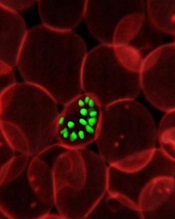
infecting a red blood cell
Credit: St Jude Hospital
High-resolution technology has revealed the structure of actin proteins in the malaria parasite Plasmodium falciparum.
Researchers found the 2 versions of the protein differ from each other more than actin proteins in any other organism.
And they were able to identify areas within these proteins that cause their different behaviors.
The team believes this discovery, published in PLOS Pathogens, may contribute to the development of tailor-made drugs against malaria.
Malaria parasites use actin to enter human cells and leave them again. Inside cells, actin confers stability, allows for cell division, and makes the movement of single cells possible.
The dynamic behavior needed for these processes is enabled by individual globular actin molecules assembling together to form thread-like structures called filaments.
The malaria parasite has 2 versions of actin, actin I and actin II, which differ substantially from each other. These structural proteins are crucial for the pathogen’s infectivity, but researchers have not been able to demonstrate filament formation in the parasite, until now.
Inari Kursula, PhD, of the University of Oulu in Finland, and her colleagues have succeeded in detecting filament assembly of the parasite actin II proteins. For this, they used electron microscopy, which overcomes the resolution limit of classical light microscopy.
Male malaria parasites from which the researchers had deleted actin II were not able to form mature germ cells and, consequently, could not reproduce and propagate.
The researchers therefore speculated that having only 1 actin variant is not sufficient for this process, but they wondered why the 2 proteins show such different behavior. To answer this question, the team deciphered the structure of the globular actin proteins using X-radiation.
“We were able to determine the structures of actin I and actin II at very high resolutions–down to 1.3 and 2.2 Ångström, respectively,” Dr Kursula said.
“With this, we are in the range of single atoms. The structures show us that the 2 variants differ more from each other than actins in any other known living organism do.”
The high resolution enabled the researchers to identify areas within the proteins that cause the different behavior.
“We now understand that Plasmodium actin filaments are very different from other actin filaments—like, for example, from those found in humans—and that they are assembled in a very different manner,” Dr Kursula said.
“Now that we know the structural basis for this, we can look for ways to specifically interfere with the parasite cytoskeleton.”
This knowledge might, in the future, aid the design of tailor-made antimalarial drugs. ![]()