User login
Melflufen-dex proves active in multi-resistant MM
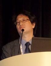
SAN DIEGO—The combination of melflufen and dexamethasone demonstrated activity in patients with multi-resistant multiple myeloma (MM) in a phase 2 trial.
Melflufen-dexamethasone produced an overall response rate of 33% in patients who had quad- or penta-refractory MM.
The combination was considered well tolerated, although 13% of patients discontinued treatment due to adverse events (AEs).
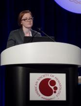
Paul Richardson, MD, of Dana-Farber Cancer Institute in Boston, Massachusetts, presented these results, from the HORIZON trial (NCT02963493), at the 2018 ASH Annual Meeting (abstract 600*).
Dr. Richardson reported results with melflufen-dexamethasone in 83 MM patients. They had a median age of 63 (range, 35-86), and 59% were male. Their median time since diagnosis was 6.5 years (range, 0.7-25) at baseline.
The patients had received a median of 5 prior lines of therapy (range, 2-13). All patients were refractory to pomalidomide or daratumumab, and 60% were refractory to both drugs. Eighty percent of patients were refractory to a monoclonal antibody, and 55% were refractory to an alkylator.
Eighty-six percent of patients were refractory to both a proteasome inhibitor and an immunomodulatory drug. Sixty percent of patients were refractory to a proteasome inhibitor, an immunomodulatory drug, and anti-CD38 therapy.
Ninety-three percent of patients were refractory to their last line of therapy. Sixty-nine percent had received a transplant, and 25% had received two transplants.
“[I]f you look at the refractoriness of these patients, 46% had used three or more prior regimens within the last 12 months before entering the trial, and I think that reflects a real challenge in these patients,” Dr. Richardson said.
Results
The patients received melflufen at 40 mg on day 1 of each 28-day cycle and dexamethasone at 40 mg weekly (20 mg for patients age 75 and older). Patients were treated until progression, consent withdrawal, or unacceptable toxicity.
The overall response rate (partial response or better) was 33%, the clinical benefit rate (minimal response or better) was 39%, and 84% of patients had stable disease or better.
Twenty seven patients responded, including one stringent complete response, nine very good partial responses, and 17 partial responses.
Five patients had a minimal response, 37 had stable disease, and 12 progressed. One patient was not evaluable, and one had data pending.
Dr. Richardson noted that melflufen-dexamethasone demonstrated activity regardless of a patient’s underlying refractory status, but serum albumin was a strong predictor of response. Specifically, patients with higher albumin levels were more likely to respond.
“We do not think it’s a mechanism-of-action issue with [melflufen], but we will be evaluating that,” Dr. Richardson said.
He went on to say that the median progression-free survival was 4.0 months overall, 6.4 months among patients with a partial response or better, and 1 month in patients with progressive disease.
Treatment-related grade 3/4 AEs occurred in 75% of patients. This included neutropenia (61%), thrombocytopenia (59%), anemia (25%), febrile neutropenia (6%), leukopenia (5%), lymphopenia (5%), infections and infestations (7%), and pneumonia (2%).
There were no treatment-related deaths. Thirteen percent of patients discontinued treatment due to AEs, most due to thrombocytopenia (8/11).
In closing, Dr. Richardson said toxicity was “generally manageable” with this treatment, which “has promising activity in multi-resistant, relapsed/refractory myeloma.”
He added that, in the phase 3 OCEAN trial (NCT03151811), researchers are comparing melflufen-dexamethasone to pomalidomide-dexamethasone in a less heavily pretreated MM population.
In the phase 1/2 ANCHOR trial (NCT03481556), researchers are testing melflufen-dexamethasone in combination with daratumumab or bortezomib.
Dr. Richardson disclosed relationships with Karyopharm Pharmaceuticals, Bristol-Myers Squibb, Janssen, Amgen, Jazz Pharmaceuticals, Takeda, Celgene, and Oncopeptides AB. The HORIZON trial is supported by Oncopeptides AB in collaboration with Precision Oncology.
*Data in the abstract differ from the presentation.
Lymphodepletion improves efficacy of CAR T cells in HL
SAN DIEGO—A phase 1 study suggests lymphodepletion can improve the efficacy of CD30-directed chimeric antigen receptor (CAR) T-cell therapy in patients with Hodgkin lymphoma (HL).
Researchers observed improved responses in HL patients treated with fludarabine and cyclophosphamide prior to CD30.CAR T-cell therapy.
This lymphodepleting regimen was also associated with increased toxicity, compared to no lymphodepletion. However, researchers consider the regimen safe.
Carlos A. Ramos, MD, of Baylor College of Medicine in Houston, Texas, presented these results at the 2018 ASH Annual Meeting (abstract 680*).
Without lymphodepletion
Dr. Ramos first discussed a previous phase 1 trial (NCT01316146), which was published in The Journal of Clinical Investigation in 2017.
In this trial, he and his colleagues had tested CD30.CAR T-cell therapy in patients with relapsed/refractory, CD30+ HL or T-cell non-Hodgkin lymphoma. None of these patients underwent lymphodepletion.
There were no dose-limiting toxicities in this trial—including no neurotoxicity or cytokine release syndrome—but responses were “limited,” according to Dr. Ramos.
Three patients achieved a complete response (CR), three had stable disease, and three progressed.
“Although we saw no significant toxicities and some good clinical responses . . ., the bottom line is that the responses were still quite limited, with several patients having, at most, stable disease or progressive disease,” Dr. Ramos said.
With lymphodepletion
Results from the previous trial prompted Dr. Ramos and his colleagues to conduct the RELY-30 trial (NCT02917083) and investigate whether lymphodepletion would improve responses to CD30.CAR T-cell therapy.
Thus far, 11 patients have been treated on this trial. All had relapsed, CD30+ HL at baseline. Six patients are male, and their median age at baseline was 30 (range, 17-69).
The patients had a median of 5 prior treatments (range, 2-9). This included PD-1 inhibitors (n=10), brentuximab vedotin (n=8), and transplant (n=6).
All patients received lymphodepletion with cyclophosphamide at 500 mg/m2 and fludarabine at 30 mg/m2 daily for 3 days. They then received CD30.CAR T-cell therapy at 2×10⁷ cells/m2 or 1×10⁸ cells/m2.
Dr. Ramos noted that CD30.CAR T-cell expansion was dose-dependent and increased by lymphodepleting chemotherapy.
“The peak expansion is much higher [with lymphodepletion], probably in the order of two to three logs higher than what we see without lymphodepleting chemotherapy,” he said. “So chemotherapy makes a difference.”
Increased CD30.CAR T-cell expansion was associated with improved response. Of the nine evaluable patients, six achieved a CR, and three progressed.
Four complete responders were still in CR at last follow-up, one of them for more than a year. However, two complete responders ultimately progressed.
In addition to improved responses, the researchers observed increased toxicity in this trial. Dr. Ramos said some of these toxicities are “probably attributable” to the lymphodepleting chemotherapy.
Toxicities included grade 1 cytokine release syndrome (no tocilizumab required), maculopapular rash, transient cytopenias, nausea, vomiting, and alopecia.
Dr. Ramos said these results suggest adoptive transfer of CD30.CAR T cells is “safe, even with chemotherapy.”
He noted that the duration of response with this treatment is unknown, but trial enrollment and follow-up are ongoing.
RELY-30 was sponsored by Baylor College of Medicine. Dr. Ramos reported relationships with Novartis, Celgene, Bluebird Bio, and Tessa Therapeutics.
*Data in the abstract differ from the presentation.
Fludarabine deemed important for CD30.CAR T-cell therapy
SAN DIEGO—Fludarabine is “very important” for lymphodepletion prior to CD30-directed chimeric antigen receptor (CAR) T-cell therapy, according to a presentation at the 2018 ASH Annual Meeting.
A phase 1/2 study showed that bendamustine alone was not sufficient as lymphodepletion.
However, adding fludarabine to bendamustine could enhance responses to CD30.CAR T-cell therapy and improve progression-free survival (PFS) in patients with Hodgkin or non-Hodgkin lymphoma.
Natalie S. Grover, MD, of the University of North Carolina in Chapel Hill, presented these results as abstract 681.*
This trial (NCT02690545) included patients with relapsed/refractory, CD30+ Hodgkin lymphoma or T-cell non-Hodgkin lymphoma.
Twenty-four adult patients have been treated thus far. Twenty-two had classical Hodgkin lymphoma, one had Sézary syndrome, and one had enteropathy-associated T-cell lymphoma.
The patients’ median age at baseline was 34.5 years (range, 23-69), and they had received a median of 7.5 prior lines of therapy (range, 3-17).
Prior treatments included brentuximab vedotin (n=23), checkpoint inhibitors (n=16), autologous transplant (n=17), and allogeneic transplant (n=7).
In this trial, patients could receive bridging therapy while their T cells were being processed. They then underwent lymphodepletion and received CAR T-cell therapy at one of two doses.
Bendamustine alone
Eight patients received lymphodepletion with 2 days of bendamustine at 90 mg/m2. Three of these patients received CD30.CAR T-cell therapy at 1×10⁸ cells/m2, and all three progressed.
Of the five patients who received CAR T-cell therapy at a dose of 2×10⁸ cells/m2, one progressed, one had stable disease, and three had a complete response (CR).
However, all three complete responders were in CR prior to lymphodepletion as a result of bridging therapy.
“Responses were more modest than what we were hoping for with lymphodepletion,” Dr. Grover noted. “We looked at the cytokine levels in patients getting bendamustine lymphodepletion and saw that bendamustine wasn’t supporting an ideal cytokine milieu. IL-7 and IL-15 are important for T-cell expansion, and these levels were not increased in patients post-bendamustine.”
When the researchers added fludarabine to the lymphodepleting regimen, they observed an increase in T-cell expansion.
Bendamustine plus fludarabine
Sixteen patients received bendamustine plus fludarabine prior to CAR T-cell therapy. The regimen consisted of 3 days of bendamustine at 70 mg/m2 and fludarabine at 30 mg/m2.
All 16 patients received CAR T cells at 2×10⁸ cells/m2, which was the recommended phase 2 dose.
“Responses were more impressive in the bendamustine-fludarabine cohort,” Dr. Grover noted.
Twelve of the 16 patients achieved a CR, although two patients were already in CR prior to lymphodepletion.
Two patients had a partial response, one had stable disease, and one progressed.
PFS and toxicity
Dr. Grover and her colleagues also assessed PFS. At a median follow-up of 100 days, the median PFS was 164 days for the entire cohort, excluding patients who were in CR prior to lymphodepletion.
The median PFS was 396 days for the bendamustine-fludarabine cohort and 55 days for patients in the bendamustine-alone cohort (P=0.001).
There was no neurotoxicity in this trial.
Three patients developed cytokine release syndrome (CRS). Two patients had grade 1 CRS that resolved spontaneously, and one patient had grade 2 CRS, which responded to tocilizumab. Two of the patients with CRS had T-cell lymphoma. The Sézary patient had grade 2 CRS.
Eight patients had a mild rash, one of whom had a rash at baseline.
“CAR T cells against CD30 preceded by lymphodepletion with bendamustine and fludarabine have promising efficacy and a good safety profile in treating patients with relapsed/refractory, CD30+ lymphomas,” Dr. Grover said in closing.
“Fludarabine is very important in enhancing cytokines for improved growth and persistence of CAR T cells.”
This trial was sponsored by UNC Lineberger Comprehensive Cancer Center. Dr. Grover reported consulting for Seattle Genetics.
*Data in the abstract differ from the presentation.
MetS after HSCT linked to CV events, second cancers
SAN DIEGO—Patients who develop metabolic syndrome (MetS) after hematopoietic stem cell transplant (HSCT) have an increased risk of cardiovascular (CV) events and second malignancies, according to research presented at the 2018 ASH Annual Meeting.
Researchers found that HSCT recipients had a higher prevalence of MetS than the general population, and the incidence of MetS increased with age.
In addition, MetS was a predictor of CV events, and these events were associated with second malignancy.
“Our data support metabolic syndrome being an age-related late effect of transplant that is strongly associated with not only cardiovascular events but also the occurrence of second cancers, and this is a novel finding,” said Diana M. Greenfield, RN, PhD, of Sheffield Teaching Hospitals NHS Foundation Trust in Sheffield, United Kingdom.
Dr. Greenfield presented the data at ASH as abstract 251.
She noted that previous studies have reported rates of MetS after HSCT ranging from 31% to 49%. Intensive chemotherapy and radiation have been associated with MetS, and proposed mechanisms of MetS after HSCT include:
- Conditioning regimen-mediated damage to the neuro-hormonal system and vascular endothelium
- Immunological and inflammatory effects of allo-grafting, including graft-vs-host disease (GVHD) and treatment.
“Those with metabolic syndrome in the general population are already known to be twice as likely to develop cardiovascular disease as those without metabolic syndrome,” Dr. Greenfield noted. “The risk of cardiovascular hospitalizations and mortality is 3.6-fold higher in transplant patients vs the general population.”
With all this in mind, she and her colleagues set out to determine the prevalence of MetS and associated complications among HSCT recipients treated at 9 European centers in Belgium, France, Germany, Turkey, and the United Kingdom.
The researchers analyzed 462 patients—375 who had received an allogeneic HSCT and 87 who had undergone autologous HSCT. All patients were at least 18 years of age and at least 2 years post-HSCT.
The median age at transplant was 43 (interquartile range, 32-53), and 57.4% of patients were male.
Patients’ diagnoses included acute leukemia (37.9%), lymphoma (27.3%), myelodysplastic syndromes/myeloproliferative neoplasms (13%), chronic leukemia (12.3%), plasma cell disorders (5%), bone marrow failure (4.1%), solid tumors (0.2%), and inherited disorders (0.2%).
Results
The prevalence of MetS in this population was 30.4%. This is higher than the prevalence in the general population in Europe, which is 25%, according to the World Health Organization.
There was no significant difference in MetS prevalence between allogeneic and autologous HSCT recipients—29% and 35.6%, respectively.
However, there was a significant difference in MetS prevalence by age (P<0.001 with increasing age).
Among allogeneic HSCT recipients, there was no relationship between MetS prevalence and the presence or degree of acute or chronic GVHD, current use of immunosuppressive therapy, or conditioning intensity.
There was a significantly higher prevalence of CV events in patients with MetS than in patients without the syndrome—22.6% and 10.7%, respectively (P=0.006).
And logistic regression analysis confirmed that MetS is a predictor of CV events (odds ratio [OR]=4.72; 95% confidence interval [CI], 2.11-10.57).
The researchers also found that increasing age influenced the prevalence of MetS (OR=7.3; 95% CI, 3.2-16.8) and CV events (OR=3; 95% CI, 0.8-11.32) for patients older than 50, compared to patients in the 18-29 age group.
And CV events were associated with second malignancy (OR=7.93; 95% CI, 2.91-21.61).
“Early intervention of reversible features of metabolic syndrome with lifestyle and medical management may reduce the risk of cardiovascular events,” Dr. Greenfield said. “Meanwhile, screening and management should be robustly integrated within routine transplant long-term follow-up care.”
She added that the CIBMTR and EBMT guidelines on MetS and CV disease after HSCT should help in that regard.
Dr. Greenfield reported no conflicts of interest. However, her fellow researchers reported relationships with a range of pharmaceutical companies.
Early switch to dasatinib offers clinical benefit to CML patients
SAN DIEGO—Early results of the DASCERN trial indicate that patients with chronic myeloid leukemia (CML) in chronic phase who have a suboptimal response to imatinib as a first-line treatment benefit from switching to dasatinib at 3 months.
Twenty-nine percent of dasatinib-treated patients achieved a major molecular response (MMR) at 12 months, compared to 13% of patients who remained on imatinib (P=0.005).
Dasatinib-treated patients also attained MMR much faster than those on imatinib, at a median of 14 months, compared to 20 months for those treated with imatinib.
DASCERN is the first study, according to investigators, to explore the significance of an early switch for patients who have not achieved an early molecular response (EMR) with imatinib.
“[EMRs] are important because they correlate with the outcome of patients, certainly with progression-free survival and overall survival,” explained Jorge E. Cortes, MD, of The University of Texas MD Anderson Cancer Center in Houston.
“[T]he possibility of changing to dasatinib appears, with these early results, to suggest that there may be a benefit to switching these patients to achieve better long-term outcomes. But also, those patients that have these early molecular responses have a better probability of achieving deep molecular responses that we desire for treatment-free remission.”
Dr. Cortes elaborated on the DASCERN data at the 2018 ASH Annual Meeting (abstract 788*).
Study design
DASCERN (NCT01593254) is a randomized, open-label, international, phase 2b trial in adult patients with chronic-phase CML who had achieved a complete hematologic response but still had more than 10% BCR-ABL1 transcripts at 3 months.
Patients were initially treated with imatinib at 400 mg daily, and 1,126 patients had a molecular assessment at 3 months.
Those who did not achieve an MMR (n=260) were randomized in a 2:1 fashion to 100 mg daily of dasatinib (n=174) or 400 mg daily or twice daily of imatinib (n=86).
If patients subsequently failed imatinib treatment by European LeukemiaNet standards, they could cross over to dasatinib.
“Importantly, there was a window of enrollment up to 8 weeks after the 3-month assessment, allowing for time to get response results and screen and enroll patients onto the study,” Dr. Cortes clarified.
Patients were also stratified according to Sokal risk score and time from molecular assessment to randomization.
Dr. Cortes noted that about 40% of patients were started on treatment within 4 weeks of the 3-month assessment. And another 58% were enrolled between 4 and 8 weeks from the 3-month assessment.
The primary endpoint is the achievement of MMR at 12 months from the first day of imatinib treatment; that is, at about 9 months from the start of the protocol treatment, in both trial arms.
Patient characteristics
Seventy-eight percent of patients were male, and 95% were younger than 65.
“This is a relatively younger patient population,” Dr. Cortes noted. “This has to do with the fact that this was an international study with a significant representation of patients that were from Asia (73%).”
The Asian patients were primarily from China, Dr. Cortes said, “and we know that, in some parts of the world, including Asia, patients seem to be younger.”
“This is also associated with a higher percentage of patients with high-risk Sokal scores, more than 20%,” he added. “That is different than what’s seen, for example, in the U.S. or in Europe.”
The prevalence of male patients, he said, broadly represents the distribution of patients in other parts of the world.
Patient disposition
Patients were followed for a median of 30 months.
Of the randomized patients—the intent-to-treat (ITT) population—143 (84%) in the dasatinib arm and 72 (84%) in the imatinib arm continued on treatment.
Study drug toxicity was the most common reason for discontinuing treatment and occurred in 9 patients (5%) in the dasatinib arm and 3 (4%) in the imatinib arm.
Nearly half the imatinib patients (n=42, 49%) crossed over to dasatinib at a median of 9 months.
The median duration of treatment was 22 months (range, 1 – 44) for patients on imatinib who did not cross over and 15 months (range, <1 – 38) for patients on dasatinib after crossing over from imatinib.
Response
In the ITT population, the rate of MMR at 12 months was 29% in the dasatinib arm and 13% in the imatinib arm (P=0.005).
The median time to MMR was significantly shorter for patients who received dasatinib compared with imatinib—14 months and 20 months, respectively (P=0.053).
“Over 60% of patients on the dasatinib arm achieved a major molecular response,” Dr. Cortes said. “This compares to about 55% of patients on the imatinib arm, even when you consider the crossover of a significant number of these patients.”
A few patients achieved a molecular response of a 4.5-log reduction in BCR-ABL1 transcripts (MR4.5).
“Of course, the follow-up is short, but we had twice as many patients [on dasatinib] at 12 months with MR4.5— 5% with dasatinib versus 2% with imatinib,” Dr. Cortes said.
Survival outcomes
Both overall and progression-free survival “look very good with both treatment approaches,” Dr. Cortes pointed out.
At a median follow-up of 30 months, the overall survival in the ITT population was 98.8% in each treatment arm.
Progression-free survival was 96.9% in the dasatinib arm and 97.6% in the imatinib arm.
“It is important to note that no patient in either treatment arm has transformed to accelerated or blast phase,” Dr. Cortes said.
Safety
Treatment was well tolerated with both agents, Dr. Cortes observed, with very few grade 3 adverse events (AEs) noted to date.
“The one that stands out here, and it’s only 2% of patients, is headache with dasatinib,” he said.
The headache did not lead to treatment discontinuation, and the patients were managed with dose adjustments.
The investigators observed no new safety signals with either drug.
Treatment-emergent AEs of any grade occurring in 15% or fewer dasatinib- or imatinib-treated patients, respectively, in the ITT population included headache (15%, 9%), diarrhea (9%, 8%), nausea (9%, 8%), eyelid edema (1%, 9%), hypocalcemia (1%, 7%), and muscle spasms (1%, 8%).
Patients who crossed over to dasatinib had similar rates of AEs to those documented for imatinib.
“They are typical for what we know of both of these tyrosine kinase inhibitors,” Dr. Cortes stated.
Investigators observed pleural effusion in 9 (5%) patients on dasatinib, and only one grade 3. That patient discontinued therapy due to the AE.
Of the patients who were on imatinib and crossed over to dasatinib, 3 (7%) experienced pleural effusion, most of them grade 1 or 2. One patient with grade 4 discontinued therapy with dasatinib due to the AE.
“Hematologic toxicity has been mild,” Dr. Cortes said, and in keeping with the known toxicities of dasatinib and imatinib.
Grade 3/4 treatment-emergent AEs in the dasatinib and imatinib arms, respectively, in the ITT population included anemia (5%, 4%), neutropenia (11%, 16%), thrombocytopenia (11%, 11%), and leukopenia (1%, 1%).
Patients who crossed over to dasatinib experienced more of these AEs than patients who did not cross over, Dr. Cortes clarified, but they are still within the expected range with these agents.
“We acknowledge the results are early and we need to continue following for both safety and efficacy, as it will be important to see if those rates of MR4.5 continue to increase with the same difference in favor of dasatinib that we are starting to see very early on,” Dr. Cortes said.
He disclosed serving as a consultant for Pfizer, Daiichi Sankyo, Astellas Pharma, Novartis, and Bristol-Myers Squibb, and he received research funding from Pfizer, Daiichi Sankyo, Arog Pharmaceuticals, Astellas Pharma, Novartis, and Bristol-Myers Squibb.
The study was supported by Bristol-Myers Squibb.
*Data in the abstract differ from the presentation.
Emapalumab found safe, effective in primary HLH
SAN DIEGO—Emapalumab, an interferon gamma-blocking antibody, controls disease activity and has a favorable safety profile in pediatric patients with primary hemophagocytic lymphohistiocytosis (HLH), according to research presented at the 2018 ASH Annual Meeting.
Investigators believe emapalumab, which was recently approved to treat HLH in the United States, should be considered a new therapeutic option for this rare and life-threatening syndrome because of the drug’s targeted mode of action.
Multiple lines of evidence have pointed to interferon gamma as a “rational target” in HLH, and elevated levels of interferon gamma are consistently observed in patients with HLH, said Franco Locatelli, MD, of Ospedale Pediatrico Bambino Gesù in Rome, Italy.
Emapalumab binds to its target with high affinity, recognizing both free and receptor-bound interferon gamma, he added.
Dr. Locatelli described trial results with emapalumab in children with HLH during the late-breaking abstracts session at ASH (abstract LBA-6).
This phase 2/3 study (NCT01818492) included 34 children with primary HLH—7 who were treatment-naïve and 27 who had failed conventional HLH therapy.
The patients received emapalumab intravenously with concomitant dexamethasone for up to 8 weeks or extended to the point of allogeneic hematopoietic stem cell transplant if needed.
The study met its primary endpoint of overall response rate higher than 40%, Dr. Locatelli reported.
The overall response rate was 64.7% for all 34 treated patients (95% confidence interval [CI], 46% to 80%; P=0.0031) and 63% for the 27 patients who had failed prior therapy (95% CI, 42% to 81%; P=0.0134).
Response was rapid, occurring at a median of 8 days after starting emapalumab, and patients were in response for a median of 75% of days during treatment, Dr. Locatelli said.
Ninety-four percent of patients had at least one adverse event (AE), and 63% had serious AEs. Common AEs included infections (56%), hypertension (35%), infusion-related reactions (27%), and pyrexia (24%).
One patient had disseminated histoplasmosis that led to discontinuation of emapalumab but resolved with appropriate treatment.
This study was sponsored by Novimmune. Investigators provided disclosures related to Sobi, Novimmune, Rocket Pharmaceuticals, Inc., AB2Bio, Novartis, Eli Lilly, Sanofi, UCB, Pfizer, and AbbVie. Two investigators reported employment with Novimmune.
Inhibitor can improve symptoms of systemic mastocytosis
SAN DIEGO—The KIT D816V inhibitor avapritinib can improve symptoms of systemic mastocytosis (SM), according to researchers.
Patients treated with avapritinib in a phase 1 trial had an overall response rate (ORR) of 83%, a 41% mean reduction in mastocytosis symptoms from baseline, and a 58% mean reduction in the most bothersome symptom domain.
Most adverse events (AEs) in this trial were grade 1 or 2. However, 66% of patients did have grade 3 or higher treatment-related AEs that necessitated dose reductions.
Jason R. Gotlib, MD, of Stanford University School of Medicine in California, presented these results, from the EXPLORER trial (NCT02561988), at the 2018 ASH Annual Meeting (abstract 351).
Patients and treatment
The trial enrolled 67 patients—90% with advanced SM and 10% with indolent or smoldering SM. The patients’ median age was 62 (range, 34 to 83), and 49% were female.
Patients had received a median of 3 (range, 0 to 3) prior therapies. Sixty percent of patients had received any prior therapy, and 23% had received midostaurin. Thirty-three percent were on steroid therapy at baseline.
Eighty-four percent of patients had the KIT D816V mutation, and 1% had the KIT D816Y mutation. Forty-five percent of patients had mutations in SRSF2, ASXL1, and RUNX1.
In the dose-escalation portion of the trial, patients received avapritinib at 30 mg to 400 mg once daily in continuous 28-day cycles. In the expansion portion of the trial, patients received avapritinib at 200 mg or 300 mg once daily.
Response
There were 29 patients evaluable for response. The ORR was 83% (n=24). The rate of complete response (CR) was 10% (n=3), the rate of CR with partial hematologic recovery (CRh) was 14% (n=4), and the partial response rate was 48% (n=14).
Ten percent (n=3) of patients had clinical improvement, 17% (n=5) had stable disease, and none of the patients progressed.
Among the 10 patients treated at a dose of 200 mg or lower, the ORR was 90% (n=9). The CR rate was 30% (n=3), the rate of CRh was 20% (n=2), and the partial response rate was 30% (n=3). Ten percent (n=1) of patients each had clinical improvement or stable disease.
Dr. Gotlib noted that responses have been durable and deepened over time.
At a median follow-up of 14 months, the median duration of response had not been reached. The 12-month response rate is 76%.
The median time to initial response is 2 months, and the median time to CR/CRh is 9 months.
Safety
Seventy-eight percent of patients (52/67) were still on treatment at last follow-up. Four percent (4/67) discontinued treatment due to related AEs, and 66% (44/67) had grade 3 or higher AEs that prompted dose reductions.
AEs prompting discontinuation included refractory ascites, encephalopathy, and intracranial bleeding. AEs necessitating dose reductions were largely hematologic events.
The most common AEs were periorbital edema (67%), anemia (52%), fatigue (37%), nausea (36%), diarrhea (34%), peripheral edema (34%), thrombocytopenia (31%) vomiting (28%), cognitive effects (28%), and hair color changes (25%).
The most common grade 3/4 AEs were anemia (26%), thrombocytopenia (17%), and neutropenia (10%). There were no treatment-related deaths.
Symptoms
For symptom assessment, patients completed the Advanced Systemic Mastocytosis Symptom Assessment Form (AdvSM-SAF), a patient-reported outcomes (PRO) tool that included eight symptoms:
- Abdominal pain
- Diarrhea
- Nausea
- Vomiting
- Spots
- Itching
- Flushing
- Fatigue.
Symptoms were scored on a scale of 1 to 10. Results were analyzed as a total symptom score (TSS) combining all eight items, as a gastrointestinal domain (combining nausea, vomiting, diarrhea, and abdominal pain), and as a skin domain (combining itching, flushing, and spots). Analyses were based on 7-day average scores.
Among the 32 evaluable patients, there was a 41% reduction in TSS from baseline (P=0.043). Among the 16 most symptomatic patients, there was a 46% reduction in TSS from baseline (P=0.038).
In all 32 evaluable patients, there was a 58% reduction from baseline in the score for most bothersome symptom domain (gastrointestinal or skin; P=0.0034). In the 16 most symptomatic patients, there was 63% reduction from baseline (P=0.0038).
“This is the first advanced SM-specific PRO to demonstrate significant improvements in total symptom score,” Dr. Gotlib said. “The clinical activity and initial PRO data do support further evaluation of avapritinib in both advanced and indolent disease.”
Dr. Gotlib noted that the PATHFINDER trial (NCT03580655), a study of avapritinib in advanced SM, is now enrolling. And the PIONEER trial (NCT03731260), a study of avapritinib in patients with indolent or smoldering SM, is scheduled to begin at the end of the year.
The EXPLORER trial was sponsored by Blueprint Medicines Corporation. Dr. Gotlib reported relationships with Blueprint Medicines, Celgene, Incyte, Novartis, Deciphera, Gilead, Promedior, and Kartos.
Algorithm uncovers DS in AML patients on IDH inhibitors
SAN DIEGO—An algorithm has proven effective for identifying differentiation syndrome (DS) in patients taking ivosidenib or enasidenib, according to a speaker at the 2018 ASH Annual Meeting.
The U.S. Food and Drug Administration (FDA) recently announced that DS is going unnoticed in some patients with acute myeloid leukemia (AML) who are taking the IDH2 inhibitor enasidenib (Idhifa) or the IDH1 inhibitor ivosidenib (Tibsovo).
Though both drug labels include boxed warnings detailing the risk of DS, the FDA found evidence to suggest that DS is underdiagnosed, which can result in fatalities.
The FDA performed a systematic analysis of DS in AML patients taking either drug to determine if an algorithm could uncover a higher incidence of DS than was previously reported.
Kelly J. Norsworthy, MD, of the FDA, described the results of this analysis at ASH as abstract 288.
The analysis included patients with relapsed/refractory AML treated on a phase 1 study of ivosidenib (NCT02074839, AG120-C-001) and a phase 1/2 study of enasidenib (NCT01915498, AG221-C-001).
There were 179 patients treated with the approved dose of ivosidenib and 214 treated with the approved dose of enasidenib.
The researchers searched for DS events in the first 90 days of therapy. Patients were categorized as having DS if they had at least one investigator-reported DS event (IDH DS or retinoic acid syndrome) or if they had at least two signs or symptoms of DS, according to revised Montesinos criteria, within 7 days.
The signs/symptoms included:
- Dyspnea
- Unexplained fever
- Weight gain
- Unexplained hypotension
- Acute renal failure
- Pulmonary infiltrates or pleuropericardial effusion
- Multiple organ dysfunction.
“We added an event for multiple organ dysfunction since this adverse event could satisfy multiple Montesinos criteria,” Dr. Norsworthy said.
“Although leukocytosis is not a diagnostic criterion for DS, it is frequently seen in association with DS, so we performed an additional query for concomitant leukocytosis,” she added.
The researchers looked for adverse events of leukocytosis, hyperleukocytosis, white blood cell count increase, and leukocyte count greater than 10 Gi/L within 7 days of clinical signs/symptoms.
DS incidence
The algorithm suggested 40% of patients in each treatment group had potential DS—72 of 179 patients treated with ivosidenib and 86 of 214 patients treated with enasidenib.
“We reviewed case narratives and laboratory data from the algorithmically defined cases of DS to adjudicate whether cases were DS or unlikely DS due to an alternative explanation, most commonly due to a clinical course inconsistent with DS or confirmed infection,” Dr. Norsworthy said.
The reviewer-adjudicated incidence of DS was 19% in both groups—34 patients on ivosidenib and 41 patients on enasidenib.
“This contrasts with the DS incidence of 11% to 14% reported by investigators,” Dr. Norsworthy said. “Thus, there was a subset of patients where the syndrome was not recognized by investigators.”
Characteristics of DS
The median time to DS onset in this analysis was 20 days (range, 1 to 78) in the ivosidenib group and 19 days in the enasidenib group (range, 1 to 86).
In both treatment groups, most patients had moderate DS—71% (n=24) in the ivosidenib group and 80% (n=33) in the enasidenib group. Moderate DS was defined as meeting two to three of the aforementioned criteria for DS.
Fewer patients had severe DS (four or more criteria)—24% (n=8) in the ivosidenib group and 12% (n=5) in the enasidenib group.
For the remaining patients, DS severity could not be determined—6% (n=2) in the ivosidenib group and 10% (n=4) in the enasidenib group. These were investigator-reported cases of DS.
Most DS cases in the ivosidenib and enasidenib groups—68% (n=23) and 66% (n=27), respectively— included grade 3 or higher adverse reactions.
Two patients in each group died of DS—6% and 5%, respectively. Only one of these cases was recognized as DS and treated with steroids, Dr. Norsworthy noted.
She also pointed out that most patients with DS had leukocytosis—79% (n=27) in the ivosidenib group and 61% (n=25) in the enasidenib group.
In addition, rates of complete response (CR) and CR with incomplete hematologic recovery (CRh) were numerically lower among patients with DS, although the confidence intervals (CI) overlap.
Among patients on ivosidenib, the CR/CRh rate was 18% (95% CI, 7-35) in those with DS and 36% (95% CI, 28-45) in those without DS.
Among patients on enasidenib, the CR/CRh rate was 18% (95% CI, 7-33) in those with DS and 25% (95% CI, 18-32) in those without DS.
“[F]irm conclusions regarding the impact on response cannot be inferred based on this post-hoc subgroup analysis,” Dr. Norsworthy stressed.
Predicting DS
Dr. Norsworthy noted that baseline patient and disease characteristics were similar between patients with and without DS.
The researchers did see a trend toward higher blasts in the marrow and peripheral blood as well as higher white blood cell counts at baseline among patients with DS.
“However, there did not appear to be a distinct baseline white blood cell count or absolute blast cell count cutoff above which DS was more common,” Dr. Norsworthy said.
She added that the patient numbers are small, so it’s not possible to make firm conclusions about prognostic factors for DS.
In closing, Dr. Norsworthy said the algorithmic approach used here “led to the recognition of additional cases of DS not identified by investigators or review committee determination for patients treated with the IDH inhibitors ivo and ena.”
“Increased recognition of the signs and symptoms of DS through the framework of the Montesinos criteria may lead to early diagnosis and treatment, which may decrease severe complications and mortality. Furthermore, integration of the algorithm into clinical trials of differentiating therapies, in a prospective fashion, may help to systematically monitor the incidence and severity of DS.”
Dr. Norsworthy declared no conflicts of interest.
Mutation confers resistance to venetoclax in CLL
SAN DIEGO—A recurrent mutation in BCL2, the therapeutic target of venetoclax, appears to be a major contributor to drug resistance in patients with chronic lymphocytic leukemia (CLL), investigators reported.
The mutation has been detected in some patients with CLL up to 2 years before resistance to venetoclax actually develops, according to Piers Blombery, MBBS, of the Peter MacCallum Cancer Center in Melbourne, Victoria, Australia.
“We have identified the first acquired BCL2 mutation developed in patients clinically treated with venetoclax,” he said during the late-breaking abstracts session at the 2018 ASH Annual Meeting.
The mutation, which the investigators have labeled BCL2 Gly101Val, “is a recurrent and frequent mediator of resistance and may be detected years before clinical relapse occurs,” Dr. Blombery added.
A paper on the mutation was published in Cancer Discovery to coincide with the presentation at ASH (abstract LBA-7).
Despite the demonstrated efficacy of venetoclax as continuous therapy in patients with relapsed or refractory CLL, the majority of patients experience disease progression, prompting the investigators to explore molecular mechanisms of secondary resistance.
To do this, they analyzed paired samples from 15 patients with CLL, enrolled in clinical trials of venetoclax, collected both before the start of venetoclax therapy and at the time of disease progression.
In seven patients, the investigators identified a novel mutation that showed up at the time of progression but was absent from the pre-venetoclax samples.
The mutation first became detectable from about 19 to 42 months after the start of therapy and preceded clinical progression by as much as 25 months, the investigators found.
They pinned the mutation down to the BH3-binding groove on BCL2, the same molecular site targeted by venetoclax. They found the mutation was not present in samples from 96 patients with venetoclax-naive CLL nor in any other B-cell malignancies.
Searches for references to the mutation in both a cancer database (COSMIC) and a population database (gnomAD) came up empty.
In other experiments, the investigators determined that cell lines overexpressing BCL2 Gly101Val are resistant to venetoclax, and, in the presence of venetoclax in vitro, BCL2 Gly101Val-expressing cells have a growth advantage compared with wild-type cells.
Additionally, they showed that the mutation results in impaired venetoclax binding in vitro.
“BCL2 Gly101Val is observed subclonally, implicating multiple mechanisms of venetoclax resistance in the same patient,” Dr. Blombery said.
He added that the identification of the resistance mutation is a strong rationale for using combination therapy to treat patients with relapsed or refractory CLL to help prevent or attenuate selection pressures that lead to resistance.
Dr. Blombery reported having no relevant disclosures. The investigators were supported by the Wilson Center for Lymphoma Genomics, Snowdome Foundation, National Health Medical Research Council, Leukemia and Lymphoma Society, Leukemia Foundation, Cancer Council of Victoria, and Australian Cancer Research Foundation.
Dasatinib re-challenge feasible as 2nd attempt at TKI discontinuation
SAN DIEGO—Preliminary trial results suggest re-treatment with dasatinib is feasible and safe for a second attempt at tyrosine kinase inhibitor (TKI) discontinuation in chronic myeloid leukemia (CML) patients who fail to achieve treatment-free remission (TFR) after discontinuing imatinib.
However, investigators reported the rate of second TFR (TFR2) was 21% at 6 months, which was not enough to confirm, at this time, that dasatinib could improve the TFR2 rate after imatinib discontinuation failure.
Dennis Kim, MD, of the University of Toronto in Ontario, Canada, presented these results at the 2018 ASH Annual Meeting (abstract 787).
The design of this trial (NCT02268370) includes three phases: the imatinib discontinuation phase, the dasatinib re-challenge phase to achieve a molecular response of ≥ 4.5-log reduction in BCR-ABL1 transcripts (MR4.5), and the dasatinib discontinuation phase.
The primary objective of the trial is to determine the proportion of patients who remain in deep molecular remission (> MR4.5) after discontinuing dasatinib following a failed attempt at discontinuation of imatinib.
If patients had a confirmed molecular relapse after discontinuing imatinib, they were started on 100 mg of dasatinib daily, and, after achieving MR4.5 or greater for 12 months, they discontinued dasatinib for a try at the second TFR.
Investigators defined loss of molecular response, or relapse, as a loss of a major molecular response (MMR) once or loss of MR4.0 on two consecutive occasions.
Patient characteristics
The 131 enrolled CML patients were a median age of 61 (range, 21 to 84), and 62% were male.
Patients had a median 9.36 years of disease duration, 9.18 years of imatinib treatment, 6.82 years of MR4 duration, and 5.08 years of MR4.5 duration.
“The cohort has a very long history of imatinib treatment as well as MR4 duration,” Dr. Kim pointed out, “which also can affect our TFR1 rate, and I think, also, it can affect our TFR2 rate.”
TFR1 and TFR2 rates
As of October 25, the TFR1 rate using loss of MMR as the measure was 69.9% at 12 months from imatinib discontinuation. Relapse-free survival was 57.2% at that time.
Of the 53 patients who lost response, 51 patients received dasatinib. At 3 months of treatment, 97.7% achieved an MMR, 89.9% achieved MR4, and 84.6% achieved MR4.5.
Twenty-five of 51 patients treated with dasatinib attained MR4.5 for 12 months or longer and discontinued treatment for a second attempt at TFR.
Twenty-one patients are still receiving dasatinib and have attained MR4.5, but not for the 12-month duration yet.
Dr. Kim noted that the median time to achievement of molecular response after dasatinib re-challenge ranged from 2.76 months for MR4.5 to 1.71 months for MMR.
Twenty-one of 25 patients (84.0%) who discontinued dasatinib lost their molecular response at a median of 3.7 months.
The estimated TFR2 rate after dasatinib discontinuation is 21.0% to 24.4% at 6 months, which means the investigators cannot reject the null hypothesis of 28% or more patients remaining in remission.
Patients who lost response rapidly after dasatinib discontinuation also tended to lose response rapidly after imatinib discontinuation, Dr. Kim pointed out.
“However, you see some patients who do not lose their response after dasatinib discontinuation or who lose the response but later after the dasatinib discontinuation, they tend to lose their imatinib response also in a later time point,” he said. “So we started to look at the risk factors.”
Risk factor analysis
Out of seven potential risk factors, the investigators were able to demonstrate that time to molecular relapse after imatinib discontinuation, molecular relapse pattern after imatinib discontinuation, and BCR-ABL1 quantitative polymerase chain reaction (qPCR) value prior to dasatinib discontinuation “seemed to be very significant,” Dr. Kim said.
Time to molecular relapse after discontinuation of imatinib correlates with TFR2. The group of patients who relapsed in 3 to 6 months of stopping imatinib had a significantly longer TFR2 than patients who relapsed within 3 months of stopping imatinib (P=0.018).
The molecular relapse pattern also correlates with TFR2. The group with a single loss of MMR after imatinib discontinuation had a significantly shorter TFR2 than those who lost MR4 twice after imatinib discontinuation (P=0.043).
And 0% of the patients who had qPCR transcript levels between a 4.5 and 5.4 log reduction maintained TFR2 at 6 months. However, 28.7% who had qPCR deeper than 5.5 logs prior to dasatinib discontinuation had TFR2 at 6 months (P=0.017).
The risk factor analysis shed light, in part, on the reason the trial thus far failed to satisfy the null hypothesis.
“In other words, because we have selected a really good-risk group for TFR1, the remaining patients are actually a high-risk group for TFR2,” Dr. Kim said. “Because of that, the TFR2 rate might be somewhat lower than we had expected.”
“Or is it related to our conservative treatment with dasatinib, which is 12 months after achieving MR4.5 or deeper response? That may affect our TFR2 rate. We still have to think about that.”
Dr. Kim suggested stricter criteria be considered for attempting TFR2, such as achieving a 5.5 log reduction or deeper in BCR-ABL1 qPCR levels prior to the second TKI discontinuation attempt, and/or an MR4 duration of more than 12 months.
Dr. Kim disclosed receiving honoraria and research funding from Novartis and Bristol-Myers Squibb and serving as a consultant for Pfizer, Paladin, Novartis, and Bristol-Myers Squibb.
