User login
Uncommon Locations for Brain Herniations Into Arachnoid Granulations: 5 Cases and Literature Review
The circulation of cerebrospinal fluid (CSF) is crucial for maintaining homeostasis for the optimal functioning of the multiple complex activities of the brain and spinal cord, including the disposal of metabolic waste products of brain and spinal cord activity into the cerebral venous drainage. Throughout the brain, the arachnoid mater forms small outpouchings or diverticula that penetrate the dura mater and communicate with the dural venous sinuses. These outpuchings are called arachnoid granulations or arachnoid villi, and most are found within the dural sinuses, primarily in the transverse sinuses and superior sagittal sinus, but can occasionally be seen extending into the inner table of the calvarium.1,2
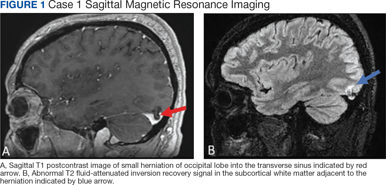
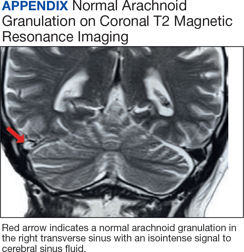
The amount of arachnoid granulations seen in bone, particularly around the superior sagittal sinus, may increase with age.2 Arachnoid granulations are generally small but the largest ones can be seen on gross examination during intracranial procedures or autopsy.3 Magnetic resonance imaging (MRI) can detect arachnoid granulations, which are characterized as T1 hypointense and T2 hyperintense (CSF isointense), well-circumscribed, small, nonenhancing masses within the dural sinuses or in the diploic space (Figure 1). Even small arachnoid granulations < 1 mm in length can be detected.2
Smaller arachnoid granulations have been described histologically as entirely covered by a dural membrane, thus creating a subdural space that separates the body of the arachnoid granulation from the lumen of the accompanying venous sinus.4 However, larger arachnoid granulations may not be completely covered by a dural membrane, thus creating a point of contact between the arachnoid granulation and the venous sinus.4 Larger arachnoid granulations are normally filled with CSF, and their signal characteristics are similar to CSF on imaging.5,6 Arachnoid granulations also often contain vessels draining into the adjacent venous sinus.5,6
When larger arachnoid granulations are present, they may permit the protrusion of herniated brain tissue. There has been an increasing number of reports of these brain herniations into arachnoid granulations (BHAGs) in the literature.7-10 While these herniations have been associated with nonspecific neurologic symptoms like tinnitus and idiopathic intracranial hypertension, their true clinical significance remains undetermined.10,11 This article presents 5 cases of BHAG, discusses their clinical presentations and image findings, and reviews the current literature.
Case 1
A 30-year-old male with a history of multiple traumatic brain injuries presented for evaluation of seizures. The patient described the semiology of the seizures as a bright, colorful light in his right visual field, followed by loss of vision, then loss of awareness and full body convulsion. The semiology of this patient’s seizures was consistent with left temporo-occipital lobe seizure. The only abnormality seen in the brain MRI was the herniation of brain parenchyma originating from the occipital lobe into the transverse sinus, presumably through an arachnoid granulation (Figure 1). An electroencephalogram (EEG) was unremarkable, though the semiology of the seizure historically described by the patient was localized to the area of BHAG. The patient is currently taking antiseizure medications and has experienced no additional seizures.
Case 2
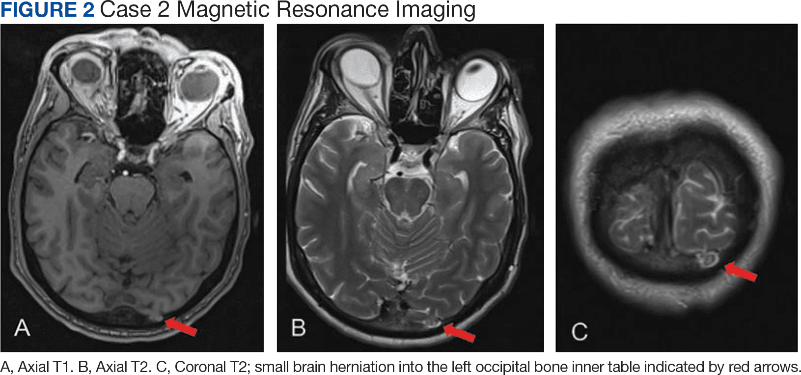
A male aged 53 years with a history of peripheral artery disease presented with a 6-month history of headaches and dizziness. The patient reported the onset of visual aura to his right visual field, starting as a fingernail-sized scintillating kaleidoscope light that would gradually increase in size to a round shape with fading kaleidoscope colors. This episode would last for a few minutes and was immediately followed by a headache. There was no alteration of consciousness during visual aura, although sometimes the patient would have right-sided scalp tingling. These episodes were often unprovoked, but occasionally triggered by bright lights. A single routine EEG was unremarkable. The patient reported headaches without aura, but not aura without headaches, which made occipital lobe seizure less likely. MRI demonstrated a small herniation of brain parenchyma into the inner table of the left occipital bone (Figure 2). The patient was diagnosed with migraine with aura, and the semiology of the visual aura corresponded to the location of the herniation in the left occipital region.
Case 3
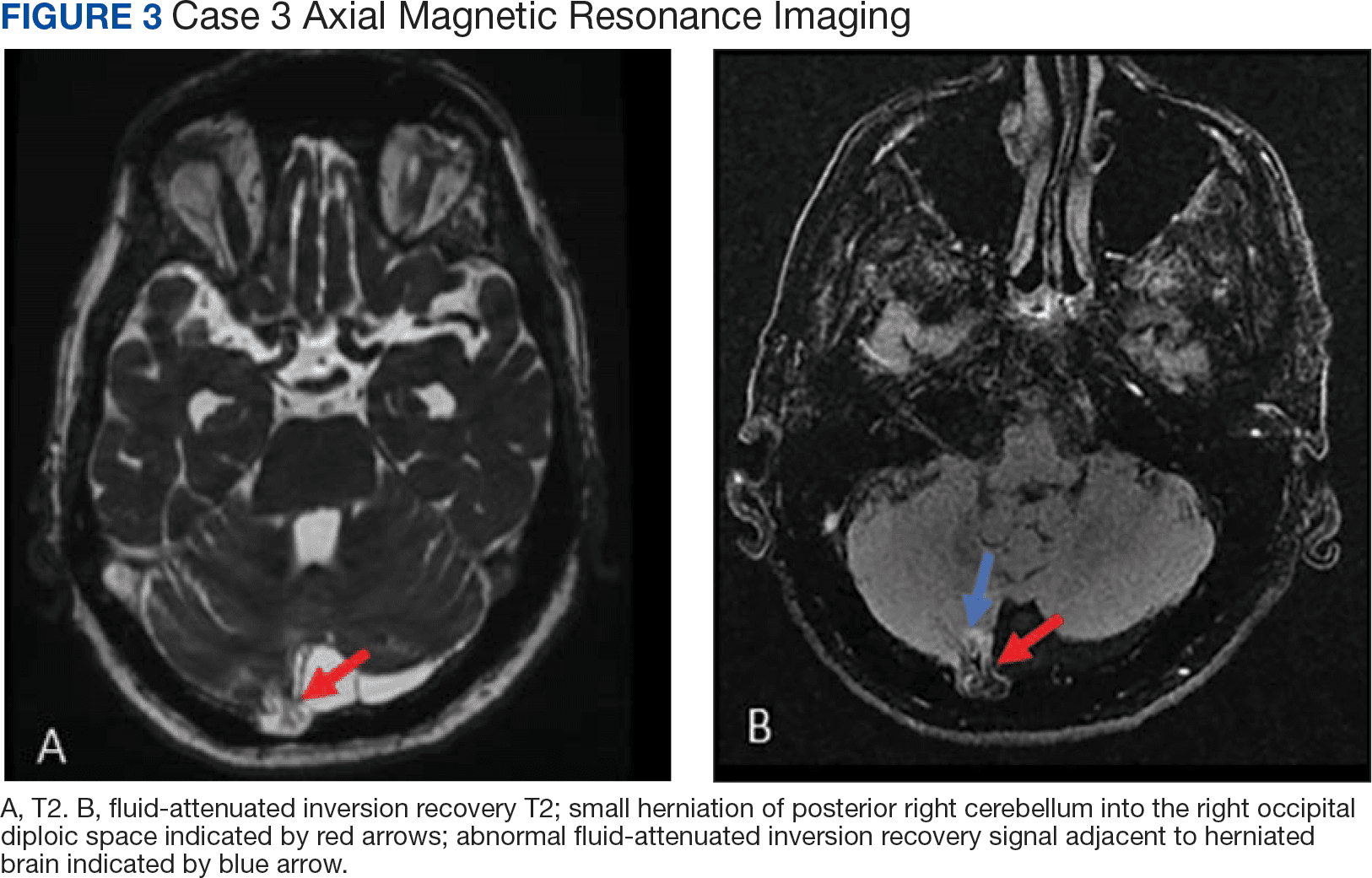
A 77-year-old male with a history of left ear diving injury presented with left-sided asymmetric hearing loss and word recognition difficulty for several years. MRI obtained as part of his work-up to evaluate for possible schwannoma of the eighth left cranial nerve instead demonstrated an incidental right cerebellar herniation within an arachnoid granulation into the diploic space of the occipital bone (Figure 3). The BHAG for this patient appeared to be an incidental finding unrelated to his asymmetric hearing loss.
Case 4
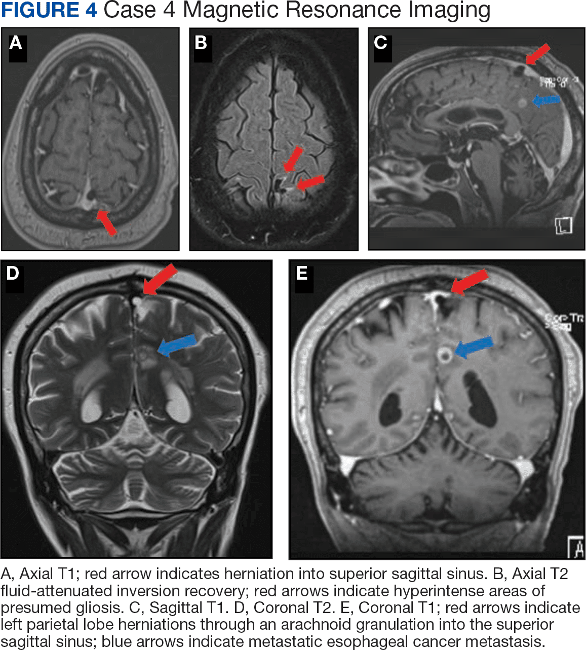
A male aged 62 years with a history of metastatic esophageal cancer, substance abuse, and a prior presumed alcohol withdrawal seizure underwent an MRI for evaluation of brain metastasis after presenting to the hospital with confusion 1 day after starting chemotherapy (Figure 4). Nine years prior, the patient had an isolated generalized tonic-clonic seizure approximately 72 hours following a period of alcohol cessation. The MRI demonstrated an incidental left parasagittal herniation of left parietal lobe tissue through an arachnoid granulation into the superior sagittal sinus, in addition to metastatic brain lesions. An EEG showed mild encephalopathy without evidence of seizures. It was determined that the patient's confusion was most likely due to toxic-metabolic encephalopathy from chemotherapy.
Case 5
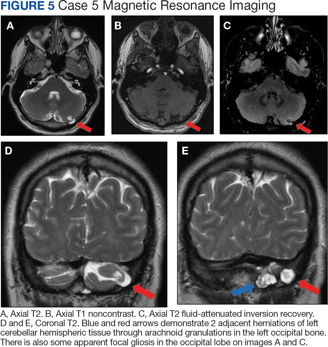
A 51-year-old male presented with worsening headache severity and frequency. He had a history of chronic headaches for about 20 years that occurred annually, but were now occurring twice weekly. The headaches often started with a left eye visual aura followed by pressure in the left eye, left frontal region, and left ear, with at times a cervicogenic component. No cervical spine imaging was available. An MRI revealed 2 small adjacent areas of cerebellar herniation into arachnoid granulations in the left occipital bone (Figure 5).
Discussion
Arachnoid granulations appear very early in life, although they are uncommon before age 2 years.2 Classically, they have been understood to act as 1-way valves permitting the outflow of CSF from the subarachnoid space to the dural venous sinuses. However, increasing evidence shows they may only play a minor role in that process.12 The structure of arachnoid granulations is being reexamined. A recent microscopy study demonstrated structural heterogeneity with a fine, porous lining that permits flow.13 Additionally, associated immune components in the microenvironment suggests that arachnoid granulations may function similarly to lymph nodes as part of a central nervous system lymphatic network.13 Evidence is lacking for arachnoid granulations being the primary route of CSF outflow, and newer models include CSF exit pathways along the cranial nerves and drainage through lymphatics within the dura mater.12
New MRI systems have demonstrated that the prevalence of arachnoid granulations increases with age. One study found that all subjects in the aged 40 years cohort had detectable arachnoid granulations on images obtained with a 3T MRI system, with the main site being the superior sagittal sinus.2 The prevalence increased until age 40 years and then noticeably decreased. Not only did the prevalence increase in this pattern, but the total number of detectable arachnoid granulations followed a similar pattern.2 In addition, the detectable arachnoid granulations tend to be larger in older patients. Arachnoid granulations are very common in adults, but little is known about when and why brain tissue herniates through these structures.
This case series illustrates how a small amount of adult cerebral or cerebellar matter in large arachnoid granulations can herniate into the dural sinuses and diploic space. Although arachnoid granulations extending into the dural sinuses and diploic space are a relatively common finding on MRI, BHAGs are rare in these locations.1,2,8 Improved spatial resolution afforded by newer high-field scanners with thinner sections, such as very thin (1 mm) T1- and heavily T2-weighted 3 dimensional sequences may lead to increased detection of BHAG. Some of these herniations are small and may be easily missed or confused for normal arachnoid granulations on 3 to 5 mm thickness MRIs.
Despite increased recognition, it is still uncertain to what degree these herniations contribute to the clinical presentations. Associated neurologic symptoms may include seizures, headaches, tinnitus, syncope, and increased intracranial pressure.7-10
Three cases presented in this article demonstrated abnormal signals adjacent to the herniated brain, presumably due to dysplasia of gliotic tissue. In 1 study, parenchymal signal and structural changes occurred in about one-half of the reported BHAG, all of which were cerebellar herniations.7 In Case 1, the herniation and adjacent abnormal MRI signal corresponded to localization of the seizure semiology as obtained from patient history, strongly suggesting the BHAG played a role in the presentation. Signal abnormality accompanying an adjacent BHAG may suggest a higher likelihood that the BHAG has clinical relevance. However, the patient in Case 2 had a visual aura that corresponded to the BHAG location, so a signal abnormality may not be necessary for a patient to develop symptoms. Case 1 also included a history of documented traumatic brain injuries, suggesting that perhaps head trauma may facilitate BHAG development. Regardless, there is likely also a congenital component to their formation, as BHAG has been observed in the pediatric population.14
The patient's asymmetric left-sided hearing loss in Case 3 appeared unrelated to the BHAG as its location was in the contralateral cerebellar region and did not correspond to the patient’s clinical findings. The patient in Case 4 had a limited history regarding localization details of their prior presumed alcohol withdrawal seizure, such as head movements, eye deviation, or lateralized onset of convulsions. Given this limited data, it is unclear whether their prior seizure could have been related to BHAG or not. The patient in case 5 reported worsening headaches on the left side of his head, which corresponded to BHAG occurring on the left side. However, given that the increased T2 signal occurred in the left cerebellar hemisphere with BHAG in the left occipital bone, the occipital cortex was not involved. In this case, the BHAG would not explain the patient’s visual aura as such a lesion would have been expected in the right occipital cortex rather than its actual location in this patient’s left cerebellar hemisphere.
CONCLUSIONS
Understanding the clinical impact of brain herniations is important because they are probably more common than previously thought. Improved MRI capabilities suggest that more BHAG will be detected moving forward as radiologists interpret images with higher resolution and thinner slices. Until its significance is fully understood, BHAG will continue to complicate the diagnosis of patients with neurologic complaints whose brain MRIs and EEGs are otherwise unremarkable.
There have been no cases of surgical BHAG intervention and pathology analysis that would help determine their clinical significance. A related entity, temporal lobe encephalocele, has been linked to focal temporal lobe epilepsy, which has demonstrated significant symptom improvement following surgical correction.15 However, encephaloceles have been distinguished from BHAG in part because they do not necessarily herniate through an arachnoid granulation.8 BHAG has only begun to be characterized in detail over the last decade, so more research is needed to understand how it develops and what clinical significance it truly holds.
1. Ikushima I, Korogi Y, Makita O, et al. MRI of arachnoid granulations within the dural sinuses using a FLAIR pulse sequence. Br J Radiol. 1999;72(863):1046-1051. doi:10.1259/bjr.72.863.10700819
2. Rados M, Zivko M, Perisa A, Oreskovic D, Klarica M. No arachnoid granulations-no problems: number, size, and distribution of arachnoid granulations from birth to 80 years of age. Front Aging Neurosci. 2021;13:698865. doi:10.3389/fnagi.2021.698865
3. Grossman CB, Potts DG. Arachnoid granulations: radiology and anatomy. Radiology. 1974;113(1):95-100. doi:10.1148/113.1.95
4. Wolpow ER, Schaumburg HH. Structure of the human arachnoid granulation. J Neurosurg. 1972;37(6):724-727. doi:10.3171/jns.1972.37.6.0724
5. Leach JL, Jones BV, Tomsick TA, Stewart CA, Balko MG. Normal appearance of arachnoid granulations on contrast-enhanced CT and MR of the brain: differentiation from dural sinus disease. AJNR Am J Neuroradiol. 1996;17(8):1523-1532.
6. Roche J, Warner D. Arachnoid granulations in the transverse and sigmoid sinuses: CT, MR, and MR angiographic appearance of a normal anatomic variation. AJNR Am J Neuroradiol. 1996;17(4):677-683.
7. Malekzadehlashkariani S, Wanke I, Rufenacht DA, San Millan D. Brain herniations into arachnoid granulations: about 68 cases in 38 patients and review of the literature. Neuroradiology. 2016;58(5):443-457. doi:10.1007/s00234-016-1662-5
8. Battal B, Castillo M. Brain herniations into the dural venous sinuses or calvarium: MRI of a recently recognized entity. Neuroradiol J. 2014;27(1):55-62. doi:10.15274/NRJ-2014-10006
9. Liebo GB, Lane JJ, Van Gompel JJ, Eckel LJ, Schwartz KM, Lehman VT. Brain herniation into arachnoid granulations: clinical and neuroimaging features. J Neuroimaging. 2016;26(6):592-598. doi:10.1111/jon.12366
10. Smith ER, Caton MT, Villanueva-Meyer JE, et al. Brain herniation (encephalocele) into arachnoid granulations: Prevalence and association with pulsatile tinnitus and idiopathic intracranial hypertension. Neuroradiology. 2022;64(9):1747-1754.
11. Battal B, Hamcan S, Akgun V, et al. Brain herniations into the dural venous sinus or calvarium: MRI findings, possible causes and clinical significance. Eur Radiol. 2016;26(6):1723-1731.
12. Proulx ST. Cerebrospinal fluid outflow: A review of the historical and contemporary evidence for arachnoid villi, perineural routes, and dural lymphatics. Cell Mol Life Sci. 2021;78(6):2429-2457.
13. Shah T, Leurgans SE, Mehta RI, et al. Arachnoid granulations are lymphatic conduits that communicate with bone marrow and dura-arachnoid stroma. J Exp Med. 2023;220(2).
14. Sade R, Ogul H, Polat G, Pirimoglu B, Kantarci M. Brain herniation into the transverse sinuses’ arachnoid granulations in the pediatric population investigated with 3 T MRI. Acta Neurol Belg. 2019;119(2):225-231.
15. Saavalainen T, Jutila L, Mervaala E, Kalviainen R, Vanninen R, Immonen A. Temporal anteroinferior encephalocele: An underrecognized etiology of temporal lobe epilepsy? Neurology. 2015;85(17):1467-1474.
The circulation of cerebrospinal fluid (CSF) is crucial for maintaining homeostasis for the optimal functioning of the multiple complex activities of the brain and spinal cord, including the disposal of metabolic waste products of brain and spinal cord activity into the cerebral venous drainage. Throughout the brain, the arachnoid mater forms small outpouchings or diverticula that penetrate the dura mater and communicate with the dural venous sinuses. These outpuchings are called arachnoid granulations or arachnoid villi, and most are found within the dural sinuses, primarily in the transverse sinuses and superior sagittal sinus, but can occasionally be seen extending into the inner table of the calvarium.1,2
The amount of arachnoid granulations seen in bone, particularly around the superior sagittal sinus, may increase with age.2 Arachnoid granulations are generally small but the largest ones can be seen on gross examination during intracranial procedures or autopsy.3 Magnetic resonance imaging (MRI) can detect arachnoid granulations, which are characterized as T1 hypointense and T2 hyperintense (CSF isointense), well-circumscribed, small, nonenhancing masses within the dural sinuses or in the diploic space (Figure 1). Even small arachnoid granulations < 1 mm in length can be detected.2
Smaller arachnoid granulations have been described histologically as entirely covered by a dural membrane, thus creating a subdural space that separates the body of the arachnoid granulation from the lumen of the accompanying venous sinus.4 However, larger arachnoid granulations may not be completely covered by a dural membrane, thus creating a point of contact between the arachnoid granulation and the venous sinus.4 Larger arachnoid granulations are normally filled with CSF, and their signal characteristics are similar to CSF on imaging.5,6 Arachnoid granulations also often contain vessels draining into the adjacent venous sinus.5,6
When larger arachnoid granulations are present, they may permit the protrusion of herniated brain tissue. There has been an increasing number of reports of these brain herniations into arachnoid granulations (BHAGs) in the literature.7-10 While these herniations have been associated with nonspecific neurologic symptoms like tinnitus and idiopathic intracranial hypertension, their true clinical significance remains undetermined.10,11 This article presents 5 cases of BHAG, discusses their clinical presentations and image findings, and reviews the current literature.
Case 1
A 30-year-old male with a history of multiple traumatic brain injuries presented for evaluation of seizures. The patient described the semiology of the seizures as a bright, colorful light in his right visual field, followed by loss of vision, then loss of awareness and full body convulsion. The semiology of this patient’s seizures was consistent with left temporo-occipital lobe seizure. The only abnormality seen in the brain MRI was the herniation of brain parenchyma originating from the occipital lobe into the transverse sinus, presumably through an arachnoid granulation (Figure 1). An electroencephalogram (EEG) was unremarkable, though the semiology of the seizure historically described by the patient was localized to the area of BHAG. The patient is currently taking antiseizure medications and has experienced no additional seizures.
Case 2
A male aged 53 years with a history of peripheral artery disease presented with a 6-month history of headaches and dizziness. The patient reported the onset of visual aura to his right visual field, starting as a fingernail-sized scintillating kaleidoscope light that would gradually increase in size to a round shape with fading kaleidoscope colors. This episode would last for a few minutes and was immediately followed by a headache. There was no alteration of consciousness during visual aura, although sometimes the patient would have right-sided scalp tingling. These episodes were often unprovoked, but occasionally triggered by bright lights. A single routine EEG was unremarkable. The patient reported headaches without aura, but not aura without headaches, which made occipital lobe seizure less likely. MRI demonstrated a small herniation of brain parenchyma into the inner table of the left occipital bone (Figure 2). The patient was diagnosed with migraine with aura, and the semiology of the visual aura corresponded to the location of the herniation in the left occipital region.
Case 3
A 77-year-old male with a history of left ear diving injury presented with left-sided asymmetric hearing loss and word recognition difficulty for several years. MRI obtained as part of his work-up to evaluate for possible schwannoma of the eighth left cranial nerve instead demonstrated an incidental right cerebellar herniation within an arachnoid granulation into the diploic space of the occipital bone (Figure 3). The BHAG for this patient appeared to be an incidental finding unrelated to his asymmetric hearing loss.
Case 4
A male aged 62 years with a history of metastatic esophageal cancer, substance abuse, and a prior presumed alcohol withdrawal seizure underwent an MRI for evaluation of brain metastasis after presenting to the hospital with confusion 1 day after starting chemotherapy (Figure 4). Nine years prior, the patient had an isolated generalized tonic-clonic seizure approximately 72 hours following a period of alcohol cessation. The MRI demonstrated an incidental left parasagittal herniation of left parietal lobe tissue through an arachnoid granulation into the superior sagittal sinus, in addition to metastatic brain lesions. An EEG showed mild encephalopathy without evidence of seizures. It was determined that the patient's confusion was most likely due to toxic-metabolic encephalopathy from chemotherapy.
Case 5
A 51-year-old male presented with worsening headache severity and frequency. He had a history of chronic headaches for about 20 years that occurred annually, but were now occurring twice weekly. The headaches often started with a left eye visual aura followed by pressure in the left eye, left frontal region, and left ear, with at times a cervicogenic component. No cervical spine imaging was available. An MRI revealed 2 small adjacent areas of cerebellar herniation into arachnoid granulations in the left occipital bone (Figure 5).
Discussion
Arachnoid granulations appear very early in life, although they are uncommon before age 2 years.2 Classically, they have been understood to act as 1-way valves permitting the outflow of CSF from the subarachnoid space to the dural venous sinuses. However, increasing evidence shows they may only play a minor role in that process.12 The structure of arachnoid granulations is being reexamined. A recent microscopy study demonstrated structural heterogeneity with a fine, porous lining that permits flow.13 Additionally, associated immune components in the microenvironment suggests that arachnoid granulations may function similarly to lymph nodes as part of a central nervous system lymphatic network.13 Evidence is lacking for arachnoid granulations being the primary route of CSF outflow, and newer models include CSF exit pathways along the cranial nerves and drainage through lymphatics within the dura mater.12
New MRI systems have demonstrated that the prevalence of arachnoid granulations increases with age. One study found that all subjects in the aged 40 years cohort had detectable arachnoid granulations on images obtained with a 3T MRI system, with the main site being the superior sagittal sinus.2 The prevalence increased until age 40 years and then noticeably decreased. Not only did the prevalence increase in this pattern, but the total number of detectable arachnoid granulations followed a similar pattern.2 In addition, the detectable arachnoid granulations tend to be larger in older patients. Arachnoid granulations are very common in adults, but little is known about when and why brain tissue herniates through these structures.
This case series illustrates how a small amount of adult cerebral or cerebellar matter in large arachnoid granulations can herniate into the dural sinuses and diploic space. Although arachnoid granulations extending into the dural sinuses and diploic space are a relatively common finding on MRI, BHAGs are rare in these locations.1,2,8 Improved spatial resolution afforded by newer high-field scanners with thinner sections, such as very thin (1 mm) T1- and heavily T2-weighted 3 dimensional sequences may lead to increased detection of BHAG. Some of these herniations are small and may be easily missed or confused for normal arachnoid granulations on 3 to 5 mm thickness MRIs.
Despite increased recognition, it is still uncertain to what degree these herniations contribute to the clinical presentations. Associated neurologic symptoms may include seizures, headaches, tinnitus, syncope, and increased intracranial pressure.7-10
Three cases presented in this article demonstrated abnormal signals adjacent to the herniated brain, presumably due to dysplasia of gliotic tissue. In 1 study, parenchymal signal and structural changes occurred in about one-half of the reported BHAG, all of which were cerebellar herniations.7 In Case 1, the herniation and adjacent abnormal MRI signal corresponded to localization of the seizure semiology as obtained from patient history, strongly suggesting the BHAG played a role in the presentation. Signal abnormality accompanying an adjacent BHAG may suggest a higher likelihood that the BHAG has clinical relevance. However, the patient in Case 2 had a visual aura that corresponded to the BHAG location, so a signal abnormality may not be necessary for a patient to develop symptoms. Case 1 also included a history of documented traumatic brain injuries, suggesting that perhaps head trauma may facilitate BHAG development. Regardless, there is likely also a congenital component to their formation, as BHAG has been observed in the pediatric population.14
The patient's asymmetric left-sided hearing loss in Case 3 appeared unrelated to the BHAG as its location was in the contralateral cerebellar region and did not correspond to the patient’s clinical findings. The patient in Case 4 had a limited history regarding localization details of their prior presumed alcohol withdrawal seizure, such as head movements, eye deviation, or lateralized onset of convulsions. Given this limited data, it is unclear whether their prior seizure could have been related to BHAG or not. The patient in case 5 reported worsening headaches on the left side of his head, which corresponded to BHAG occurring on the left side. However, given that the increased T2 signal occurred in the left cerebellar hemisphere with BHAG in the left occipital bone, the occipital cortex was not involved. In this case, the BHAG would not explain the patient’s visual aura as such a lesion would have been expected in the right occipital cortex rather than its actual location in this patient’s left cerebellar hemisphere.
CONCLUSIONS
Understanding the clinical impact of brain herniations is important because they are probably more common than previously thought. Improved MRI capabilities suggest that more BHAG will be detected moving forward as radiologists interpret images with higher resolution and thinner slices. Until its significance is fully understood, BHAG will continue to complicate the diagnosis of patients with neurologic complaints whose brain MRIs and EEGs are otherwise unremarkable.
There have been no cases of surgical BHAG intervention and pathology analysis that would help determine their clinical significance. A related entity, temporal lobe encephalocele, has been linked to focal temporal lobe epilepsy, which has demonstrated significant symptom improvement following surgical correction.15 However, encephaloceles have been distinguished from BHAG in part because they do not necessarily herniate through an arachnoid granulation.8 BHAG has only begun to be characterized in detail over the last decade, so more research is needed to understand how it develops and what clinical significance it truly holds.
The circulation of cerebrospinal fluid (CSF) is crucial for maintaining homeostasis for the optimal functioning of the multiple complex activities of the brain and spinal cord, including the disposal of metabolic waste products of brain and spinal cord activity into the cerebral venous drainage. Throughout the brain, the arachnoid mater forms small outpouchings or diverticula that penetrate the dura mater and communicate with the dural venous sinuses. These outpuchings are called arachnoid granulations or arachnoid villi, and most are found within the dural sinuses, primarily in the transverse sinuses and superior sagittal sinus, but can occasionally be seen extending into the inner table of the calvarium.1,2
The amount of arachnoid granulations seen in bone, particularly around the superior sagittal sinus, may increase with age.2 Arachnoid granulations are generally small but the largest ones can be seen on gross examination during intracranial procedures or autopsy.3 Magnetic resonance imaging (MRI) can detect arachnoid granulations, which are characterized as T1 hypointense and T2 hyperintense (CSF isointense), well-circumscribed, small, nonenhancing masses within the dural sinuses or in the diploic space (Figure 1). Even small arachnoid granulations < 1 mm in length can be detected.2
Smaller arachnoid granulations have been described histologically as entirely covered by a dural membrane, thus creating a subdural space that separates the body of the arachnoid granulation from the lumen of the accompanying venous sinus.4 However, larger arachnoid granulations may not be completely covered by a dural membrane, thus creating a point of contact between the arachnoid granulation and the venous sinus.4 Larger arachnoid granulations are normally filled with CSF, and their signal characteristics are similar to CSF on imaging.5,6 Arachnoid granulations also often contain vessels draining into the adjacent venous sinus.5,6
When larger arachnoid granulations are present, they may permit the protrusion of herniated brain tissue. There has been an increasing number of reports of these brain herniations into arachnoid granulations (BHAGs) in the literature.7-10 While these herniations have been associated with nonspecific neurologic symptoms like tinnitus and idiopathic intracranial hypertension, their true clinical significance remains undetermined.10,11 This article presents 5 cases of BHAG, discusses their clinical presentations and image findings, and reviews the current literature.
Case 1
A 30-year-old male with a history of multiple traumatic brain injuries presented for evaluation of seizures. The patient described the semiology of the seizures as a bright, colorful light in his right visual field, followed by loss of vision, then loss of awareness and full body convulsion. The semiology of this patient’s seizures was consistent with left temporo-occipital lobe seizure. The only abnormality seen in the brain MRI was the herniation of brain parenchyma originating from the occipital lobe into the transverse sinus, presumably through an arachnoid granulation (Figure 1). An electroencephalogram (EEG) was unremarkable, though the semiology of the seizure historically described by the patient was localized to the area of BHAG. The patient is currently taking antiseizure medications and has experienced no additional seizures.
Case 2
A male aged 53 years with a history of peripheral artery disease presented with a 6-month history of headaches and dizziness. The patient reported the onset of visual aura to his right visual field, starting as a fingernail-sized scintillating kaleidoscope light that would gradually increase in size to a round shape with fading kaleidoscope colors. This episode would last for a few minutes and was immediately followed by a headache. There was no alteration of consciousness during visual aura, although sometimes the patient would have right-sided scalp tingling. These episodes were often unprovoked, but occasionally triggered by bright lights. A single routine EEG was unremarkable. The patient reported headaches without aura, but not aura without headaches, which made occipital lobe seizure less likely. MRI demonstrated a small herniation of brain parenchyma into the inner table of the left occipital bone (Figure 2). The patient was diagnosed with migraine with aura, and the semiology of the visual aura corresponded to the location of the herniation in the left occipital region.
Case 3
A 77-year-old male with a history of left ear diving injury presented with left-sided asymmetric hearing loss and word recognition difficulty for several years. MRI obtained as part of his work-up to evaluate for possible schwannoma of the eighth left cranial nerve instead demonstrated an incidental right cerebellar herniation within an arachnoid granulation into the diploic space of the occipital bone (Figure 3). The BHAG for this patient appeared to be an incidental finding unrelated to his asymmetric hearing loss.
Case 4
A male aged 62 years with a history of metastatic esophageal cancer, substance abuse, and a prior presumed alcohol withdrawal seizure underwent an MRI for evaluation of brain metastasis after presenting to the hospital with confusion 1 day after starting chemotherapy (Figure 4). Nine years prior, the patient had an isolated generalized tonic-clonic seizure approximately 72 hours following a period of alcohol cessation. The MRI demonstrated an incidental left parasagittal herniation of left parietal lobe tissue through an arachnoid granulation into the superior sagittal sinus, in addition to metastatic brain lesions. An EEG showed mild encephalopathy without evidence of seizures. It was determined that the patient's confusion was most likely due to toxic-metabolic encephalopathy from chemotherapy.
Case 5
A 51-year-old male presented with worsening headache severity and frequency. He had a history of chronic headaches for about 20 years that occurred annually, but were now occurring twice weekly. The headaches often started with a left eye visual aura followed by pressure in the left eye, left frontal region, and left ear, with at times a cervicogenic component. No cervical spine imaging was available. An MRI revealed 2 small adjacent areas of cerebellar herniation into arachnoid granulations in the left occipital bone (Figure 5).
Discussion
Arachnoid granulations appear very early in life, although they are uncommon before age 2 years.2 Classically, they have been understood to act as 1-way valves permitting the outflow of CSF from the subarachnoid space to the dural venous sinuses. However, increasing evidence shows they may only play a minor role in that process.12 The structure of arachnoid granulations is being reexamined. A recent microscopy study demonstrated structural heterogeneity with a fine, porous lining that permits flow.13 Additionally, associated immune components in the microenvironment suggests that arachnoid granulations may function similarly to lymph nodes as part of a central nervous system lymphatic network.13 Evidence is lacking for arachnoid granulations being the primary route of CSF outflow, and newer models include CSF exit pathways along the cranial nerves and drainage through lymphatics within the dura mater.12
New MRI systems have demonstrated that the prevalence of arachnoid granulations increases with age. One study found that all subjects in the aged 40 years cohort had detectable arachnoid granulations on images obtained with a 3T MRI system, with the main site being the superior sagittal sinus.2 The prevalence increased until age 40 years and then noticeably decreased. Not only did the prevalence increase in this pattern, but the total number of detectable arachnoid granulations followed a similar pattern.2 In addition, the detectable arachnoid granulations tend to be larger in older patients. Arachnoid granulations are very common in adults, but little is known about when and why brain tissue herniates through these structures.
This case series illustrates how a small amount of adult cerebral or cerebellar matter in large arachnoid granulations can herniate into the dural sinuses and diploic space. Although arachnoid granulations extending into the dural sinuses and diploic space are a relatively common finding on MRI, BHAGs are rare in these locations.1,2,8 Improved spatial resolution afforded by newer high-field scanners with thinner sections, such as very thin (1 mm) T1- and heavily T2-weighted 3 dimensional sequences may lead to increased detection of BHAG. Some of these herniations are small and may be easily missed or confused for normal arachnoid granulations on 3 to 5 mm thickness MRIs.
Despite increased recognition, it is still uncertain to what degree these herniations contribute to the clinical presentations. Associated neurologic symptoms may include seizures, headaches, tinnitus, syncope, and increased intracranial pressure.7-10
Three cases presented in this article demonstrated abnormal signals adjacent to the herniated brain, presumably due to dysplasia of gliotic tissue. In 1 study, parenchymal signal and structural changes occurred in about one-half of the reported BHAG, all of which were cerebellar herniations.7 In Case 1, the herniation and adjacent abnormal MRI signal corresponded to localization of the seizure semiology as obtained from patient history, strongly suggesting the BHAG played a role in the presentation. Signal abnormality accompanying an adjacent BHAG may suggest a higher likelihood that the BHAG has clinical relevance. However, the patient in Case 2 had a visual aura that corresponded to the BHAG location, so a signal abnormality may not be necessary for a patient to develop symptoms. Case 1 also included a history of documented traumatic brain injuries, suggesting that perhaps head trauma may facilitate BHAG development. Regardless, there is likely also a congenital component to their formation, as BHAG has been observed in the pediatric population.14
The patient's asymmetric left-sided hearing loss in Case 3 appeared unrelated to the BHAG as its location was in the contralateral cerebellar region and did not correspond to the patient’s clinical findings. The patient in Case 4 had a limited history regarding localization details of their prior presumed alcohol withdrawal seizure, such as head movements, eye deviation, or lateralized onset of convulsions. Given this limited data, it is unclear whether their prior seizure could have been related to BHAG or not. The patient in case 5 reported worsening headaches on the left side of his head, which corresponded to BHAG occurring on the left side. However, given that the increased T2 signal occurred in the left cerebellar hemisphere with BHAG in the left occipital bone, the occipital cortex was not involved. In this case, the BHAG would not explain the patient’s visual aura as such a lesion would have been expected in the right occipital cortex rather than its actual location in this patient’s left cerebellar hemisphere.
CONCLUSIONS
Understanding the clinical impact of brain herniations is important because they are probably more common than previously thought. Improved MRI capabilities suggest that more BHAG will be detected moving forward as radiologists interpret images with higher resolution and thinner slices. Until its significance is fully understood, BHAG will continue to complicate the diagnosis of patients with neurologic complaints whose brain MRIs and EEGs are otherwise unremarkable.
There have been no cases of surgical BHAG intervention and pathology analysis that would help determine their clinical significance. A related entity, temporal lobe encephalocele, has been linked to focal temporal lobe epilepsy, which has demonstrated significant symptom improvement following surgical correction.15 However, encephaloceles have been distinguished from BHAG in part because they do not necessarily herniate through an arachnoid granulation.8 BHAG has only begun to be characterized in detail over the last decade, so more research is needed to understand how it develops and what clinical significance it truly holds.
1. Ikushima I, Korogi Y, Makita O, et al. MRI of arachnoid granulations within the dural sinuses using a FLAIR pulse sequence. Br J Radiol. 1999;72(863):1046-1051. doi:10.1259/bjr.72.863.10700819
2. Rados M, Zivko M, Perisa A, Oreskovic D, Klarica M. No arachnoid granulations-no problems: number, size, and distribution of arachnoid granulations from birth to 80 years of age. Front Aging Neurosci. 2021;13:698865. doi:10.3389/fnagi.2021.698865
3. Grossman CB, Potts DG. Arachnoid granulations: radiology and anatomy. Radiology. 1974;113(1):95-100. doi:10.1148/113.1.95
4. Wolpow ER, Schaumburg HH. Structure of the human arachnoid granulation. J Neurosurg. 1972;37(6):724-727. doi:10.3171/jns.1972.37.6.0724
5. Leach JL, Jones BV, Tomsick TA, Stewart CA, Balko MG. Normal appearance of arachnoid granulations on contrast-enhanced CT and MR of the brain: differentiation from dural sinus disease. AJNR Am J Neuroradiol. 1996;17(8):1523-1532.
6. Roche J, Warner D. Arachnoid granulations in the transverse and sigmoid sinuses: CT, MR, and MR angiographic appearance of a normal anatomic variation. AJNR Am J Neuroradiol. 1996;17(4):677-683.
7. Malekzadehlashkariani S, Wanke I, Rufenacht DA, San Millan D. Brain herniations into arachnoid granulations: about 68 cases in 38 patients and review of the literature. Neuroradiology. 2016;58(5):443-457. doi:10.1007/s00234-016-1662-5
8. Battal B, Castillo M. Brain herniations into the dural venous sinuses or calvarium: MRI of a recently recognized entity. Neuroradiol J. 2014;27(1):55-62. doi:10.15274/NRJ-2014-10006
9. Liebo GB, Lane JJ, Van Gompel JJ, Eckel LJ, Schwartz KM, Lehman VT. Brain herniation into arachnoid granulations: clinical and neuroimaging features. J Neuroimaging. 2016;26(6):592-598. doi:10.1111/jon.12366
10. Smith ER, Caton MT, Villanueva-Meyer JE, et al. Brain herniation (encephalocele) into arachnoid granulations: Prevalence and association with pulsatile tinnitus and idiopathic intracranial hypertension. Neuroradiology. 2022;64(9):1747-1754.
11. Battal B, Hamcan S, Akgun V, et al. Brain herniations into the dural venous sinus or calvarium: MRI findings, possible causes and clinical significance. Eur Radiol. 2016;26(6):1723-1731.
12. Proulx ST. Cerebrospinal fluid outflow: A review of the historical and contemporary evidence for arachnoid villi, perineural routes, and dural lymphatics. Cell Mol Life Sci. 2021;78(6):2429-2457.
13. Shah T, Leurgans SE, Mehta RI, et al. Arachnoid granulations are lymphatic conduits that communicate with bone marrow and dura-arachnoid stroma. J Exp Med. 2023;220(2).
14. Sade R, Ogul H, Polat G, Pirimoglu B, Kantarci M. Brain herniation into the transverse sinuses’ arachnoid granulations in the pediatric population investigated with 3 T MRI. Acta Neurol Belg. 2019;119(2):225-231.
15. Saavalainen T, Jutila L, Mervaala E, Kalviainen R, Vanninen R, Immonen A. Temporal anteroinferior encephalocele: An underrecognized etiology of temporal lobe epilepsy? Neurology. 2015;85(17):1467-1474.
1. Ikushima I, Korogi Y, Makita O, et al. MRI of arachnoid granulations within the dural sinuses using a FLAIR pulse sequence. Br J Radiol. 1999;72(863):1046-1051. doi:10.1259/bjr.72.863.10700819
2. Rados M, Zivko M, Perisa A, Oreskovic D, Klarica M. No arachnoid granulations-no problems: number, size, and distribution of arachnoid granulations from birth to 80 years of age. Front Aging Neurosci. 2021;13:698865. doi:10.3389/fnagi.2021.698865
3. Grossman CB, Potts DG. Arachnoid granulations: radiology and anatomy. Radiology. 1974;113(1):95-100. doi:10.1148/113.1.95
4. Wolpow ER, Schaumburg HH. Structure of the human arachnoid granulation. J Neurosurg. 1972;37(6):724-727. doi:10.3171/jns.1972.37.6.0724
5. Leach JL, Jones BV, Tomsick TA, Stewart CA, Balko MG. Normal appearance of arachnoid granulations on contrast-enhanced CT and MR of the brain: differentiation from dural sinus disease. AJNR Am J Neuroradiol. 1996;17(8):1523-1532.
6. Roche J, Warner D. Arachnoid granulations in the transverse and sigmoid sinuses: CT, MR, and MR angiographic appearance of a normal anatomic variation. AJNR Am J Neuroradiol. 1996;17(4):677-683.
7. Malekzadehlashkariani S, Wanke I, Rufenacht DA, San Millan D. Brain herniations into arachnoid granulations: about 68 cases in 38 patients and review of the literature. Neuroradiology. 2016;58(5):443-457. doi:10.1007/s00234-016-1662-5
8. Battal B, Castillo M. Brain herniations into the dural venous sinuses or calvarium: MRI of a recently recognized entity. Neuroradiol J. 2014;27(1):55-62. doi:10.15274/NRJ-2014-10006
9. Liebo GB, Lane JJ, Van Gompel JJ, Eckel LJ, Schwartz KM, Lehman VT. Brain herniation into arachnoid granulations: clinical and neuroimaging features. J Neuroimaging. 2016;26(6):592-598. doi:10.1111/jon.12366
10. Smith ER, Caton MT, Villanueva-Meyer JE, et al. Brain herniation (encephalocele) into arachnoid granulations: Prevalence and association with pulsatile tinnitus and idiopathic intracranial hypertension. Neuroradiology. 2022;64(9):1747-1754.
11. Battal B, Hamcan S, Akgun V, et al. Brain herniations into the dural venous sinus or calvarium: MRI findings, possible causes and clinical significance. Eur Radiol. 2016;26(6):1723-1731.
12. Proulx ST. Cerebrospinal fluid outflow: A review of the historical and contemporary evidence for arachnoid villi, perineural routes, and dural lymphatics. Cell Mol Life Sci. 2021;78(6):2429-2457.
13. Shah T, Leurgans SE, Mehta RI, et al. Arachnoid granulations are lymphatic conduits that communicate with bone marrow and dura-arachnoid stroma. J Exp Med. 2023;220(2).
14. Sade R, Ogul H, Polat G, Pirimoglu B, Kantarci M. Brain herniation into the transverse sinuses’ arachnoid granulations in the pediatric population investigated with 3 T MRI. Acta Neurol Belg. 2019;119(2):225-231.
15. Saavalainen T, Jutila L, Mervaala E, Kalviainen R, Vanninen R, Immonen A. Temporal anteroinferior encephalocele: An underrecognized etiology of temporal lobe epilepsy? Neurology. 2015;85(17):1467-1474.
Why Don’t Migraine Patients Seek Treatment?
SAN DIEGO — results of a recent survey showed.
Participants cited concerns that their complaints would be dismissed, a belief that healthcare providers could offer no additional help, and a prior unsuccessful clinician visit as reasons for not seeking care. Survey respondents saw an average of four clinicians before finally receiving a diagnosis.
“I was shocked that a third of patients were reluctant to seek care,” said study investigator Elizabeth K. Seng, PhD, associate professor, Ferkauf Graduate School of Psychology, Yeshiva University, and research associate professor, department of neurology, Albert Einstein College of Medicine, both in New York City. “That just shows a much higher level of medical distress than I expected from this community of people who are obviously suffering from this significant neurologic disease.”
The findings were presented at the annual meeting of the American Headache Society.
‘Significant Disease’
The study included 500 adults with migraine (mean age, 40 years) who signed up for a patient support group sponsored by Eli Lilly and completed a comprehensive survey. Respondents were mostly female, White, non-Hispanic, and well-educated individuals.
Half of participants had episodic migraines, and half had chronic migraines; 46% reported experiencing anxiety and 33% reported depression.
Almost all respondents had initiated treatment with a first calcitonin gene-related peptide (CGRP) monoclonal antibody.
“These are people who have significant enough disease that eventually they needed our top-tier preventive medication,” Dr. Seng said.
Participants answered a variety of questions pertaining to disease factors and treatment seeking. Just over 70% said they suspected they had migraine prior to diagnosis, “which means for almost 30%, it was a surprise when they received the diagnosis,” said Dr. Seng.
Nearly 40% reported that a relative first suggested they may have migraine, and 33% suspected it themselves. Only 17.4% said a healthcare provider suggested they may have the condition.
Almost a third of respondents (30.5%) reported they were reluctant to seek medical help.
“Some said they didn’t think their physician could do anything more than they were already doing for themselves, or that they’d be taken seriously, or they had had talked to doctors before and this wasn’t helpful,” said Dr. Seng.
These responses speak to the need for better public health messaging, she said. “People have this idea that migraine attacks aren’t a big deal when, in fact, these attacks area big deal and certainly deserve treatment.”
Family and friends were participants’ most common source of information on migraine, followed by the Internet. “This highlights the importance of getting migraine-related information out there so that when people talk to their friends and family, they’re receiving accurate information,” said Dr. Seng.
When asked about the path to a diagnosis, respondents reported consulting an average of four providers before receiving an accurate diagnosis. “That’s pretty remarkable,” Dr. Seng said.
An increase in frequency or severity of migraine attacks or attacks that interfered with work or school “pushed people over the threshold to seek care,” Dr. Seng said.
A subset of patients was asked about the factors they believed could help with migraine attacks. Of these, 80% cited diet and 70% stress reduction. Supplements, exercise, and relaxation techniques were cited much less frequently, said Dr. Seng.
The mean age of respondents’ migraine diagnosis was 26 years, so there was about 18 years from the time of diagnosis to participation in the survey, which could introduce recall bias. Other potential limitations included the fact that the survey had no open-ended questions, and men and ethnic minorities were underrepresented.
Useful Data
Commenting on the study findings, Nina Riggins, MD, PhD, president, Brain Performance Center and Research Institute, and director of the Headache Center at The Neuron Clinic, San Diego, California, said the survey findings are “very useful” and highlight “significant opportunities for improvement in migraine education for clinicians and people living with migraine disease.”
The fact that participants reported consulting an average of four healthcare providers before receiving an accurate diagnosis underscores the importance of providing clinicians with tools to identify migraine, she said.
This is especially relevant as new migraine therapies that may improve efficacy and have fewer side effects become available, she added.
“It would be interesting to see in future studies if migraine recognition by non-headache specialists improved after CGRP-blocking medications for migraine management became available,” said Dr. Riggins, who is cochair of the AHS First Contact program which is aimed at improving headache management in primary care.
She added that she and her colleagues will keep these survey results in mind when creating future educational materials for clinicians.
The study was supported by Eli Lily. Dr. Seng is a consultant for GlaxoSmithKline, Theranica, and Abbvie, and receives research support from the National Institutes of Health, National Center for Complementary and Integrative Health, National Institute of Neurological Disorders and Stroke, Veterans Health Administration, Cystic Fibrosis Foundation, and the American Heart Association. Dr. Riggins reported no relevant conflicts.
A version of this article appeared on Medscape.com.
SAN DIEGO — results of a recent survey showed.
Participants cited concerns that their complaints would be dismissed, a belief that healthcare providers could offer no additional help, and a prior unsuccessful clinician visit as reasons for not seeking care. Survey respondents saw an average of four clinicians before finally receiving a diagnosis.
“I was shocked that a third of patients were reluctant to seek care,” said study investigator Elizabeth K. Seng, PhD, associate professor, Ferkauf Graduate School of Psychology, Yeshiva University, and research associate professor, department of neurology, Albert Einstein College of Medicine, both in New York City. “That just shows a much higher level of medical distress than I expected from this community of people who are obviously suffering from this significant neurologic disease.”
The findings were presented at the annual meeting of the American Headache Society.
‘Significant Disease’
The study included 500 adults with migraine (mean age, 40 years) who signed up for a patient support group sponsored by Eli Lilly and completed a comprehensive survey. Respondents were mostly female, White, non-Hispanic, and well-educated individuals.
Half of participants had episodic migraines, and half had chronic migraines; 46% reported experiencing anxiety and 33% reported depression.
Almost all respondents had initiated treatment with a first calcitonin gene-related peptide (CGRP) monoclonal antibody.
“These are people who have significant enough disease that eventually they needed our top-tier preventive medication,” Dr. Seng said.
Participants answered a variety of questions pertaining to disease factors and treatment seeking. Just over 70% said they suspected they had migraine prior to diagnosis, “which means for almost 30%, it was a surprise when they received the diagnosis,” said Dr. Seng.
Nearly 40% reported that a relative first suggested they may have migraine, and 33% suspected it themselves. Only 17.4% said a healthcare provider suggested they may have the condition.
Almost a third of respondents (30.5%) reported they were reluctant to seek medical help.
“Some said they didn’t think their physician could do anything more than they were already doing for themselves, or that they’d be taken seriously, or they had had talked to doctors before and this wasn’t helpful,” said Dr. Seng.
These responses speak to the need for better public health messaging, she said. “People have this idea that migraine attacks aren’t a big deal when, in fact, these attacks area big deal and certainly deserve treatment.”
Family and friends were participants’ most common source of information on migraine, followed by the Internet. “This highlights the importance of getting migraine-related information out there so that when people talk to their friends and family, they’re receiving accurate information,” said Dr. Seng.
When asked about the path to a diagnosis, respondents reported consulting an average of four providers before receiving an accurate diagnosis. “That’s pretty remarkable,” Dr. Seng said.
An increase in frequency or severity of migraine attacks or attacks that interfered with work or school “pushed people over the threshold to seek care,” Dr. Seng said.
A subset of patients was asked about the factors they believed could help with migraine attacks. Of these, 80% cited diet and 70% stress reduction. Supplements, exercise, and relaxation techniques were cited much less frequently, said Dr. Seng.
The mean age of respondents’ migraine diagnosis was 26 years, so there was about 18 years from the time of diagnosis to participation in the survey, which could introduce recall bias. Other potential limitations included the fact that the survey had no open-ended questions, and men and ethnic minorities were underrepresented.
Useful Data
Commenting on the study findings, Nina Riggins, MD, PhD, president, Brain Performance Center and Research Institute, and director of the Headache Center at The Neuron Clinic, San Diego, California, said the survey findings are “very useful” and highlight “significant opportunities for improvement in migraine education for clinicians and people living with migraine disease.”
The fact that participants reported consulting an average of four healthcare providers before receiving an accurate diagnosis underscores the importance of providing clinicians with tools to identify migraine, she said.
This is especially relevant as new migraine therapies that may improve efficacy and have fewer side effects become available, she added.
“It would be interesting to see in future studies if migraine recognition by non-headache specialists improved after CGRP-blocking medications for migraine management became available,” said Dr. Riggins, who is cochair of the AHS First Contact program which is aimed at improving headache management in primary care.
She added that she and her colleagues will keep these survey results in mind when creating future educational materials for clinicians.
The study was supported by Eli Lily. Dr. Seng is a consultant for GlaxoSmithKline, Theranica, and Abbvie, and receives research support from the National Institutes of Health, National Center for Complementary and Integrative Health, National Institute of Neurological Disorders and Stroke, Veterans Health Administration, Cystic Fibrosis Foundation, and the American Heart Association. Dr. Riggins reported no relevant conflicts.
A version of this article appeared on Medscape.com.
SAN DIEGO — results of a recent survey showed.
Participants cited concerns that their complaints would be dismissed, a belief that healthcare providers could offer no additional help, and a prior unsuccessful clinician visit as reasons for not seeking care. Survey respondents saw an average of four clinicians before finally receiving a diagnosis.
“I was shocked that a third of patients were reluctant to seek care,” said study investigator Elizabeth K. Seng, PhD, associate professor, Ferkauf Graduate School of Psychology, Yeshiva University, and research associate professor, department of neurology, Albert Einstein College of Medicine, both in New York City. “That just shows a much higher level of medical distress than I expected from this community of people who are obviously suffering from this significant neurologic disease.”
The findings were presented at the annual meeting of the American Headache Society.
‘Significant Disease’
The study included 500 adults with migraine (mean age, 40 years) who signed up for a patient support group sponsored by Eli Lilly and completed a comprehensive survey. Respondents were mostly female, White, non-Hispanic, and well-educated individuals.
Half of participants had episodic migraines, and half had chronic migraines; 46% reported experiencing anxiety and 33% reported depression.
Almost all respondents had initiated treatment with a first calcitonin gene-related peptide (CGRP) monoclonal antibody.
“These are people who have significant enough disease that eventually they needed our top-tier preventive medication,” Dr. Seng said.
Participants answered a variety of questions pertaining to disease factors and treatment seeking. Just over 70% said they suspected they had migraine prior to diagnosis, “which means for almost 30%, it was a surprise when they received the diagnosis,” said Dr. Seng.
Nearly 40% reported that a relative first suggested they may have migraine, and 33% suspected it themselves. Only 17.4% said a healthcare provider suggested they may have the condition.
Almost a third of respondents (30.5%) reported they were reluctant to seek medical help.
“Some said they didn’t think their physician could do anything more than they were already doing for themselves, or that they’d be taken seriously, or they had had talked to doctors before and this wasn’t helpful,” said Dr. Seng.
These responses speak to the need for better public health messaging, she said. “People have this idea that migraine attacks aren’t a big deal when, in fact, these attacks area big deal and certainly deserve treatment.”
Family and friends were participants’ most common source of information on migraine, followed by the Internet. “This highlights the importance of getting migraine-related information out there so that when people talk to their friends and family, they’re receiving accurate information,” said Dr. Seng.
When asked about the path to a diagnosis, respondents reported consulting an average of four providers before receiving an accurate diagnosis. “That’s pretty remarkable,” Dr. Seng said.
An increase in frequency or severity of migraine attacks or attacks that interfered with work or school “pushed people over the threshold to seek care,” Dr. Seng said.
A subset of patients was asked about the factors they believed could help with migraine attacks. Of these, 80% cited diet and 70% stress reduction. Supplements, exercise, and relaxation techniques were cited much less frequently, said Dr. Seng.
The mean age of respondents’ migraine diagnosis was 26 years, so there was about 18 years from the time of diagnosis to participation in the survey, which could introduce recall bias. Other potential limitations included the fact that the survey had no open-ended questions, and men and ethnic minorities were underrepresented.
Useful Data
Commenting on the study findings, Nina Riggins, MD, PhD, president, Brain Performance Center and Research Institute, and director of the Headache Center at The Neuron Clinic, San Diego, California, said the survey findings are “very useful” and highlight “significant opportunities for improvement in migraine education for clinicians and people living with migraine disease.”
The fact that participants reported consulting an average of four healthcare providers before receiving an accurate diagnosis underscores the importance of providing clinicians with tools to identify migraine, she said.
This is especially relevant as new migraine therapies that may improve efficacy and have fewer side effects become available, she added.
“It would be interesting to see in future studies if migraine recognition by non-headache specialists improved after CGRP-blocking medications for migraine management became available,” said Dr. Riggins, who is cochair of the AHS First Contact program which is aimed at improving headache management in primary care.
She added that she and her colleagues will keep these survey results in mind when creating future educational materials for clinicians.
The study was supported by Eli Lily. Dr. Seng is a consultant for GlaxoSmithKline, Theranica, and Abbvie, and receives research support from the National Institutes of Health, National Center for Complementary and Integrative Health, National Institute of Neurological Disorders and Stroke, Veterans Health Administration, Cystic Fibrosis Foundation, and the American Heart Association. Dr. Riggins reported no relevant conflicts.
A version of this article appeared on Medscape.com.
FROM AHS 2024
Intensive Lifestyle Changes May Counter Early Alzheimer’s Symptoms
, in what authors said is the first randomized controlled trial of intensive lifestyle modification for patients diagnosed with Alzheimer’s disease. Results could help physicians address patients at risk of Alzheimer’s disease who reject relevant testing because they believe nothing can forestall development of the disease, the authors added. The study was published online in Alzheimer’s Research & Therapy.
Although technology allows probable Alzheimer’s disease diagnosis years before clinical symptoms appear, wrote investigators led by Dean Ornish, MD, of the Preventive Medicine Research Institute in Sausalito, California, “many people do not want to know if they are likely to get Alzheimer’s disease if they do not believe they can do anything about it. If intensive lifestyle changes may cause improvement in cognition and function in MCI or early dementia due to Alzheimer’s disease, then it is reasonable to think that these lifestyle changes may also help to prevent MCI or early dementia due to Alzheimer’s disease.” As with cardiovascular disease, the authors added, preventing Alzheimer’s disease might require less intensive lifestyle modifications than treating it.
Study Methodology
Investigators randomized 26 patients with Montréal Cognitive Assessment scores of 18 or higher to an intensive intervention involving nutrition, exercise, and stress management techniques. To improve adherence, the protocol included participants’ spouses or caregivers.
Two patients, both in the treatment group, withdrew over logistical concerns.
After 20 weeks, treated patients exhibited statistically significant differences in several key measures versus a 25-patient usual-care control group. Scores that improved in the intervention group and worsened among controls included the following:
- Clinical Global Impression of Change (CGIC, P = .001)
- Clinical Dementia Rating-Global (CDR-Global, -0.04, P = .037)
- Clinical Dementia Rating Sum of Boxes (CDR-SB, +0.08, P = .032)
- Alzheimer’s Disease Assessment Scale (ADAS-Cog, -1.01, P = .053)
The validity of these changes in cognition and function, and possible biological mechanisms of improvement, were supported by statistically significant improvements in several clinically relevant biomarkers versus controls, the investigators wrote. These biomarkers included Abeta42/40 ratio, HbA1c, insulin, and glycoprotein acetylation. “This information may also help in predicting which patients are more likely to show improvements in cognition and function by making these intensive lifestyle changes,” the authors added.
In primary analysis, the degree of lifestyle changes required to stop progression of MCI ranged from 71.4% (ADAS-Cog) to 120.6% (CDR-SB). “This helps to explain why other studies of less intensive lifestyle interventions may not have been sufficient to stop deterioration or improve cognition and function,” the authors wrote. Moreover, they added, variable adherence might explain why in the intervention group, 10 patients improved their CGIC scores, while the rest held static or worsened.
Caveats
Alzheimer’s Association Vice President of Medical and Scientific Relations Heather M. Snyder, PhD, said, “This is an interesting paper in an important area of research and adds to the growing body of literature on how behavior or lifestyle may be related to cognitive decline. However, because this is a small phase 2 study, it is important for this or similar work to be done in larger, more diverse populations and over a longer duration of the intervention.” She was not involved with the study but was asked to comment.
Investigators chose the 20-week duration, they explained, because control-group patients likely would not refrain from trying the lifestyle intervention beyond that timeframe. Perhaps more importantly, challenges created by the COVID-19 pandemic required researchers to cut planned enrollment in half, eliminate planned MRI and amyloid PET scans, and reduce the number of cognition and function tests.
Such shortcomings limit what neurologists can glean and generalize from the study, said Dr. Snyder. “That said,” she added, “it does demonstrate the potential of an intensive behavior/lifestyle intervention, and the importance of this sort of research in Alzheimer’s and dementia.” Although the complexity of the interventions makes these studies challenging, she added, “it is important that we continue to advance larger, longer studies in more representative study populations to develop specific recommendations.”
Further Study
The Alzheimer’s Association’s U.S. POINTER study is the first large-scale study in the United States to explore the impact of comprehensive lifestyle changes on cognitive health. About 2000 older adults at risk for cognitive decline are participating, from diverse locations across the country. More than 25% of participants come from groups typically underrepresented in dementia research, said Dr. Snyder. Initial results are expected in summer 2025.
Future research also should explore reasons (beyond adherence) why some patients respond to lifestyle interventions better than others, and the potential synergy of lifestyle changes with drug therapies, wrote Dr. Ornish and colleagues.
“For now,” said Dr. Snyder, “there is an opportunity for providers to incorporate or expand messaging with their patients and families about the habits that they can incorporate into their daily lives. The Alzheimer’s Association offers 10 Healthy Habits for Your Brain — everyday actions that can make a difference for your brain health.”
Investigators received study funding from more than two dozen charitable foundations and other organizations. Dr. Snyder is a full-time employee of the Alzheimer’s Association and in this role, serves on the leadership team of the U.S. POINTER study. Her partner works for Abbott in an unrelated field.
, in what authors said is the first randomized controlled trial of intensive lifestyle modification for patients diagnosed with Alzheimer’s disease. Results could help physicians address patients at risk of Alzheimer’s disease who reject relevant testing because they believe nothing can forestall development of the disease, the authors added. The study was published online in Alzheimer’s Research & Therapy.
Although technology allows probable Alzheimer’s disease diagnosis years before clinical symptoms appear, wrote investigators led by Dean Ornish, MD, of the Preventive Medicine Research Institute in Sausalito, California, “many people do not want to know if they are likely to get Alzheimer’s disease if they do not believe they can do anything about it. If intensive lifestyle changes may cause improvement in cognition and function in MCI or early dementia due to Alzheimer’s disease, then it is reasonable to think that these lifestyle changes may also help to prevent MCI or early dementia due to Alzheimer’s disease.” As with cardiovascular disease, the authors added, preventing Alzheimer’s disease might require less intensive lifestyle modifications than treating it.
Study Methodology
Investigators randomized 26 patients with Montréal Cognitive Assessment scores of 18 or higher to an intensive intervention involving nutrition, exercise, and stress management techniques. To improve adherence, the protocol included participants’ spouses or caregivers.
Two patients, both in the treatment group, withdrew over logistical concerns.
After 20 weeks, treated patients exhibited statistically significant differences in several key measures versus a 25-patient usual-care control group. Scores that improved in the intervention group and worsened among controls included the following:
- Clinical Global Impression of Change (CGIC, P = .001)
- Clinical Dementia Rating-Global (CDR-Global, -0.04, P = .037)
- Clinical Dementia Rating Sum of Boxes (CDR-SB, +0.08, P = .032)
- Alzheimer’s Disease Assessment Scale (ADAS-Cog, -1.01, P = .053)
The validity of these changes in cognition and function, and possible biological mechanisms of improvement, were supported by statistically significant improvements in several clinically relevant biomarkers versus controls, the investigators wrote. These biomarkers included Abeta42/40 ratio, HbA1c, insulin, and glycoprotein acetylation. “This information may also help in predicting which patients are more likely to show improvements in cognition and function by making these intensive lifestyle changes,” the authors added.
In primary analysis, the degree of lifestyle changes required to stop progression of MCI ranged from 71.4% (ADAS-Cog) to 120.6% (CDR-SB). “This helps to explain why other studies of less intensive lifestyle interventions may not have been sufficient to stop deterioration or improve cognition and function,” the authors wrote. Moreover, they added, variable adherence might explain why in the intervention group, 10 patients improved their CGIC scores, while the rest held static or worsened.
Caveats
Alzheimer’s Association Vice President of Medical and Scientific Relations Heather M. Snyder, PhD, said, “This is an interesting paper in an important area of research and adds to the growing body of literature on how behavior or lifestyle may be related to cognitive decline. However, because this is a small phase 2 study, it is important for this or similar work to be done in larger, more diverse populations and over a longer duration of the intervention.” She was not involved with the study but was asked to comment.
Investigators chose the 20-week duration, they explained, because control-group patients likely would not refrain from trying the lifestyle intervention beyond that timeframe. Perhaps more importantly, challenges created by the COVID-19 pandemic required researchers to cut planned enrollment in half, eliminate planned MRI and amyloid PET scans, and reduce the number of cognition and function tests.
Such shortcomings limit what neurologists can glean and generalize from the study, said Dr. Snyder. “That said,” she added, “it does demonstrate the potential of an intensive behavior/lifestyle intervention, and the importance of this sort of research in Alzheimer’s and dementia.” Although the complexity of the interventions makes these studies challenging, she added, “it is important that we continue to advance larger, longer studies in more representative study populations to develop specific recommendations.”
Further Study
The Alzheimer’s Association’s U.S. POINTER study is the first large-scale study in the United States to explore the impact of comprehensive lifestyle changes on cognitive health. About 2000 older adults at risk for cognitive decline are participating, from diverse locations across the country. More than 25% of participants come from groups typically underrepresented in dementia research, said Dr. Snyder. Initial results are expected in summer 2025.
Future research also should explore reasons (beyond adherence) why some patients respond to lifestyle interventions better than others, and the potential synergy of lifestyle changes with drug therapies, wrote Dr. Ornish and colleagues.
“For now,” said Dr. Snyder, “there is an opportunity for providers to incorporate or expand messaging with their patients and families about the habits that they can incorporate into their daily lives. The Alzheimer’s Association offers 10 Healthy Habits for Your Brain — everyday actions that can make a difference for your brain health.”
Investigators received study funding from more than two dozen charitable foundations and other organizations. Dr. Snyder is a full-time employee of the Alzheimer’s Association and in this role, serves on the leadership team of the U.S. POINTER study. Her partner works for Abbott in an unrelated field.
, in what authors said is the first randomized controlled trial of intensive lifestyle modification for patients diagnosed with Alzheimer’s disease. Results could help physicians address patients at risk of Alzheimer’s disease who reject relevant testing because they believe nothing can forestall development of the disease, the authors added. The study was published online in Alzheimer’s Research & Therapy.
Although technology allows probable Alzheimer’s disease diagnosis years before clinical symptoms appear, wrote investigators led by Dean Ornish, MD, of the Preventive Medicine Research Institute in Sausalito, California, “many people do not want to know if they are likely to get Alzheimer’s disease if they do not believe they can do anything about it. If intensive lifestyle changes may cause improvement in cognition and function in MCI or early dementia due to Alzheimer’s disease, then it is reasonable to think that these lifestyle changes may also help to prevent MCI or early dementia due to Alzheimer’s disease.” As with cardiovascular disease, the authors added, preventing Alzheimer’s disease might require less intensive lifestyle modifications than treating it.
Study Methodology
Investigators randomized 26 patients with Montréal Cognitive Assessment scores of 18 or higher to an intensive intervention involving nutrition, exercise, and stress management techniques. To improve adherence, the protocol included participants’ spouses or caregivers.
Two patients, both in the treatment group, withdrew over logistical concerns.
After 20 weeks, treated patients exhibited statistically significant differences in several key measures versus a 25-patient usual-care control group. Scores that improved in the intervention group and worsened among controls included the following:
- Clinical Global Impression of Change (CGIC, P = .001)
- Clinical Dementia Rating-Global (CDR-Global, -0.04, P = .037)
- Clinical Dementia Rating Sum of Boxes (CDR-SB, +0.08, P = .032)
- Alzheimer’s Disease Assessment Scale (ADAS-Cog, -1.01, P = .053)
The validity of these changes in cognition and function, and possible biological mechanisms of improvement, were supported by statistically significant improvements in several clinically relevant biomarkers versus controls, the investigators wrote. These biomarkers included Abeta42/40 ratio, HbA1c, insulin, and glycoprotein acetylation. “This information may also help in predicting which patients are more likely to show improvements in cognition and function by making these intensive lifestyle changes,” the authors added.
In primary analysis, the degree of lifestyle changes required to stop progression of MCI ranged from 71.4% (ADAS-Cog) to 120.6% (CDR-SB). “This helps to explain why other studies of less intensive lifestyle interventions may not have been sufficient to stop deterioration or improve cognition and function,” the authors wrote. Moreover, they added, variable adherence might explain why in the intervention group, 10 patients improved their CGIC scores, while the rest held static or worsened.
Caveats
Alzheimer’s Association Vice President of Medical and Scientific Relations Heather M. Snyder, PhD, said, “This is an interesting paper in an important area of research and adds to the growing body of literature on how behavior or lifestyle may be related to cognitive decline. However, because this is a small phase 2 study, it is important for this or similar work to be done in larger, more diverse populations and over a longer duration of the intervention.” She was not involved with the study but was asked to comment.
Investigators chose the 20-week duration, they explained, because control-group patients likely would not refrain from trying the lifestyle intervention beyond that timeframe. Perhaps more importantly, challenges created by the COVID-19 pandemic required researchers to cut planned enrollment in half, eliminate planned MRI and amyloid PET scans, and reduce the number of cognition and function tests.
Such shortcomings limit what neurologists can glean and generalize from the study, said Dr. Snyder. “That said,” she added, “it does demonstrate the potential of an intensive behavior/lifestyle intervention, and the importance of this sort of research in Alzheimer’s and dementia.” Although the complexity of the interventions makes these studies challenging, she added, “it is important that we continue to advance larger, longer studies in more representative study populations to develop specific recommendations.”
Further Study
The Alzheimer’s Association’s U.S. POINTER study is the first large-scale study in the United States to explore the impact of comprehensive lifestyle changes on cognitive health. About 2000 older adults at risk for cognitive decline are participating, from diverse locations across the country. More than 25% of participants come from groups typically underrepresented in dementia research, said Dr. Snyder. Initial results are expected in summer 2025.
Future research also should explore reasons (beyond adherence) why some patients respond to lifestyle interventions better than others, and the potential synergy of lifestyle changes with drug therapies, wrote Dr. Ornish and colleagues.
“For now,” said Dr. Snyder, “there is an opportunity for providers to incorporate or expand messaging with their patients and families about the habits that they can incorporate into their daily lives. The Alzheimer’s Association offers 10 Healthy Habits for Your Brain — everyday actions that can make a difference for your brain health.”
Investigators received study funding from more than two dozen charitable foundations and other organizations. Dr. Snyder is a full-time employee of the Alzheimer’s Association and in this role, serves on the leadership team of the U.S. POINTER study. Her partner works for Abbott in an unrelated field.
FROM ALZHEIMER’S RESEARCH & THERAPY
Lidocaine Effective Against Pediatric Migraine
SAN DIEGO — The treatment has long been used in adults, and frequently in children on the strength of observational evidence.
Prior Research
Most of the studies have been conducted in adults, and these were often in specific settings like the emergency department for status migrainosus, while outpatient studies were generally conducted in chronic migraine, according to presenting author Christina Szperka, MD. “The assumptions were a little bit different,” Dr. Szperka, director of the pediatric headache program at Children’s Hospital of Philadelphia, said in an interview.

Retrospective studies are also fraught with bias. “We’ve tried to look at retrospective data. People don’t necessarily report how they’re doing unless they come back, and so you lose a huge portion of kids,” said Dr. Szperka, who presented the research at the annual meeting of the American Headache Society.
“From a clinical perspective, I think it gives us additional evidence that what we’re doing makes a difference, and I think that will help us in terms of insurance coverage, because that’s really been a major barrier,” said Dr. Szperka.
The study also opens other avenues for research. “Just doing the greater occipital nerves only reduces the pain so much. So what’s the next step? Do I study additional injections? Do I do a study where I compare different medications?”
She previously conducted a study of how providers were using lidocaine injections, and “there was a large amount of variability, both in terms of what nerves are being injected, what medications they were using, the patient population, et cetera,” said Dr. Szperka. Previous observational studies have suggested efficacy in pediatric populations for transition and prevention of migraine, new daily persistent headache, posttraumatic headache, and post-shunt occipital neuralgia.
A Randomized, Controlled Trial
In the new study, 58 adolescents aged 7 to 21 (mean age, 16.0 years; 44 female) were initially treated with lidocaine cream. The patients were “relatively refractory,” said Dr. Szperka, with 25 having received intravenous medications and 6 having been inpatients. After 30 minutes, if they still had pain and consented to further treatment, Dr. Szperka performed bilateral greater occipital nerve injections with lidocaine or a saline placebo, and did additional injections after 30 minutes if there wasn’t sufficient improvement.
There was no significant change in pain after the lidocaine cream treatment, and all patients proceeded to be randomized to lidocaine or placebo injections. The primary outcome of 30-minute reduction in pain score ranked 0-10 favored the lidocaine group (2.3 vs 1.1; P = .013). There was a 2-point reduction in pain scores in 69% of the lidocaine group and 34% of the saline group (P = .009) and a higher frequency of pain relief from moderate/severe to no pain or mild (52% versus 24%; P = .03). There was no significant difference in pain freedom.
After 24 hours, the treatment group was more likely to experience pain relief from moderate/severe to no pain or mild (24% vs 3%; P = .05) and to be free from associated symptoms (48% vs 21%; P = .027). Pain at the injection site was significantly higher in the placebo group (5.4 vs 3.2), prompting a change in plans for future trials. “I don’t think I would do saline again, because I think it hurt them, and I don’t want to cause them harm,” said Dr. Szperka.
Adverse events were common, with all but one patient in the study experiencing at least one. “I think this is a couple of things: One, kids don’t like needles in their head. Nerve blocks hurt. And so it was not surprising in some ways that we had a very high rate of adverse events. We also consented them, and that had a long wait period, and there’s a lot of anxiety in the room. However, most of the adverse events were mild,” said Dr. Szperka.
Important Research in an Understudied Population
Laine Greene, MD, who moderated the session, was asked for comment. “I think it’s an important study. Occipital nerve blocks have been used for a long period of time in management of migraine and other headache disorders. The quality of the evidence has always been brought into question, especially from payers, but also a very important aspect to this is that a lot of clinical trials over time have not specifically been done in children or adolescents, so any work that is done in that age category is significantly helpful to advancing therapeutics,” said Dr. Greene, associate professor of neurology at Mayo Clinic Arizona.
Dr. Szperka has consulted for AbbVie and Teva, and serves on data safety and monitoring boards for Eli Lilly and Upsher-Smith. She has been a principal investigator in trials sponsored by Abbvie, Amgen, Biohaven/Pfizer, Teva, and Theranica. Dr. Greene has no relevant financial disclosures.
SAN DIEGO — The treatment has long been used in adults, and frequently in children on the strength of observational evidence.
Prior Research
Most of the studies have been conducted in adults, and these were often in specific settings like the emergency department for status migrainosus, while outpatient studies were generally conducted in chronic migraine, according to presenting author Christina Szperka, MD. “The assumptions were a little bit different,” Dr. Szperka, director of the pediatric headache program at Children’s Hospital of Philadelphia, said in an interview.
Retrospective studies are also fraught with bias. “We’ve tried to look at retrospective data. People don’t necessarily report how they’re doing unless they come back, and so you lose a huge portion of kids,” said Dr. Szperka, who presented the research at the annual meeting of the American Headache Society.
“From a clinical perspective, I think it gives us additional evidence that what we’re doing makes a difference, and I think that will help us in terms of insurance coverage, because that’s really been a major barrier,” said Dr. Szperka.
The study also opens other avenues for research. “Just doing the greater occipital nerves only reduces the pain so much. So what’s the next step? Do I study additional injections? Do I do a study where I compare different medications?”
She previously conducted a study of how providers were using lidocaine injections, and “there was a large amount of variability, both in terms of what nerves are being injected, what medications they were using, the patient population, et cetera,” said Dr. Szperka. Previous observational studies have suggested efficacy in pediatric populations for transition and prevention of migraine, new daily persistent headache, posttraumatic headache, and post-shunt occipital neuralgia.
A Randomized, Controlled Trial
In the new study, 58 adolescents aged 7 to 21 (mean age, 16.0 years; 44 female) were initially treated with lidocaine cream. The patients were “relatively refractory,” said Dr. Szperka, with 25 having received intravenous medications and 6 having been inpatients. After 30 minutes, if they still had pain and consented to further treatment, Dr. Szperka performed bilateral greater occipital nerve injections with lidocaine or a saline placebo, and did additional injections after 30 minutes if there wasn’t sufficient improvement.
There was no significant change in pain after the lidocaine cream treatment, and all patients proceeded to be randomized to lidocaine or placebo injections. The primary outcome of 30-minute reduction in pain score ranked 0-10 favored the lidocaine group (2.3 vs 1.1; P = .013). There was a 2-point reduction in pain scores in 69% of the lidocaine group and 34% of the saline group (P = .009) and a higher frequency of pain relief from moderate/severe to no pain or mild (52% versus 24%; P = .03). There was no significant difference in pain freedom.
After 24 hours, the treatment group was more likely to experience pain relief from moderate/severe to no pain or mild (24% vs 3%; P = .05) and to be free from associated symptoms (48% vs 21%; P = .027). Pain at the injection site was significantly higher in the placebo group (5.4 vs 3.2), prompting a change in plans for future trials. “I don’t think I would do saline again, because I think it hurt them, and I don’t want to cause them harm,” said Dr. Szperka.
Adverse events were common, with all but one patient in the study experiencing at least one. “I think this is a couple of things: One, kids don’t like needles in their head. Nerve blocks hurt. And so it was not surprising in some ways that we had a very high rate of adverse events. We also consented them, and that had a long wait period, and there’s a lot of anxiety in the room. However, most of the adverse events were mild,” said Dr. Szperka.
Important Research in an Understudied Population
Laine Greene, MD, who moderated the session, was asked for comment. “I think it’s an important study. Occipital nerve blocks have been used for a long period of time in management of migraine and other headache disorders. The quality of the evidence has always been brought into question, especially from payers, but also a very important aspect to this is that a lot of clinical trials over time have not specifically been done in children or adolescents, so any work that is done in that age category is significantly helpful to advancing therapeutics,” said Dr. Greene, associate professor of neurology at Mayo Clinic Arizona.
Dr. Szperka has consulted for AbbVie and Teva, and serves on data safety and monitoring boards for Eli Lilly and Upsher-Smith. She has been a principal investigator in trials sponsored by Abbvie, Amgen, Biohaven/Pfizer, Teva, and Theranica. Dr. Greene has no relevant financial disclosures.
SAN DIEGO — The treatment has long been used in adults, and frequently in children on the strength of observational evidence.
Prior Research
Most of the studies have been conducted in adults, and these were often in specific settings like the emergency department for status migrainosus, while outpatient studies were generally conducted in chronic migraine, according to presenting author Christina Szperka, MD. “The assumptions were a little bit different,” Dr. Szperka, director of the pediatric headache program at Children’s Hospital of Philadelphia, said in an interview.
Retrospective studies are also fraught with bias. “We’ve tried to look at retrospective data. People don’t necessarily report how they’re doing unless they come back, and so you lose a huge portion of kids,” said Dr. Szperka, who presented the research at the annual meeting of the American Headache Society.
“From a clinical perspective, I think it gives us additional evidence that what we’re doing makes a difference, and I think that will help us in terms of insurance coverage, because that’s really been a major barrier,” said Dr. Szperka.
The study also opens other avenues for research. “Just doing the greater occipital nerves only reduces the pain so much. So what’s the next step? Do I study additional injections? Do I do a study where I compare different medications?”
She previously conducted a study of how providers were using lidocaine injections, and “there was a large amount of variability, both in terms of what nerves are being injected, what medications they were using, the patient population, et cetera,” said Dr. Szperka. Previous observational studies have suggested efficacy in pediatric populations for transition and prevention of migraine, new daily persistent headache, posttraumatic headache, and post-shunt occipital neuralgia.
A Randomized, Controlled Trial
In the new study, 58 adolescents aged 7 to 21 (mean age, 16.0 years; 44 female) were initially treated with lidocaine cream. The patients were “relatively refractory,” said Dr. Szperka, with 25 having received intravenous medications and 6 having been inpatients. After 30 minutes, if they still had pain and consented to further treatment, Dr. Szperka performed bilateral greater occipital nerve injections with lidocaine or a saline placebo, and did additional injections after 30 minutes if there wasn’t sufficient improvement.
There was no significant change in pain after the lidocaine cream treatment, and all patients proceeded to be randomized to lidocaine or placebo injections. The primary outcome of 30-minute reduction in pain score ranked 0-10 favored the lidocaine group (2.3 vs 1.1; P = .013). There was a 2-point reduction in pain scores in 69% of the lidocaine group and 34% of the saline group (P = .009) and a higher frequency of pain relief from moderate/severe to no pain or mild (52% versus 24%; P = .03). There was no significant difference in pain freedom.
After 24 hours, the treatment group was more likely to experience pain relief from moderate/severe to no pain or mild (24% vs 3%; P = .05) and to be free from associated symptoms (48% vs 21%; P = .027). Pain at the injection site was significantly higher in the placebo group (5.4 vs 3.2), prompting a change in plans for future trials. “I don’t think I would do saline again, because I think it hurt them, and I don’t want to cause them harm,” said Dr. Szperka.
Adverse events were common, with all but one patient in the study experiencing at least one. “I think this is a couple of things: One, kids don’t like needles in their head. Nerve blocks hurt. And so it was not surprising in some ways that we had a very high rate of adverse events. We also consented them, and that had a long wait period, and there’s a lot of anxiety in the room. However, most of the adverse events were mild,” said Dr. Szperka.
Important Research in an Understudied Population
Laine Greene, MD, who moderated the session, was asked for comment. “I think it’s an important study. Occipital nerve blocks have been used for a long period of time in management of migraine and other headache disorders. The quality of the evidence has always been brought into question, especially from payers, but also a very important aspect to this is that a lot of clinical trials over time have not specifically been done in children or adolescents, so any work that is done in that age category is significantly helpful to advancing therapeutics,” said Dr. Greene, associate professor of neurology at Mayo Clinic Arizona.
Dr. Szperka has consulted for AbbVie and Teva, and serves on data safety and monitoring boards for Eli Lilly and Upsher-Smith. She has been a principal investigator in trials sponsored by Abbvie, Amgen, Biohaven/Pfizer, Teva, and Theranica. Dr. Greene has no relevant financial disclosures.
FROM AHS 2024
Genetic Test Combo May Help Identify Global Development Delay
, a new study suggests.
Researchers, led by Jiamei Zhang, MS, Department of Rehabilitation Medicine, Third Affiliated Hospital of Zhengzhou University, Zhengzhou, China, in a multicenter, prospective cohort study enrolled patients ages 12 to 60 months with GDD from six centers in China from July 2020 through August 2023. Participants underwent trio whole exome sequencing (trio-WES) paired with copy number variation sequencing (CNV-seq).
“To the best of our knowledge, this study represents the largest prospective examination of combined genetic testing methods in a GDD cohort,” the authors reported in JAMA Network Open.
GDD is a common neurodevelopmental disorder, marked by cognitive impairment, and affects about 1% of children, the paper states. Most children with GDD develop intellectual disability (ID) after 5 years of age, with implications for quality of life, their physical abilities, and social functioning. Early and accurate diagnosis followed by appropriately targeted treatment is critical, but lacking. Researchers note that there is lack of consensus among health care professionals on whether genetic testing is necessary.
Genetics are known to play a significant role in pathogenesis of GDD, but definitive biomarkers have been elusive.
Positive Detection Rate of 61%
In this study, the combined use of trio-WES with CNV-seq in children with early-stage GDD resulted in a positive detection rate of 61%, a significant improvement over performing individual tests, “enhancing the positive detection rate by 18%-40%,” the researchers wrote. The combined approach also saves families time and costs, they note, while leading to more comprehensive genetic analysis and fewer missed diagnoses.
The combined approach also addressed the limitations of trio-WES and CNV-seq used alone, the authors wrote. Because of technological constraints, trio-WES may miss 55% of CNV variations, and CNV-seq has a missed diagnosis rate of 3%.
The study included 434 patients with GDD (60% male; average age, 25 months) with diverse degrees of cognitive impairment: mild (23%); moderate (32%); severe (28%); and profound (17%).
Three characteristics were linked with higher likelihood of having genetic variants: Craniofacial abnormalities (odds ratio [OR], 2.27; 95% confidence interval [CI], 1.45-3.56); moderate or severe cognitive impairment (OR, 1.69; 95% CI, 1.05-2.70); and age between 12 and 24 months (OR, 1.57; 95% CI, 1.05-2.35).
Dopaminergic Pathway Promising for Treatment
Researchers also discovered that GDD-related genes were primarily enriched in lysosome, dopaminergic synapse, and lysine degradation pathways. Dopaminergic synapse emerged as a significant pathway linked with GDD.
“In this cohort study, our findings support the correlation between dopaminergic synapse and cognitive impairment, as substantiated by prior research and animal models. Therefore, targeting the dopaminergic pathway holds promise for treating GDD and ID,” the authors wrote.
However, the authors note in the limitations that they used only a subset of 100 patients with GDD to measure dopamine concentration.
“Expanding the sample size and conducting in vivo and in vitro experiments are necessary steps to verify whether dopamine can be targeted for clinical precision medical intervention in patients with GDD,” they wrote.
The authors reported no relevant financial relationships.
, a new study suggests.
Researchers, led by Jiamei Zhang, MS, Department of Rehabilitation Medicine, Third Affiliated Hospital of Zhengzhou University, Zhengzhou, China, in a multicenter, prospective cohort study enrolled patients ages 12 to 60 months with GDD from six centers in China from July 2020 through August 2023. Participants underwent trio whole exome sequencing (trio-WES) paired with copy number variation sequencing (CNV-seq).
“To the best of our knowledge, this study represents the largest prospective examination of combined genetic testing methods in a GDD cohort,” the authors reported in JAMA Network Open.
GDD is a common neurodevelopmental disorder, marked by cognitive impairment, and affects about 1% of children, the paper states. Most children with GDD develop intellectual disability (ID) after 5 years of age, with implications for quality of life, their physical abilities, and social functioning. Early and accurate diagnosis followed by appropriately targeted treatment is critical, but lacking. Researchers note that there is lack of consensus among health care professionals on whether genetic testing is necessary.
Genetics are known to play a significant role in pathogenesis of GDD, but definitive biomarkers have been elusive.
Positive Detection Rate of 61%
In this study, the combined use of trio-WES with CNV-seq in children with early-stage GDD resulted in a positive detection rate of 61%, a significant improvement over performing individual tests, “enhancing the positive detection rate by 18%-40%,” the researchers wrote. The combined approach also saves families time and costs, they note, while leading to more comprehensive genetic analysis and fewer missed diagnoses.
The combined approach also addressed the limitations of trio-WES and CNV-seq used alone, the authors wrote. Because of technological constraints, trio-WES may miss 55% of CNV variations, and CNV-seq has a missed diagnosis rate of 3%.
The study included 434 patients with GDD (60% male; average age, 25 months) with diverse degrees of cognitive impairment: mild (23%); moderate (32%); severe (28%); and profound (17%).
Three characteristics were linked with higher likelihood of having genetic variants: Craniofacial abnormalities (odds ratio [OR], 2.27; 95% confidence interval [CI], 1.45-3.56); moderate or severe cognitive impairment (OR, 1.69; 95% CI, 1.05-2.70); and age between 12 and 24 months (OR, 1.57; 95% CI, 1.05-2.35).
Dopaminergic Pathway Promising for Treatment
Researchers also discovered that GDD-related genes were primarily enriched in lysosome, dopaminergic synapse, and lysine degradation pathways. Dopaminergic synapse emerged as a significant pathway linked with GDD.
“In this cohort study, our findings support the correlation between dopaminergic synapse and cognitive impairment, as substantiated by prior research and animal models. Therefore, targeting the dopaminergic pathway holds promise for treating GDD and ID,” the authors wrote.
However, the authors note in the limitations that they used only a subset of 100 patients with GDD to measure dopamine concentration.
“Expanding the sample size and conducting in vivo and in vitro experiments are necessary steps to verify whether dopamine can be targeted for clinical precision medical intervention in patients with GDD,” they wrote.
The authors reported no relevant financial relationships.
, a new study suggests.
Researchers, led by Jiamei Zhang, MS, Department of Rehabilitation Medicine, Third Affiliated Hospital of Zhengzhou University, Zhengzhou, China, in a multicenter, prospective cohort study enrolled patients ages 12 to 60 months with GDD from six centers in China from July 2020 through August 2023. Participants underwent trio whole exome sequencing (trio-WES) paired with copy number variation sequencing (CNV-seq).
“To the best of our knowledge, this study represents the largest prospective examination of combined genetic testing methods in a GDD cohort,” the authors reported in JAMA Network Open.
GDD is a common neurodevelopmental disorder, marked by cognitive impairment, and affects about 1% of children, the paper states. Most children with GDD develop intellectual disability (ID) after 5 years of age, with implications for quality of life, their physical abilities, and social functioning. Early and accurate diagnosis followed by appropriately targeted treatment is critical, but lacking. Researchers note that there is lack of consensus among health care professionals on whether genetic testing is necessary.
Genetics are known to play a significant role in pathogenesis of GDD, but definitive biomarkers have been elusive.
Positive Detection Rate of 61%
In this study, the combined use of trio-WES with CNV-seq in children with early-stage GDD resulted in a positive detection rate of 61%, a significant improvement over performing individual tests, “enhancing the positive detection rate by 18%-40%,” the researchers wrote. The combined approach also saves families time and costs, they note, while leading to more comprehensive genetic analysis and fewer missed diagnoses.
The combined approach also addressed the limitations of trio-WES and CNV-seq used alone, the authors wrote. Because of technological constraints, trio-WES may miss 55% of CNV variations, and CNV-seq has a missed diagnosis rate of 3%.
The study included 434 patients with GDD (60% male; average age, 25 months) with diverse degrees of cognitive impairment: mild (23%); moderate (32%); severe (28%); and profound (17%).
Three characteristics were linked with higher likelihood of having genetic variants: Craniofacial abnormalities (odds ratio [OR], 2.27; 95% confidence interval [CI], 1.45-3.56); moderate or severe cognitive impairment (OR, 1.69; 95% CI, 1.05-2.70); and age between 12 and 24 months (OR, 1.57; 95% CI, 1.05-2.35).
Dopaminergic Pathway Promising for Treatment
Researchers also discovered that GDD-related genes were primarily enriched in lysosome, dopaminergic synapse, and lysine degradation pathways. Dopaminergic synapse emerged as a significant pathway linked with GDD.
“In this cohort study, our findings support the correlation between dopaminergic synapse and cognitive impairment, as substantiated by prior research and animal models. Therefore, targeting the dopaminergic pathway holds promise for treating GDD and ID,” the authors wrote.
However, the authors note in the limitations that they used only a subset of 100 patients with GDD to measure dopamine concentration.
“Expanding the sample size and conducting in vivo and in vitro experiments are necessary steps to verify whether dopamine can be targeted for clinical precision medical intervention in patients with GDD,” they wrote.
The authors reported no relevant financial relationships.
FROM JAMA NETWORK OPEN
What Toxic Stress Can Do to Health
We recently shared a clinical case drawn from a family medicine practice about the effect of adverse childhood experiences (ACEs) on health. The widespread epidemiology and significant health consequences require a focus on the prevention and management of ACEs.
The Centers for Disease Control and Prevention published an important monograph on ACEs in 2019. Although it is evidence based, most of the interventions recommended to reduce ACEs and their sequelae are larger policy and public health efforts that go well beyond the clinician’s office. Important highlights from these recommended strategies to reduce ACEs include:
- Strengthen economic support for families through policies such as the earned income tax credit and child tax credit.
- Establish routine parental work/shift times to optimize cognitive outcomes in children.
- Promote social norms for healthy families through public health campaigns and legislative efforts to reduce corporal punishment of children. Bystander training that targets boys and men has also proven effective in reducing sexual violence.
- Facilitate early in-home visitation for at-risk families as well as high-quality childcare.
- Employ social-emotional learning approaches for children and adolescents, which can improve aggressive or violent behavior, rates of substance use, and academic success.
- Connect youth to after-school programs featuring caring adults.
But clinicians still play a vital role in the prevention and management of ACEs among their patients. Akin to gathering a patient’s past medical history or family history is initiating universal ACE screening in practice and exploring related topics in conversation.
The ACEs Aware initiative in California provides a comprehensive ACE screening clinical workflow to help implement these conversations in practice, including the assessment of associated health conditions and their appropriate clinical follow-up. While it is encouraged to universally screen patients, the key screenings to prioritize for the pediatric population are “parental depression, severe stress, unhealthy drug use, domestic violence, harsh punishment, [and] food insecurity.” Moreover, a systematic review by Steen and colleagues shared insight into newer interpretations of ACE screening which relate trauma to “[...] community violence, poverty, housing instability, structural racism, environmental blight, and climate change.”
These exposures are now being investigated for a connection to the toxic stress response. In the long term, this genetic regulatory mechanism can be affected by “high doses of cumulative adversity experienced during critical and sensitive periods of early life development — without the buffering protections of trusted, nurturing caregivers and safe, stable environments.” This micro and macro lens fosters a deeper clinician understanding of a patient’s trauma origin and can better guide appropriate clinical follow-up.
ACE-associated health conditions can be neurologic, endocrine, metabolic, or immune system–related. Early diagnosis and treatment of these conditions can help prevent long-term health care complications, costly for both patient and the health care system.
The ACEs Aware Stress Buster wheel highlights seven targets to strategize stress regulation. This wheel can be used to identify existing protective factors for patients and track treatment progress, which may buffer the negative impact of stressors and contribute to health and resilience.
The burden of universal screenings in primary care is high. Without ACE screening, however, the opportunity to address downstream health effects from toxic stress may be lost. Dubowitz and colleagues suggest ways to successfully incorporate ACE screenings in clinical workflow:
- Utilize technology to implement a streamlined referral processing/tracking system.
- Train clinicians to respond competently to positive ACE screens.
- Gather in-network and community-based resources for patients.
In addition, prioritize screening for families with children younger than 6 years of age to begin interventions as early as possible. Primary care clinicians have the unique opportunity to provide appropriate intervention over continual care. An intervention as simple as encouraging pediatric patient involvement in after-school programs may mitigate toxic stress and prevent the development of an ACE-associated health condition.
Dr. Vega, Health Sciences Clinical Professor, Family Medicine, University of California, Irvine, disclosed ties with McNeil Pharmaceuticals. Alejandra Hurtado, MD candidate, University of California, Irvine School of Medicine, has disclosed no relevant financial relationships.
A version of this article appeared on Medscape.com.
We recently shared a clinical case drawn from a family medicine practice about the effect of adverse childhood experiences (ACEs) on health. The widespread epidemiology and significant health consequences require a focus on the prevention and management of ACEs.
The Centers for Disease Control and Prevention published an important monograph on ACEs in 2019. Although it is evidence based, most of the interventions recommended to reduce ACEs and their sequelae are larger policy and public health efforts that go well beyond the clinician’s office. Important highlights from these recommended strategies to reduce ACEs include:
- Strengthen economic support for families through policies such as the earned income tax credit and child tax credit.
- Establish routine parental work/shift times to optimize cognitive outcomes in children.
- Promote social norms for healthy families through public health campaigns and legislative efforts to reduce corporal punishment of children. Bystander training that targets boys and men has also proven effective in reducing sexual violence.
- Facilitate early in-home visitation for at-risk families as well as high-quality childcare.
- Employ social-emotional learning approaches for children and adolescents, which can improve aggressive or violent behavior, rates of substance use, and academic success.
- Connect youth to after-school programs featuring caring adults.
But clinicians still play a vital role in the prevention and management of ACEs among their patients. Akin to gathering a patient’s past medical history or family history is initiating universal ACE screening in practice and exploring related topics in conversation.
The ACEs Aware initiative in California provides a comprehensive ACE screening clinical workflow to help implement these conversations in practice, including the assessment of associated health conditions and their appropriate clinical follow-up. While it is encouraged to universally screen patients, the key screenings to prioritize for the pediatric population are “parental depression, severe stress, unhealthy drug use, domestic violence, harsh punishment, [and] food insecurity.” Moreover, a systematic review by Steen and colleagues shared insight into newer interpretations of ACE screening which relate trauma to “[...] community violence, poverty, housing instability, structural racism, environmental blight, and climate change.”
These exposures are now being investigated for a connection to the toxic stress response. In the long term, this genetic regulatory mechanism can be affected by “high doses of cumulative adversity experienced during critical and sensitive periods of early life development — without the buffering protections of trusted, nurturing caregivers and safe, stable environments.” This micro and macro lens fosters a deeper clinician understanding of a patient’s trauma origin and can better guide appropriate clinical follow-up.
ACE-associated health conditions can be neurologic, endocrine, metabolic, or immune system–related. Early diagnosis and treatment of these conditions can help prevent long-term health care complications, costly for both patient and the health care system.
The ACEs Aware Stress Buster wheel highlights seven targets to strategize stress regulation. This wheel can be used to identify existing protective factors for patients and track treatment progress, which may buffer the negative impact of stressors and contribute to health and resilience.
The burden of universal screenings in primary care is high. Without ACE screening, however, the opportunity to address downstream health effects from toxic stress may be lost. Dubowitz and colleagues suggest ways to successfully incorporate ACE screenings in clinical workflow:
- Utilize technology to implement a streamlined referral processing/tracking system.
- Train clinicians to respond competently to positive ACE screens.
- Gather in-network and community-based resources for patients.
In addition, prioritize screening for families with children younger than 6 years of age to begin interventions as early as possible. Primary care clinicians have the unique opportunity to provide appropriate intervention over continual care. An intervention as simple as encouraging pediatric patient involvement in after-school programs may mitigate toxic stress and prevent the development of an ACE-associated health condition.
Dr. Vega, Health Sciences Clinical Professor, Family Medicine, University of California, Irvine, disclosed ties with McNeil Pharmaceuticals. Alejandra Hurtado, MD candidate, University of California, Irvine School of Medicine, has disclosed no relevant financial relationships.
A version of this article appeared on Medscape.com.
We recently shared a clinical case drawn from a family medicine practice about the effect of adverse childhood experiences (ACEs) on health. The widespread epidemiology and significant health consequences require a focus on the prevention and management of ACEs.
The Centers for Disease Control and Prevention published an important monograph on ACEs in 2019. Although it is evidence based, most of the interventions recommended to reduce ACEs and their sequelae are larger policy and public health efforts that go well beyond the clinician’s office. Important highlights from these recommended strategies to reduce ACEs include:
- Strengthen economic support for families through policies such as the earned income tax credit and child tax credit.
- Establish routine parental work/shift times to optimize cognitive outcomes in children.
- Promote social norms for healthy families through public health campaigns and legislative efforts to reduce corporal punishment of children. Bystander training that targets boys and men has also proven effective in reducing sexual violence.
- Facilitate early in-home visitation for at-risk families as well as high-quality childcare.
- Employ social-emotional learning approaches for children and adolescents, which can improve aggressive or violent behavior, rates of substance use, and academic success.
- Connect youth to after-school programs featuring caring adults.
But clinicians still play a vital role in the prevention and management of ACEs among their patients. Akin to gathering a patient’s past medical history or family history is initiating universal ACE screening in practice and exploring related topics in conversation.
The ACEs Aware initiative in California provides a comprehensive ACE screening clinical workflow to help implement these conversations in practice, including the assessment of associated health conditions and their appropriate clinical follow-up. While it is encouraged to universally screen patients, the key screenings to prioritize for the pediatric population are “parental depression, severe stress, unhealthy drug use, domestic violence, harsh punishment, [and] food insecurity.” Moreover, a systematic review by Steen and colleagues shared insight into newer interpretations of ACE screening which relate trauma to “[...] community violence, poverty, housing instability, structural racism, environmental blight, and climate change.”
These exposures are now being investigated for a connection to the toxic stress response. In the long term, this genetic regulatory mechanism can be affected by “high doses of cumulative adversity experienced during critical and sensitive periods of early life development — without the buffering protections of trusted, nurturing caregivers and safe, stable environments.” This micro and macro lens fosters a deeper clinician understanding of a patient’s trauma origin and can better guide appropriate clinical follow-up.
ACE-associated health conditions can be neurologic, endocrine, metabolic, or immune system–related. Early diagnosis and treatment of these conditions can help prevent long-term health care complications, costly for both patient and the health care system.
The ACEs Aware Stress Buster wheel highlights seven targets to strategize stress regulation. This wheel can be used to identify existing protective factors for patients and track treatment progress, which may buffer the negative impact of stressors and contribute to health and resilience.
The burden of universal screenings in primary care is high. Without ACE screening, however, the opportunity to address downstream health effects from toxic stress may be lost. Dubowitz and colleagues suggest ways to successfully incorporate ACE screenings in clinical workflow:
- Utilize technology to implement a streamlined referral processing/tracking system.
- Train clinicians to respond competently to positive ACE screens.
- Gather in-network and community-based resources for patients.
In addition, prioritize screening for families with children younger than 6 years of age to begin interventions as early as possible. Primary care clinicians have the unique opportunity to provide appropriate intervention over continual care. An intervention as simple as encouraging pediatric patient involvement in after-school programs may mitigate toxic stress and prevent the development of an ACE-associated health condition.
Dr. Vega, Health Sciences Clinical Professor, Family Medicine, University of California, Irvine, disclosed ties with McNeil Pharmaceuticals. Alejandra Hurtado, MD candidate, University of California, Irvine School of Medicine, has disclosed no relevant financial relationships.
A version of this article appeared on Medscape.com.
Anticoagulation Shows No Benefit in Preventing Second Stroke
BOSTON — Patients who have had a stroke are thought to be at a higher risk for another one, but oral anticoagulation with edoxaban led to no discernible reduction in the risk for a second stroke, and the risk for major bleeding was more than quadruple the risk with no anticoagulation, a subanalysis of a major European trial has shown.
“There is no interaction between prior stroke or TIA [transient ischemic attack] and the treatment effect, and this is true for the primary outcome and the safety outcome,” Paulus Kirchoff, MD, director of cardiology at the University Heart and Vascular Center in Hamburg, Germany, said during his presentation of a subanalysis of the NOAH-AFNET 6 trial at the annual meeting of the Heart Rhythm Society (HRS) 2024. However, “there is a signal for more safety events in patients randomized to anticoagulation with a prior stroke.”
The subanalysis involved 253 patients who had had a stroke or TIA and who had device-detected atrial fibrillation (AF) from the overall NOAH-AFNET 6 population of 2536 patients, which enrolled patients 65 years and older with at least one additional CHA2DS-VASc risk factor and patients 75 years and older with device-detected subclinical AF episodes of at least 6 minutes. Patients were randomized to either edoxaban or no anticoagulation, but 53.9% of the no-anticoagulation group was taking aspirin at trial enrollment. Anticoagulation with edoxaban was shown to have no significant impact on stroke rates or other cardiovascular outcomes.
Subanalysis Results
In the subanalysis, a composite of stroke, systemic embolism, and cardiovascular death — the primary outcome — was similar in the edoxaban and no-anticoagulation groups (14/122 patients [11.5%] vs 16/131 patients [12.2%]; 5.7% vs 6.3% per patient-year).
The rate of recurrent stroke was also similar in the edoxaban and no-anticoagulation groups (4 of 122 patients [3.3%] vs 6 of 131 patients [4.6%]; 1.6% vs 2.3% per patient-year). And there were eight cardiovascular deaths in each group.
However, edoxaban patients had significantly higher rates of major bleeding.
“This is a subanalysis, so what we see in terms of the number of patients with events is not powered for a definitive answer, but we do see that there were 10 major bleeds in the group of patients with a prior stroke or TIA in NOAH,” Dr. Kirchoff reported. “Eight of those 10 major bleeds occurred in patients randomized to edoxaban.”
Results from the NOAH-AFNET 6 trial have been compared with those from the ARTESiA trial, which compared apixaban anticoagulation with aspirin in patients with subclinical AF and was also presented at HRS 2024. ARTESiA showed that apixaban significantly lowered the risk for stroke and systemic embolism.
“In ARTESiA, everyone was on aspirin when they were randomized to no anticoagulation; in NOAH, only about half were on aspirin,” Dr. Kirchoff said.
Both studies had similar outcomes for cardiovascular death in the anticoagulation and no-anticoagulation groups. “It’s not significant; it may be chance, but it’s definitely not the reduction in death that we have seen in the anticoagulant trials,” Dr. Kirchoff said. “When you look at the meta-analyses of the early anticoagulation trials, there’s a one third reduction in death, and here we’re talking about a smaller reduction.”
This research points to a need for a better way to evaluate stroke risk. “We need new markers,” Dr. Kirchoff said. “Some of them may be in the blood or imaging, genetics maybe, and one thing that really emerges from my perspective is that we now have the first evidence to suggest that patients with a very low atrial fibrillation burden have a low stroke rate.”
More research is needed to better understand AF characteristics and stroke risk, he said.
AF Care Enters a ‘Gray Zone’
The NOAH-AFNET 6 results, coupled with those from ARTESiA, are changing the paradigm for anticoagulation in patients with stroke, said Taya Glotzer, MD, an electrophysiologist at the Hackensack University Medical Center in Hackensack, New Jersey, who compiled her own analysis of the studies’ outcomes.
“In ARTESiA, the stroke reduction was only 0.44% a year, with a number needed to treat of 250,” she said. “In the NOAH-AFNET 6 main trial, the stroke reduction was 0.2%, with the number needed to treat of 500, and in the NOAH prior stroke patients, there was a 0.7% reduction, with a number needed to treat of 143.”
None of these trials would meet the standard for a class 1 recommendation for anticoagulation with a reduction of even 1%-2% per year, she noted, but they do show that the stroke rate “is very, very low” in prior patients with stroke.
“Prior to 2024, we knew what was black and white; we knew who to anticoagulate and who not to anticoagulate. And now we are in a gray zone, trying to balance the risk of stroke and bleeding. We have to individualize or hope for substudies, perhaps using the CHA2DS-VASc score or other information about the left atrium, to help us make decisions in these patients. It’s not just going to be black and white,” she said.
Dr. Kirchoff had no relevant financial relationships to disclose. Dr. Glotzer disclosed financial relationships with Medtronic, Abbott, Boston Scientific, and MediaSphere Medical.
A version of this article first appeared on Medscape.com.
BOSTON — Patients who have had a stroke are thought to be at a higher risk for another one, but oral anticoagulation with edoxaban led to no discernible reduction in the risk for a second stroke, and the risk for major bleeding was more than quadruple the risk with no anticoagulation, a subanalysis of a major European trial has shown.
“There is no interaction between prior stroke or TIA [transient ischemic attack] and the treatment effect, and this is true for the primary outcome and the safety outcome,” Paulus Kirchoff, MD, director of cardiology at the University Heart and Vascular Center in Hamburg, Germany, said during his presentation of a subanalysis of the NOAH-AFNET 6 trial at the annual meeting of the Heart Rhythm Society (HRS) 2024. However, “there is a signal for more safety events in patients randomized to anticoagulation with a prior stroke.”
The subanalysis involved 253 patients who had had a stroke or TIA and who had device-detected atrial fibrillation (AF) from the overall NOAH-AFNET 6 population of 2536 patients, which enrolled patients 65 years and older with at least one additional CHA2DS-VASc risk factor and patients 75 years and older with device-detected subclinical AF episodes of at least 6 minutes. Patients were randomized to either edoxaban or no anticoagulation, but 53.9% of the no-anticoagulation group was taking aspirin at trial enrollment. Anticoagulation with edoxaban was shown to have no significant impact on stroke rates or other cardiovascular outcomes.
Subanalysis Results
In the subanalysis, a composite of stroke, systemic embolism, and cardiovascular death — the primary outcome — was similar in the edoxaban and no-anticoagulation groups (14/122 patients [11.5%] vs 16/131 patients [12.2%]; 5.7% vs 6.3% per patient-year).
The rate of recurrent stroke was also similar in the edoxaban and no-anticoagulation groups (4 of 122 patients [3.3%] vs 6 of 131 patients [4.6%]; 1.6% vs 2.3% per patient-year). And there were eight cardiovascular deaths in each group.
However, edoxaban patients had significantly higher rates of major bleeding.
“This is a subanalysis, so what we see in terms of the number of patients with events is not powered for a definitive answer, but we do see that there were 10 major bleeds in the group of patients with a prior stroke or TIA in NOAH,” Dr. Kirchoff reported. “Eight of those 10 major bleeds occurred in patients randomized to edoxaban.”
Results from the NOAH-AFNET 6 trial have been compared with those from the ARTESiA trial, which compared apixaban anticoagulation with aspirin in patients with subclinical AF and was also presented at HRS 2024. ARTESiA showed that apixaban significantly lowered the risk for stroke and systemic embolism.
“In ARTESiA, everyone was on aspirin when they were randomized to no anticoagulation; in NOAH, only about half were on aspirin,” Dr. Kirchoff said.
Both studies had similar outcomes for cardiovascular death in the anticoagulation and no-anticoagulation groups. “It’s not significant; it may be chance, but it’s definitely not the reduction in death that we have seen in the anticoagulant trials,” Dr. Kirchoff said. “When you look at the meta-analyses of the early anticoagulation trials, there’s a one third reduction in death, and here we’re talking about a smaller reduction.”
This research points to a need for a better way to evaluate stroke risk. “We need new markers,” Dr. Kirchoff said. “Some of them may be in the blood or imaging, genetics maybe, and one thing that really emerges from my perspective is that we now have the first evidence to suggest that patients with a very low atrial fibrillation burden have a low stroke rate.”
More research is needed to better understand AF characteristics and stroke risk, he said.
AF Care Enters a ‘Gray Zone’
The NOAH-AFNET 6 results, coupled with those from ARTESiA, are changing the paradigm for anticoagulation in patients with stroke, said Taya Glotzer, MD, an electrophysiologist at the Hackensack University Medical Center in Hackensack, New Jersey, who compiled her own analysis of the studies’ outcomes.
“In ARTESiA, the stroke reduction was only 0.44% a year, with a number needed to treat of 250,” she said. “In the NOAH-AFNET 6 main trial, the stroke reduction was 0.2%, with the number needed to treat of 500, and in the NOAH prior stroke patients, there was a 0.7% reduction, with a number needed to treat of 143.”
None of these trials would meet the standard for a class 1 recommendation for anticoagulation with a reduction of even 1%-2% per year, she noted, but they do show that the stroke rate “is very, very low” in prior patients with stroke.
“Prior to 2024, we knew what was black and white; we knew who to anticoagulate and who not to anticoagulate. And now we are in a gray zone, trying to balance the risk of stroke and bleeding. We have to individualize or hope for substudies, perhaps using the CHA2DS-VASc score or other information about the left atrium, to help us make decisions in these patients. It’s not just going to be black and white,” she said.
Dr. Kirchoff had no relevant financial relationships to disclose. Dr. Glotzer disclosed financial relationships with Medtronic, Abbott, Boston Scientific, and MediaSphere Medical.
A version of this article first appeared on Medscape.com.
BOSTON — Patients who have had a stroke are thought to be at a higher risk for another one, but oral anticoagulation with edoxaban led to no discernible reduction in the risk for a second stroke, and the risk for major bleeding was more than quadruple the risk with no anticoagulation, a subanalysis of a major European trial has shown.
“There is no interaction between prior stroke or TIA [transient ischemic attack] and the treatment effect, and this is true for the primary outcome and the safety outcome,” Paulus Kirchoff, MD, director of cardiology at the University Heart and Vascular Center in Hamburg, Germany, said during his presentation of a subanalysis of the NOAH-AFNET 6 trial at the annual meeting of the Heart Rhythm Society (HRS) 2024. However, “there is a signal for more safety events in patients randomized to anticoagulation with a prior stroke.”
The subanalysis involved 253 patients who had had a stroke or TIA and who had device-detected atrial fibrillation (AF) from the overall NOAH-AFNET 6 population of 2536 patients, which enrolled patients 65 years and older with at least one additional CHA2DS-VASc risk factor and patients 75 years and older with device-detected subclinical AF episodes of at least 6 minutes. Patients were randomized to either edoxaban or no anticoagulation, but 53.9% of the no-anticoagulation group was taking aspirin at trial enrollment. Anticoagulation with edoxaban was shown to have no significant impact on stroke rates or other cardiovascular outcomes.
Subanalysis Results
In the subanalysis, a composite of stroke, systemic embolism, and cardiovascular death — the primary outcome — was similar in the edoxaban and no-anticoagulation groups (14/122 patients [11.5%] vs 16/131 patients [12.2%]; 5.7% vs 6.3% per patient-year).
The rate of recurrent stroke was also similar in the edoxaban and no-anticoagulation groups (4 of 122 patients [3.3%] vs 6 of 131 patients [4.6%]; 1.6% vs 2.3% per patient-year). And there were eight cardiovascular deaths in each group.
However, edoxaban patients had significantly higher rates of major bleeding.
“This is a subanalysis, so what we see in terms of the number of patients with events is not powered for a definitive answer, but we do see that there were 10 major bleeds in the group of patients with a prior stroke or TIA in NOAH,” Dr. Kirchoff reported. “Eight of those 10 major bleeds occurred in patients randomized to edoxaban.”
Results from the NOAH-AFNET 6 trial have been compared with those from the ARTESiA trial, which compared apixaban anticoagulation with aspirin in patients with subclinical AF and was also presented at HRS 2024. ARTESiA showed that apixaban significantly lowered the risk for stroke and systemic embolism.
“In ARTESiA, everyone was on aspirin when they were randomized to no anticoagulation; in NOAH, only about half were on aspirin,” Dr. Kirchoff said.
Both studies had similar outcomes for cardiovascular death in the anticoagulation and no-anticoagulation groups. “It’s not significant; it may be chance, but it’s definitely not the reduction in death that we have seen in the anticoagulant trials,” Dr. Kirchoff said. “When you look at the meta-analyses of the early anticoagulation trials, there’s a one third reduction in death, and here we’re talking about a smaller reduction.”
This research points to a need for a better way to evaluate stroke risk. “We need new markers,” Dr. Kirchoff said. “Some of them may be in the blood or imaging, genetics maybe, and one thing that really emerges from my perspective is that we now have the first evidence to suggest that patients with a very low atrial fibrillation burden have a low stroke rate.”
More research is needed to better understand AF characteristics and stroke risk, he said.
AF Care Enters a ‘Gray Zone’
The NOAH-AFNET 6 results, coupled with those from ARTESiA, are changing the paradigm for anticoagulation in patients with stroke, said Taya Glotzer, MD, an electrophysiologist at the Hackensack University Medical Center in Hackensack, New Jersey, who compiled her own analysis of the studies’ outcomes.
“In ARTESiA, the stroke reduction was only 0.44% a year, with a number needed to treat of 250,” she said. “In the NOAH-AFNET 6 main trial, the stroke reduction was 0.2%, with the number needed to treat of 500, and in the NOAH prior stroke patients, there was a 0.7% reduction, with a number needed to treat of 143.”
None of these trials would meet the standard for a class 1 recommendation for anticoagulation with a reduction of even 1%-2% per year, she noted, but they do show that the stroke rate “is very, very low” in prior patients with stroke.
“Prior to 2024, we knew what was black and white; we knew who to anticoagulate and who not to anticoagulate. And now we are in a gray zone, trying to balance the risk of stroke and bleeding. We have to individualize or hope for substudies, perhaps using the CHA2DS-VASc score or other information about the left atrium, to help us make decisions in these patients. It’s not just going to be black and white,” she said.
Dr. Kirchoff had no relevant financial relationships to disclose. Dr. Glotzer disclosed financial relationships with Medtronic, Abbott, Boston Scientific, and MediaSphere Medical.
A version of this article first appeared on Medscape.com.
FROM HRS 2024
GLP-1s Reduced Secondary Stroke Risk in Patients With Diabetes, Obesity
, according to authors of a recent meta-analysis. With benefits across administration routes, dosing regimens, type 2 diabetes status, and total and nonfatal strokes, the findings could improve GLP-1 RA implementation by stroke specialists in patients with stroke history and concurrent type 2 diabetes or obesity, authors said. The study was published online in the International Journal of Stoke.
Extending Longevity
Agents including GLP-1 RAs that have been found to reduce cardiovascular events among patients with type 2 diabetes and patients who are overweight or obese also reduce risk of recurrent stroke among patients with a history of stroke who are overweight, obese, or have metabolic disease, said American Heart Association (AHA) Chief Clinical Science Officer Mitchell S. V. Elkind, MD, who was not involved with the study but was asked to comment.
“Stroke is a leading cause of mortality and the leading cause of serious long-term disability,” he added, “so medications that help to reduce that risk can play an important role in improving overall health and well-being and hopefully reducing premature mortality.”
Investigators Anastasia Adamou, MD, an internal medicine resident at AHEPA University Hospital in Thessaloniki, Greece, and colleagues searched MEDLINE and Scopus for cardiovascular outcome trials involving adults randomly assigned to GLP-1 RAs or placebo through November 2023, ultimately analyzing 11 randomized controlled trials (RCTs).
Among 60,380 participants in the nine studies that assessed total strokes, 2.5% of the GLP-1 RA group experienced strokes during follow-up, versus 3% in the placebo group (relative risk [RR] 0.85, 95% confidence interval [CI] 0.77-0.93). Regarding secondary outcomes, the GLP-1 RA group showed a significantly lower rate of nonfatal strokes versus patients on placebo (RR 0.87, 95% CI 0.79-0.95). Conversely, investigators observed no significant risk difference among the groups regarding fatal strokes, probably due to the low rate of events — 0.3% and 0.4% for treated and untreated patients, respectively.
Subgroup analyses revealed no interaction between dosing frequency and total, nonfatal, or fatal strokes. The investigators observed no difference in nonfatal strokes among participants by type 2 diabetes status and medication administration route (oral versus subcutaneous).
“The oral administration route could provide the advantage of lower local ecchymoses and allergic reactions due to subcutaneous infusions,” Dr. Adamou said in an interview. But because oral administration demands daily intake, she added, treatment adherence might be affected. “For this reason, our team performed another subgroup analysis to compare the once-a-day to the once-a-month administration. No interaction effect was again presented between the two subgroups. This outcome allows for personalization of the administration method for each patient.”

Addressing Underutilization
Despite more than 2 decades of widespread use and well-established effects on body weight, HbA1c, and cardiovascular risk, GLP-1 RAs remain underutilized, authors wrote. This is especially true in primary care, noted one study published in Clinical Diabetes.
“GLP-1 RAs have been used for many years to treat diabetic patients,” said Dr. Adamou. But because their impact on cardiovascular health regardless of diabetic status is only recently known, she said, physicians are exercising caution when prescribing this medication to patients without diabetes. “This is why more studies need to be available, especially RCTs.”
Most neurologists traditionally have left management of type 2 diabetes and other metabolic disorders to primary care doctors, said Dr. Elkind. “However, these medications are increasingly important to vascular risk reduction and should be considered part of the stroke specialist’s armamentarium.”
Vascular neurologists can play an important role in managing metabolic disease and obesity by recommending GLP-1 RAs for patients with a history of stroke, or by initiating these medications themselves, Dr. Elkind said. “These drugs are likely to become an important part of stroke patients’ medication regimens, along with antithrombotic agents, blood pressure control, and statins. Neurologists are well-positioned to educate other physicians about the important connections among brain, heart, and metabolic health.”
To that end, he said, the AHA will update guidelines for both primary and secondary stroke prevention as warranted by evidence supporting GLP-1 RAs and other medications that could impact stroke risk in type 2 diabetes and related metabolic disorders. However, no guidelines concerning use of GLP-1 RAs for secondary stroke prevention in obesity exist. Here, said Dr. Elkind, the AHA will continue building on its innovative Cardiovascular-Kidney Metabolic Health program, which includes clinical suggestions and may include more formal clinical practice guidelines as the evidence evolves.
Among the main drivers of the initiative, he said, is the recognition that cardiovascular disease — including stroke — is the major cause of death and morbidity among patients with obesity, type 2 diabetes, and metabolic disorders. “Stroke should be considered an important part of overall cardiovascular risk, and the findings that these drugs can help to reduce the risk of stroke specifically is an important additional reason for their use.”
Dr. Elkind and Dr. Adamou reported no conflicting interests. The authors received no financial support for the study.
, according to authors of a recent meta-analysis. With benefits across administration routes, dosing regimens, type 2 diabetes status, and total and nonfatal strokes, the findings could improve GLP-1 RA implementation by stroke specialists in patients with stroke history and concurrent type 2 diabetes or obesity, authors said. The study was published online in the International Journal of Stoke.
Extending Longevity
Agents including GLP-1 RAs that have been found to reduce cardiovascular events among patients with type 2 diabetes and patients who are overweight or obese also reduce risk of recurrent stroke among patients with a history of stroke who are overweight, obese, or have metabolic disease, said American Heart Association (AHA) Chief Clinical Science Officer Mitchell S. V. Elkind, MD, who was not involved with the study but was asked to comment.
“Stroke is a leading cause of mortality and the leading cause of serious long-term disability,” he added, “so medications that help to reduce that risk can play an important role in improving overall health and well-being and hopefully reducing premature mortality.”
Investigators Anastasia Adamou, MD, an internal medicine resident at AHEPA University Hospital in Thessaloniki, Greece, and colleagues searched MEDLINE and Scopus for cardiovascular outcome trials involving adults randomly assigned to GLP-1 RAs or placebo through November 2023, ultimately analyzing 11 randomized controlled trials (RCTs).
Among 60,380 participants in the nine studies that assessed total strokes, 2.5% of the GLP-1 RA group experienced strokes during follow-up, versus 3% in the placebo group (relative risk [RR] 0.85, 95% confidence interval [CI] 0.77-0.93). Regarding secondary outcomes, the GLP-1 RA group showed a significantly lower rate of nonfatal strokes versus patients on placebo (RR 0.87, 95% CI 0.79-0.95). Conversely, investigators observed no significant risk difference among the groups regarding fatal strokes, probably due to the low rate of events — 0.3% and 0.4% for treated and untreated patients, respectively.
Subgroup analyses revealed no interaction between dosing frequency and total, nonfatal, or fatal strokes. The investigators observed no difference in nonfatal strokes among participants by type 2 diabetes status and medication administration route (oral versus subcutaneous).
“The oral administration route could provide the advantage of lower local ecchymoses and allergic reactions due to subcutaneous infusions,” Dr. Adamou said in an interview. But because oral administration demands daily intake, she added, treatment adherence might be affected. “For this reason, our team performed another subgroup analysis to compare the once-a-day to the once-a-month administration. No interaction effect was again presented between the two subgroups. This outcome allows for personalization of the administration method for each patient.”
Addressing Underutilization
Despite more than 2 decades of widespread use and well-established effects on body weight, HbA1c, and cardiovascular risk, GLP-1 RAs remain underutilized, authors wrote. This is especially true in primary care, noted one study published in Clinical Diabetes.
“GLP-1 RAs have been used for many years to treat diabetic patients,” said Dr. Adamou. But because their impact on cardiovascular health regardless of diabetic status is only recently known, she said, physicians are exercising caution when prescribing this medication to patients without diabetes. “This is why more studies need to be available, especially RCTs.”
Most neurologists traditionally have left management of type 2 diabetes and other metabolic disorders to primary care doctors, said Dr. Elkind. “However, these medications are increasingly important to vascular risk reduction and should be considered part of the stroke specialist’s armamentarium.”
Vascular neurologists can play an important role in managing metabolic disease and obesity by recommending GLP-1 RAs for patients with a history of stroke, or by initiating these medications themselves, Dr. Elkind said. “These drugs are likely to become an important part of stroke patients’ medication regimens, along with antithrombotic agents, blood pressure control, and statins. Neurologists are well-positioned to educate other physicians about the important connections among brain, heart, and metabolic health.”
To that end, he said, the AHA will update guidelines for both primary and secondary stroke prevention as warranted by evidence supporting GLP-1 RAs and other medications that could impact stroke risk in type 2 diabetes and related metabolic disorders. However, no guidelines concerning use of GLP-1 RAs for secondary stroke prevention in obesity exist. Here, said Dr. Elkind, the AHA will continue building on its innovative Cardiovascular-Kidney Metabolic Health program, which includes clinical suggestions and may include more formal clinical practice guidelines as the evidence evolves.
Among the main drivers of the initiative, he said, is the recognition that cardiovascular disease — including stroke — is the major cause of death and morbidity among patients with obesity, type 2 diabetes, and metabolic disorders. “Stroke should be considered an important part of overall cardiovascular risk, and the findings that these drugs can help to reduce the risk of stroke specifically is an important additional reason for their use.”
Dr. Elkind and Dr. Adamou reported no conflicting interests. The authors received no financial support for the study.
, according to authors of a recent meta-analysis. With benefits across administration routes, dosing regimens, type 2 diabetes status, and total and nonfatal strokes, the findings could improve GLP-1 RA implementation by stroke specialists in patients with stroke history and concurrent type 2 diabetes or obesity, authors said. The study was published online in the International Journal of Stoke.
Extending Longevity
Agents including GLP-1 RAs that have been found to reduce cardiovascular events among patients with type 2 diabetes and patients who are overweight or obese also reduce risk of recurrent stroke among patients with a history of stroke who are overweight, obese, or have metabolic disease, said American Heart Association (AHA) Chief Clinical Science Officer Mitchell S. V. Elkind, MD, who was not involved with the study but was asked to comment.
“Stroke is a leading cause of mortality and the leading cause of serious long-term disability,” he added, “so medications that help to reduce that risk can play an important role in improving overall health and well-being and hopefully reducing premature mortality.”
Investigators Anastasia Adamou, MD, an internal medicine resident at AHEPA University Hospital in Thessaloniki, Greece, and colleagues searched MEDLINE and Scopus for cardiovascular outcome trials involving adults randomly assigned to GLP-1 RAs or placebo through November 2023, ultimately analyzing 11 randomized controlled trials (RCTs).
Among 60,380 participants in the nine studies that assessed total strokes, 2.5% of the GLP-1 RA group experienced strokes during follow-up, versus 3% in the placebo group (relative risk [RR] 0.85, 95% confidence interval [CI] 0.77-0.93). Regarding secondary outcomes, the GLP-1 RA group showed a significantly lower rate of nonfatal strokes versus patients on placebo (RR 0.87, 95% CI 0.79-0.95). Conversely, investigators observed no significant risk difference among the groups regarding fatal strokes, probably due to the low rate of events — 0.3% and 0.4% for treated and untreated patients, respectively.
Subgroup analyses revealed no interaction between dosing frequency and total, nonfatal, or fatal strokes. The investigators observed no difference in nonfatal strokes among participants by type 2 diabetes status and medication administration route (oral versus subcutaneous).
“The oral administration route could provide the advantage of lower local ecchymoses and allergic reactions due to subcutaneous infusions,” Dr. Adamou said in an interview. But because oral administration demands daily intake, she added, treatment adherence might be affected. “For this reason, our team performed another subgroup analysis to compare the once-a-day to the once-a-month administration. No interaction effect was again presented between the two subgroups. This outcome allows for personalization of the administration method for each patient.”
Addressing Underutilization
Despite more than 2 decades of widespread use and well-established effects on body weight, HbA1c, and cardiovascular risk, GLP-1 RAs remain underutilized, authors wrote. This is especially true in primary care, noted one study published in Clinical Diabetes.
“GLP-1 RAs have been used for many years to treat diabetic patients,” said Dr. Adamou. But because their impact on cardiovascular health regardless of diabetic status is only recently known, she said, physicians are exercising caution when prescribing this medication to patients without diabetes. “This is why more studies need to be available, especially RCTs.”
Most neurologists traditionally have left management of type 2 diabetes and other metabolic disorders to primary care doctors, said Dr. Elkind. “However, these medications are increasingly important to vascular risk reduction and should be considered part of the stroke specialist’s armamentarium.”
Vascular neurologists can play an important role in managing metabolic disease and obesity by recommending GLP-1 RAs for patients with a history of stroke, or by initiating these medications themselves, Dr. Elkind said. “These drugs are likely to become an important part of stroke patients’ medication regimens, along with antithrombotic agents, blood pressure control, and statins. Neurologists are well-positioned to educate other physicians about the important connections among brain, heart, and metabolic health.”
To that end, he said, the AHA will update guidelines for both primary and secondary stroke prevention as warranted by evidence supporting GLP-1 RAs and other medications that could impact stroke risk in type 2 diabetes and related metabolic disorders. However, no guidelines concerning use of GLP-1 RAs for secondary stroke prevention in obesity exist. Here, said Dr. Elkind, the AHA will continue building on its innovative Cardiovascular-Kidney Metabolic Health program, which includes clinical suggestions and may include more formal clinical practice guidelines as the evidence evolves.
Among the main drivers of the initiative, he said, is the recognition that cardiovascular disease — including stroke — is the major cause of death and morbidity among patients with obesity, type 2 diabetes, and metabolic disorders. “Stroke should be considered an important part of overall cardiovascular risk, and the findings that these drugs can help to reduce the risk of stroke specifically is an important additional reason for their use.”
Dr. Elkind and Dr. Adamou reported no conflicting interests. The authors received no financial support for the study.
FROM THE INTERNATIONAL JOURNAL OF STROKE
Solving Restless Legs: Largest Genetic Study to Date May Help
For decades, scientists have been trying to unravel the mysteries of restless legs syndrome (RLS), a poorly understood and underdiagnosed neurological disorder causing itching, crawling, and aching sensations in the limbs that can only be relieved with movement.
A sweeping new genetic study, coauthored by an international team of 70 — including the world’s leading RLS experts — marks a significant advance in that pursuit. Published in Nature Genetics, it is the largest genetic study of the disease to date.
“It’s a huge step forward for patients as well as the scientific community,” said lead author Juliane Winkelmann, MD, a neurologist and geneticist with the Technical University of Munich, Munich, Germany, who’s been studying and treating patients with RLS for 30 years. “We believe it will allow us to better predict the likelihood of developing RLS and investigate new ways to prevent and modify it.”
The common condition, affecting about 1 in 10 adults, was first described centuries ago — by English physician Thomas Willis in the late 1600s. And while we know a lot more about it today — it’s familial in about half of all patients and has been linked to iron deficiency, among other conditions — its exact cause remains unknown.
With preferred drugs long prescribed to quell symptoms shown in recent years to actually worsen the disorder over time, doctors and patients are hungry for alternatives to treat or prevent the sleep-sabotaging condition.
“The main treatments that everybody continues to use are actually making people worse,” said Andrew Berkowski, MD, a Michigan-based neurologist and RLS specialist not involved in the study. These drugs — dopamine agonists such as levodopa and pramipexole — can also potentially cause drug dependence, Dr. Berkowski said.
How This Could Lead to New Treatments
In the new study, the group analyzed three genome-wide association studies, collectively including genetic information from 116,647 patients with RLS and more than 1.5 million people without it.
They identified 161 gene regions believed to contribute to RLS, about a dozen of which are already targets for existing drugs for other conditions. Previously, scientists knew of only 22 associated genes.
“It’s useful in that it identifies new genes we haven’t looked at yet and reinforces the science behind some of the older genes,” said Dr. Berkowski. “It’s given us some ideas for different things we should look into more closely.”
Among the top candidates are genes that influence glutamate — a key chemical messenger that helps move signals between nerve cells in the brain.
Several anticonvulsant and antiseizure drugs, including perampanel, lamotrigine, and gabapentin, target glutamate receptors. And at least one small study has shown perampanel prescribed off-label can improve RLS symptoms.
“Compared to starting at the beginning and developing an entirely new chemical entity, we could run clinical trials using these alternatives in RLS patients,” said the study’s first author, Steven Bell, PhD, an epidemiologist with the University of Cambridge, Cambridge, England.
The study also confirmed the MIES1 gene, which is related to dopamine expression and iron homeostasis, as a key genetic contributor to RLS risk. Low levels of iron in the blood have long been thought to trigger RLS.
The Role of Gene-Environment Interactions
Through additional data analysis, the team confirmed that many of the genes associated with RLS play a role in development of the central nervous system.
“This strongly supports the hypothesis that restless legs syndrome is a neurodevelopmental disorder that develops during the embryo stage but doesn’t clinically manifest until later in life,” said Dr. Winkelmann.
About half of people with RLS report some family history of it.
But not all with a genetic predisposition will develop symptoms.
For instance, the study found that while the same gene regions seem to be associated with risk in both men and women, in practice, RLS is twice as common among women. This suggests that something about women’s lives — menstruation, childbirth, metabolism — may switch a preexisting risk into a reality.
“We know that genetic factors play an important role in making people susceptible to the disease,” said Dr. Winkelmann, “but in the end, it is the interaction between genetic and environmental factors that may lead to its manifestation.”
The study also found associations between RLS and depression and suggests that RLS may increase the risk for type 2 diabetes.
Improving RLS Care
A potentially useful tool coming out of the study was a “polygenic risk score,” which the researchers developed based on the genes identified. When they tested how accurately the score could predict whether someone would develop RLS within the next 5 years, the model got it right about 90% of the time.
Dr. Winkelmann imagines a day when someone could use such a polygenic risk score to flag the high risk for RLS early enough to take action to try to prevent it. More research is necessary to determine precisely what that action would be.
As for treatments, Dr. Berkowski thinks it’s unlikely that doctors will suddenly begin using existing, glutamate-targeting drugs off-label to treat RLS, as many are prohibitively expensive and wouldn’t be covered by insurance. But he’s optimistic that the study can spawn new research that could ultimately help fill the treatment gap.
Shalini Paruthi, MD, an adjunct professor at Saint Louis University, St. Louis, Missouri, and chair of the Restless Legs Syndrome Foundation’s board of directors, sees another benefit.
“The associations found in this study between RLS and other medical disorders may help patients and their physicians take RLS more seriously,” Dr. Paruthi said, “as treating RLS can lead to multiple other downstream improvements in their health.”
A version of this article appeared on Medscape.com.
For decades, scientists have been trying to unravel the mysteries of restless legs syndrome (RLS), a poorly understood and underdiagnosed neurological disorder causing itching, crawling, and aching sensations in the limbs that can only be relieved with movement.
A sweeping new genetic study, coauthored by an international team of 70 — including the world’s leading RLS experts — marks a significant advance in that pursuit. Published in Nature Genetics, it is the largest genetic study of the disease to date.
“It’s a huge step forward for patients as well as the scientific community,” said lead author Juliane Winkelmann, MD, a neurologist and geneticist with the Technical University of Munich, Munich, Germany, who’s been studying and treating patients with RLS for 30 years. “We believe it will allow us to better predict the likelihood of developing RLS and investigate new ways to prevent and modify it.”
The common condition, affecting about 1 in 10 adults, was first described centuries ago — by English physician Thomas Willis in the late 1600s. And while we know a lot more about it today — it’s familial in about half of all patients and has been linked to iron deficiency, among other conditions — its exact cause remains unknown.
With preferred drugs long prescribed to quell symptoms shown in recent years to actually worsen the disorder over time, doctors and patients are hungry for alternatives to treat or prevent the sleep-sabotaging condition.
“The main treatments that everybody continues to use are actually making people worse,” said Andrew Berkowski, MD, a Michigan-based neurologist and RLS specialist not involved in the study. These drugs — dopamine agonists such as levodopa and pramipexole — can also potentially cause drug dependence, Dr. Berkowski said.
How This Could Lead to New Treatments
In the new study, the group analyzed three genome-wide association studies, collectively including genetic information from 116,647 patients with RLS and more than 1.5 million people without it.
They identified 161 gene regions believed to contribute to RLS, about a dozen of which are already targets for existing drugs for other conditions. Previously, scientists knew of only 22 associated genes.
“It’s useful in that it identifies new genes we haven’t looked at yet and reinforces the science behind some of the older genes,” said Dr. Berkowski. “It’s given us some ideas for different things we should look into more closely.”
Among the top candidates are genes that influence glutamate — a key chemical messenger that helps move signals between nerve cells in the brain.
Several anticonvulsant and antiseizure drugs, including perampanel, lamotrigine, and gabapentin, target glutamate receptors. And at least one small study has shown perampanel prescribed off-label can improve RLS symptoms.
“Compared to starting at the beginning and developing an entirely new chemical entity, we could run clinical trials using these alternatives in RLS patients,” said the study’s first author, Steven Bell, PhD, an epidemiologist with the University of Cambridge, Cambridge, England.
The study also confirmed the MIES1 gene, which is related to dopamine expression and iron homeostasis, as a key genetic contributor to RLS risk. Low levels of iron in the blood have long been thought to trigger RLS.
The Role of Gene-Environment Interactions
Through additional data analysis, the team confirmed that many of the genes associated with RLS play a role in development of the central nervous system.
“This strongly supports the hypothesis that restless legs syndrome is a neurodevelopmental disorder that develops during the embryo stage but doesn’t clinically manifest until later in life,” said Dr. Winkelmann.
About half of people with RLS report some family history of it.
But not all with a genetic predisposition will develop symptoms.
For instance, the study found that while the same gene regions seem to be associated with risk in both men and women, in practice, RLS is twice as common among women. This suggests that something about women’s lives — menstruation, childbirth, metabolism — may switch a preexisting risk into a reality.
“We know that genetic factors play an important role in making people susceptible to the disease,” said Dr. Winkelmann, “but in the end, it is the interaction between genetic and environmental factors that may lead to its manifestation.”
The study also found associations between RLS and depression and suggests that RLS may increase the risk for type 2 diabetes.
Improving RLS Care
A potentially useful tool coming out of the study was a “polygenic risk score,” which the researchers developed based on the genes identified. When they tested how accurately the score could predict whether someone would develop RLS within the next 5 years, the model got it right about 90% of the time.
Dr. Winkelmann imagines a day when someone could use such a polygenic risk score to flag the high risk for RLS early enough to take action to try to prevent it. More research is necessary to determine precisely what that action would be.
As for treatments, Dr. Berkowski thinks it’s unlikely that doctors will suddenly begin using existing, glutamate-targeting drugs off-label to treat RLS, as many are prohibitively expensive and wouldn’t be covered by insurance. But he’s optimistic that the study can spawn new research that could ultimately help fill the treatment gap.
Shalini Paruthi, MD, an adjunct professor at Saint Louis University, St. Louis, Missouri, and chair of the Restless Legs Syndrome Foundation’s board of directors, sees another benefit.
“The associations found in this study between RLS and other medical disorders may help patients and their physicians take RLS more seriously,” Dr. Paruthi said, “as treating RLS can lead to multiple other downstream improvements in their health.”
A version of this article appeared on Medscape.com.
For decades, scientists have been trying to unravel the mysteries of restless legs syndrome (RLS), a poorly understood and underdiagnosed neurological disorder causing itching, crawling, and aching sensations in the limbs that can only be relieved with movement.
A sweeping new genetic study, coauthored by an international team of 70 — including the world’s leading RLS experts — marks a significant advance in that pursuit. Published in Nature Genetics, it is the largest genetic study of the disease to date.
“It’s a huge step forward for patients as well as the scientific community,” said lead author Juliane Winkelmann, MD, a neurologist and geneticist with the Technical University of Munich, Munich, Germany, who’s been studying and treating patients with RLS for 30 years. “We believe it will allow us to better predict the likelihood of developing RLS and investigate new ways to prevent and modify it.”
The common condition, affecting about 1 in 10 adults, was first described centuries ago — by English physician Thomas Willis in the late 1600s. And while we know a lot more about it today — it’s familial in about half of all patients and has been linked to iron deficiency, among other conditions — its exact cause remains unknown.
With preferred drugs long prescribed to quell symptoms shown in recent years to actually worsen the disorder over time, doctors and patients are hungry for alternatives to treat or prevent the sleep-sabotaging condition.
“The main treatments that everybody continues to use are actually making people worse,” said Andrew Berkowski, MD, a Michigan-based neurologist and RLS specialist not involved in the study. These drugs — dopamine agonists such as levodopa and pramipexole — can also potentially cause drug dependence, Dr. Berkowski said.
How This Could Lead to New Treatments
In the new study, the group analyzed three genome-wide association studies, collectively including genetic information from 116,647 patients with RLS and more than 1.5 million people without it.
They identified 161 gene regions believed to contribute to RLS, about a dozen of which are already targets for existing drugs for other conditions. Previously, scientists knew of only 22 associated genes.
“It’s useful in that it identifies new genes we haven’t looked at yet and reinforces the science behind some of the older genes,” said Dr. Berkowski. “It’s given us some ideas for different things we should look into more closely.”
Among the top candidates are genes that influence glutamate — a key chemical messenger that helps move signals between nerve cells in the brain.
Several anticonvulsant and antiseizure drugs, including perampanel, lamotrigine, and gabapentin, target glutamate receptors. And at least one small study has shown perampanel prescribed off-label can improve RLS symptoms.
“Compared to starting at the beginning and developing an entirely new chemical entity, we could run clinical trials using these alternatives in RLS patients,” said the study’s first author, Steven Bell, PhD, an epidemiologist with the University of Cambridge, Cambridge, England.
The study also confirmed the MIES1 gene, which is related to dopamine expression and iron homeostasis, as a key genetic contributor to RLS risk. Low levels of iron in the blood have long been thought to trigger RLS.
The Role of Gene-Environment Interactions
Through additional data analysis, the team confirmed that many of the genes associated with RLS play a role in development of the central nervous system.
“This strongly supports the hypothesis that restless legs syndrome is a neurodevelopmental disorder that develops during the embryo stage but doesn’t clinically manifest until later in life,” said Dr. Winkelmann.
About half of people with RLS report some family history of it.
But not all with a genetic predisposition will develop symptoms.
For instance, the study found that while the same gene regions seem to be associated with risk in both men and women, in practice, RLS is twice as common among women. This suggests that something about women’s lives — menstruation, childbirth, metabolism — may switch a preexisting risk into a reality.
“We know that genetic factors play an important role in making people susceptible to the disease,” said Dr. Winkelmann, “but in the end, it is the interaction between genetic and environmental factors that may lead to its manifestation.”
The study also found associations between RLS and depression and suggests that RLS may increase the risk for type 2 diabetes.
Improving RLS Care
A potentially useful tool coming out of the study was a “polygenic risk score,” which the researchers developed based on the genes identified. When they tested how accurately the score could predict whether someone would develop RLS within the next 5 years, the model got it right about 90% of the time.
Dr. Winkelmann imagines a day when someone could use such a polygenic risk score to flag the high risk for RLS early enough to take action to try to prevent it. More research is necessary to determine precisely what that action would be.
As for treatments, Dr. Berkowski thinks it’s unlikely that doctors will suddenly begin using existing, glutamate-targeting drugs off-label to treat RLS, as many are prohibitively expensive and wouldn’t be covered by insurance. But he’s optimistic that the study can spawn new research that could ultimately help fill the treatment gap.
Shalini Paruthi, MD, an adjunct professor at Saint Louis University, St. Louis, Missouri, and chair of the Restless Legs Syndrome Foundation’s board of directors, sees another benefit.
“The associations found in this study between RLS and other medical disorders may help patients and their physicians take RLS more seriously,” Dr. Paruthi said, “as treating RLS can lead to multiple other downstream improvements in their health.”
A version of this article appeared on Medscape.com.
‘Shockingly High’ Rate of TBI in Older Adults
TOPLINE:
, a new study showed.
METHODOLOGY:
- Researchers analyzed data from approximately 9200 Medicare enrollees who were part of the Health and Retirement Study (HRS), aged 65 years and older, from 2000 to 2018.
- The baseline date was the date of the first age eligible HRS core interview in the community in 2000 or later.
- Incident TBI cases came from an updated list of the International Classification of Diseases (ICD), 9th and 10th edition codes, from the Defense and Veterans Brain Injury Center and the Armed Forces Health Surveillance Branch for TBI surveillance.
- Codes corresponded with emergency department, CT, and/or fMRI visits.
TAKEAWAY:
- Almost 13% of older individuals (n = 797) experienced TBI during the study, highlighting its significant prevalence in this population.
- Older adults (mean age at baseline, 75 years) who experienced TBI during the study period were more likely to be women and White individuals as well as individuals having higher levels of education and normal cognition (P < .001), challenging previous assumptions about risk factors.
- The study underscored the need for targeted interventions and research focused on TBI prevention and postdischarge care in older adults.
IN PRACTICE:
“The number of people 65 and older with TBI is shockingly high,” senior author Raquel Gardner, MD, said in a press release. “We need evidence-based guidelines to inform postdischarge care of this very large Medicare population and more research on post-TBI dementia prevention and repeat injury prevention.”
SOURCE:
The study was led by Erica Kornblith, PhD, of the University of California, San Francisco. It was published online in JAMA Network Open.
LIMITATIONS:
The study’s reliance on ICD codes for TBI identification may not capture the full spectrum of TBI severity. Self-reported data on sociodemographic factors may have introduced bias, affecting the accuracy of associations with TBI incidence. In addition, the findings’ generalizability may be limited due to the study’s focus on Medicare enrollees, potentially excluding those from diverse socioeconomic backgrounds.
DISCLOSURES:
The study was funded by the Alzheimer’s Association, the US Department of Veterans Affairs, the National Institute on Aging, and the Department of Defense. Disclosures are noted in the original study.
This article was created using several editorial tools, including AI, as part of the process. Human editors reviewed this content before publication.
A version of this article appeared on Medscape.com.
TOPLINE:
, a new study showed.
METHODOLOGY:
- Researchers analyzed data from approximately 9200 Medicare enrollees who were part of the Health and Retirement Study (HRS), aged 65 years and older, from 2000 to 2018.
- The baseline date was the date of the first age eligible HRS core interview in the community in 2000 or later.
- Incident TBI cases came from an updated list of the International Classification of Diseases (ICD), 9th and 10th edition codes, from the Defense and Veterans Brain Injury Center and the Armed Forces Health Surveillance Branch for TBI surveillance.
- Codes corresponded with emergency department, CT, and/or fMRI visits.
TAKEAWAY:
- Almost 13% of older individuals (n = 797) experienced TBI during the study, highlighting its significant prevalence in this population.
- Older adults (mean age at baseline, 75 years) who experienced TBI during the study period were more likely to be women and White individuals as well as individuals having higher levels of education and normal cognition (P < .001), challenging previous assumptions about risk factors.
- The study underscored the need for targeted interventions and research focused on TBI prevention and postdischarge care in older adults.
IN PRACTICE:
“The number of people 65 and older with TBI is shockingly high,” senior author Raquel Gardner, MD, said in a press release. “We need evidence-based guidelines to inform postdischarge care of this very large Medicare population and more research on post-TBI dementia prevention and repeat injury prevention.”
SOURCE:
The study was led by Erica Kornblith, PhD, of the University of California, San Francisco. It was published online in JAMA Network Open.
LIMITATIONS:
The study’s reliance on ICD codes for TBI identification may not capture the full spectrum of TBI severity. Self-reported data on sociodemographic factors may have introduced bias, affecting the accuracy of associations with TBI incidence. In addition, the findings’ generalizability may be limited due to the study’s focus on Medicare enrollees, potentially excluding those from diverse socioeconomic backgrounds.
DISCLOSURES:
The study was funded by the Alzheimer’s Association, the US Department of Veterans Affairs, the National Institute on Aging, and the Department of Defense. Disclosures are noted in the original study.
This article was created using several editorial tools, including AI, as part of the process. Human editors reviewed this content before publication.
A version of this article appeared on Medscape.com.
TOPLINE:
, a new study showed.
METHODOLOGY:
- Researchers analyzed data from approximately 9200 Medicare enrollees who were part of the Health and Retirement Study (HRS), aged 65 years and older, from 2000 to 2018.
- The baseline date was the date of the first age eligible HRS core interview in the community in 2000 or later.
- Incident TBI cases came from an updated list of the International Classification of Diseases (ICD), 9th and 10th edition codes, from the Defense and Veterans Brain Injury Center and the Armed Forces Health Surveillance Branch for TBI surveillance.
- Codes corresponded with emergency department, CT, and/or fMRI visits.
TAKEAWAY:
- Almost 13% of older individuals (n = 797) experienced TBI during the study, highlighting its significant prevalence in this population.
- Older adults (mean age at baseline, 75 years) who experienced TBI during the study period were more likely to be women and White individuals as well as individuals having higher levels of education and normal cognition (P < .001), challenging previous assumptions about risk factors.
- The study underscored the need for targeted interventions and research focused on TBI prevention and postdischarge care in older adults.
IN PRACTICE:
“The number of people 65 and older with TBI is shockingly high,” senior author Raquel Gardner, MD, said in a press release. “We need evidence-based guidelines to inform postdischarge care of this very large Medicare population and more research on post-TBI dementia prevention and repeat injury prevention.”
SOURCE:
The study was led by Erica Kornblith, PhD, of the University of California, San Francisco. It was published online in JAMA Network Open.
LIMITATIONS:
The study’s reliance on ICD codes for TBI identification may not capture the full spectrum of TBI severity. Self-reported data on sociodemographic factors may have introduced bias, affecting the accuracy of associations with TBI incidence. In addition, the findings’ generalizability may be limited due to the study’s focus on Medicare enrollees, potentially excluding those from diverse socioeconomic backgrounds.
DISCLOSURES:
The study was funded by the Alzheimer’s Association, the US Department of Veterans Affairs, the National Institute on Aging, and the Department of Defense. Disclosures are noted in the original study.
This article was created using several editorial tools, including AI, as part of the process. Human editors reviewed this content before publication.
A version of this article appeared on Medscape.com.