User login
AACS: Less is more when using liquid silicone for lip enhancement
HOLLYWOOD, FL. – Less may be more when it comes to liquid silicone for lip enhancement.
Tiny droplets of silicone, placed judiciously and built up gradually, can give a soft, natural, permanent effect, according to Dr. Robert Shumway, president of the American Academy of Cosmetic Surgery.
“Liquid silicone is one of the most awesome fillers you can use, as long as you use it correctly,” said Dr. Shumway at the annual meeting of the American Academy of Cosmetic Surgery. He discussed silicone lip enhancement at the meeting jointly with Dr. Salvador Renteria, his colleague in private practice in La Jolla, Calif.
Using strategically placed microdroplets can produce “permanent, soft, and amazing” results with the correct technique, said Dr. Shumway. The only equipment needed is a 27 gauge needle and a 1 cc syringe, together with the purified liquid silicone material, technically termed polydimethylsiloxane. Silicone is also relatively cost effective for all involved, he said.
Free liquid silicone is only currently approved by the FDA for ophthalmic use, and it’s this formulation that is also used off-label for cosmetic procedures. The fact that cosmetic injection of liquid silicone is off label can present a marketing barrier, and physicians must be careful to adhere to state regulations regarding advertising off-label procedures.
For Dr. Renteria and Dr. Shumway, it also means that a special patient consent form must be used, and carefully reviewed with patients. “We go over this form with them about all the complications that can occur, that it is permanent, that it’s going to require more than one session,” said Dr. Renteria.
The injection technique involves beginning at the lateral commissures and proceeding toward the philtrum in an outline fashion with microdroplets, then adding some volume to the lips after the outline has been enhanced. “In particular, you can augment the philtrum, which gives anterior projection,” said Dr. Renteria. The total amount of liquid silicone injected per session is usually about 0.5 to 0.8 cc, he said.
Managing patient expectations is also important, said Dr. Renteria. “We have a fairly lengthy discussion about what their desires are,” he said.
The interval between injection sessions should be six to eight weeks, and multiple sessions may be required. This interval between treatment sessions allows fibroplasia to occur, so the tissue surrounding the silicone has a similar texture to the injected material. “You’re never going to try to achieve the goal in one session,” said Dr. Shumway.
“Each droplet’s going to grow, which is why you give it 6 to 8 weeks for that growth to occur and encapsulate around each droplet. That’s important, because you don’t get the final result for at least 2 months,” added Dr. Renteria.
Patient compliance with this schedule is essential: “You are the one who has to be in control” of the injection schedule, said Dr. Shumway. “If you’re patient, and your patient is patient, you can get excellent results.”
Dr. Shumway and Dr. Renteria reported no relevant financial disclosures.
On Twitter @karioakes
EXPERT ANALYSIS FROM THE AACS ANNUAL MEETING
OK to mix energy and dermal fillers on the face
HOLLYWOOD, FLA. – Many physicians use microfocused ultrasound (MFU-V) or other energy devices along with neurotoxins and injectable fillers, but how these therapies should be sequenced, and how closely they can be grouped, remains an open question. One physician’s look at his own and colleagues’ data found a surprising answer: it doesn’t seem to matter.
“We have had this ongoing debate for years and years on when you should use an energy device, on when you should inject a toxin, on when you should inject a filler – and maybe more importantly, on when you shouldn’t use a toxin or a filler in proximity to the energy device,” said Dr. Phillip Werschler, a cosmetic surgeon in private practice in Spokane, Wash.
There are theoretical concerns that applying heat energy to recently placed fillers or neurotoxins may alter the protein structure of the injected material, and also about injecting skin recently treated by ultrasound or other energy-based therapies, Dr. Werschler said at the annual meeting of the American Academy of Cosmetic Surgery.
Citing a paucity of evidence for best practices, Dr. Werschler and his colleagues conducted a small retrospective study – a multicenter chart review – to examine whether microfocused ultrasound (MFU-V) is safe when used within 6 months of the administration of neurotoxins and/or dermal fillers.
The study’s primary outcome measure was the incidence of adverse events in patients who were treated with both MFU-V and toxins or fillers within 6 months’ proximity. Dr. Werschler and his colleagues examined the charts of 101 patients who had received MFU-V and either a neurotoxin or a dermal filler in the previous 2 years, and for whom the injectable therapy was administered within 6 months of the energy-based treatment. “One subject did not provide gender data. 101 subjects were enrolled. 4 males, 96 females, & 1 without gender provided.”
Men and women aged 25-70 years were included in the review. Mean age was 55.3 years (range, 32-72); 4 males, and 96 females were enrolled (gender information was not provided for one patient).
All 101 patients received MFU-V treatments, with transducer depth during treatment at 1.5 mm (n = 69 treatments), 3.0 mm (n = 99), or 4.5 mm (n = 84). Some patients received treatments in more than one area, so 48 subjects received a full-face treatment, 58 received neck treatment, 45 received partial facial or other area treatment, and 7 received décolleté treatment.
Filler treatment was administered to 83 subjects, with 57 of those (69%) receiving hyaluronic acid, and 26 (31%) receiving calcium hydroxylapatite. Depth of injection varied: 59 areas had intradermal injections, 56 had subdermal injections, and 11 injections targeted the periosteal layer.
Of the 83 patients receiving fillers, 16 received MFU-V on the same treatment day, and one patient had neurotoxin.
Twenty patients received neurotoxin treatment at the muscle (48 areas) and intradermal (10 areas) layers. Of these, four also received MFU-V on the same day.
The majority of patients treated with multiple modalities received toxin or filler within 90 days before or after the MFU-V treatment (n = 48 for filler + MFU-V and n = 12 for filler + toxin).
Of the 101 enrollees, seven patients each had one adverse event. Four of these events resolved without sequelae, while the outcomes for the other three are unknown. Most of the adverse events were mild bruising, purpura, or swelling; one patient had a herpes simplex virus outbreak, one patient had paresthesias, and one patient reported moderate bruising and swelling.
“What we didn’t see is … for example, granuloma formation in those patients that were treated previously with a filler, either a particulate or a natural filler, and then heat energy was applied over it in the form of microfocused ultrasound. We didn’t see melting of the fillers.”
The retrospective chart review supports the safety of combination treatments, said Dr. Werschler. “So what does all this mean to your practice? It’s another piece of evidence – a small piece – that it’s OK to put energy over toxins and fillers,” said Dr. Werschler.
Dr. Werschler disclosed that he is a consultant, investigator and speaker for Ulthera, a division of Merz Aesthetics. He did not receive compensation for this study.
On Twitter @karioakes
AT THE AACS ANNUAL MEETING
Key clinical point: Microfocused ultrasound (MFU-V), dermal fillers, and neurotoxins may be used in close temporal proximity.
Major finding: Adverse events were mild with no filler complications when MFU-V and injectables were used within 6 months of each other.
Data source: Multicenter retrospective chart review of 101 patients who received MFU-V within 6 months of neurotoxin and/or dermal filler injections.
Disclosures: Dr. Werschler disclosed that he is a consultant, investigator, and speaker for Ulthera, a division of Merz Aesthetics. He did not receive compensation for this study.
Clinical experience fine-tunes tips, tricks for deoxycholic acid
HOLLYWOOD, FL. – Pick the right patient, set realistic expectations, and use a cautious technique for the best result with deoxycholic acid, advised Joe Niamtu, DMD, a cosmetic facial surgeon in private practice in Midlothian, Va.
Speaking at the annual meeting of the American Academy of Cosmetic Surgery, Dr. Niamtu shared tips and techniques for best results using deoxycholic acid (Kybella) injection for reduction of submental fat, based on several months of real world use since it was approved in 2015.
Deoxycholic acid, a bile acid, causes breakdown of adipocytes when injected into fatty tissue. Since the resulting dissolution of fat cells is permanent, once the desired aesthetic effect is achieved, retreatment should not be needed, he said.
Currently, deoxycholic acid injection is only approved for the treatment of submental fat. Dr. Niamtu reviewed results of the pivotal phase III clinical trials for this indication, which characterized 68.2% of those receiving deoxycholic acid as treatment responders, compared with 20.5% of patients receiving placebo injections. Response was assessed by validated physician and patient assessment measures.
In clinical trials, the most common side effects were edema, hematoma, and pain, as well as numbness, erythema, and induration.
Whether it makes sense for an individual practice to offer deoxycholic acid injections “depends on the flavor of your practice,” Dr. Niamtu said. “I have a surgical practice, and my patients expect a big bang,” so only a limited number of his patients would be attracted to this procedure. By contrast, in a minimally invasive practice, this procedure might be more popular.
Overall, deoxycholic acid is an attractive option for patients who wish to avoid surgical procedures but still are seeking modest improvement in submental fat, he said. “It all depends what kind of practice you have, what kind of patients you attract, what their expectations are.”
The components of a youthful and fit-appearing neckline include a distinct mandibular border and subhyoid depression, a visible thyroid cartilage bulge, a visible anterior sternocleidomastoid line, and the cervicomental angle.
“There’s one thing about youth, and it’s that nice, defined cervicomental angle,” said Dr. Niamtu. “It’s important to understand what a youthful neck is, because that’s your endpoint.” When a double chin” appears, that angle droops and a crisp profile is lost.
Appropriate candidates for deoxycholic injection are “young people that have isolated fat deposits without skin excess,” said Dr. Niamtu. “If you inject Kybella into somebody who has a lot of extra skin, and they get a bad result, this stuff can come back to haunt you,” he said, recommending not to “sugar-coat predicted results and recovery.”
“When you are dealing with fat…there are some things you have to monitor,” he pointed out. He recommended taking very careful before and after pictures, and also weighing patients and calculating body mass index at each session. He also recommended taking pictures while the patient is smiling and puckering, to identify any preexisting paresis or asymmetry.
When injecting deoxycholic acid, the target is the preplatysmal fat of the submental triangle. “You’ve got to know where to put this stuff. The marginal mandibular nerve is vulnerable,” Dr. Niamtu observed. It can be tempting to treat the jowl, he said, but it’s important to give the nerve a wide margin, especially at a point about 2 cm lateral to the oral commissure, because that’s where the marginal mandibular nerve’s course becomes quite superficial.
“When you’re anterior to the mandibular notch, that nerve runs close to the mandibular border,” he said, noting that a number of mandibular nerve palsies were caused during the trials of deoxycholic acid. The resulting paresis gives an asymmetric smile, with less depression of the lower lip on the affected side since the depressor anguli oris and depressor labii are affected. In clinical trials, all episodes of paresis resolved spontaneously after a mean of 44 days.
To identify the targeted fat tissue, the operator can pinch the submental fat, or have the patient clench his or her teeth or tense the platysma. Though some apply topical anesthesia before deoxycholic acid injection, Dr. Niamtu, who uses a 32 gauge needle, does not. He prefers to use 2% lidocaine with epinephrine once the deoxycholic acid is going in and starts to burn, in order to avoid obscuring the fat target while planning the injection, and also to minimize any possibility of diluting the deoxycholic acid.
The product comes a wettable template that transfers a temporary tattoo of injection sites to the submental triangle, with 1 cm spacing between the dots to indicate where to inject. Each site receives 0.2 mL of deoxycholic acid. The mandibular border is marked, and another line is drawn about a finger breadth below the mandibular border. This second line forms the outer bound of the injection area. “That’s our no-fly zone. We don’t want to inject there. That’s where the marginal mandibular nerve lives,” said Dr. Niamtu.
He uses about 4-6 mL of deoxycholic acid per patient session. In treatment planning, the dose can be calculated once the tattoo is applied, multiplying the number of dots within the treatment zone by 0.2 mL per site. It’s important to remember to stay superficial when injecting, a technique that’s different than that used in many other injectables, Dr. Niamtu said.
Patients experience swelling, so they should be prepared for significant edema and submental fullness and some discomfort, bruising, and numbness in the few days after the procedure. However, Dr. Niamtu said all of his patients have been able to return to work the next day.
In clinical trials, most patients received two to four treatment sessions, which can get expensive for the patient, he said. “I could do a facelift for what it’s going to cost sometimes.”
Dr. Niamtu is a trainer for Allergan, the manufacturer of Kybella.
On Twitter @karioakes
EXPERT ANALYSIS FROM THE AACS ANNUAL MEETING

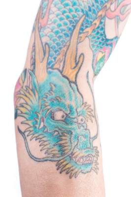
Novel oval ruby laser zaps blue-green tattoo pigments
HOLLYWOOD, FL. – Tattoos are common, but so are regrets. As tattoos become acceptable for the American mainstream, physicians are seeing more people who, for personal or professional reasons, wish to have a tattoo removed. And those who perform removal procedures benefit from having tools in their toolkit to eradicate an unwanted tattoo with minimal lasting skin damage.
Even with laser therapy – a common and effective tattoo removal technique – blue and green pigments can be especially difficult to eradicate.

At the annual meeting of the American Academy of Cosmetic Surgery, Dr. Robert H. Burke, director of the Michigan Center for Cosmetic Surgery, Ann Arbor, shared results from a pilot study of removal of blue-green pigments with a unique oval-shaped beam from a ruby laser.
Lasers, which preferentially heat chromophores, work well for tattoo removal since a selected wavelength is preferentially absorbed by a particular chromophore, targeting tattooed tissue and sparing non-inked tissue.
Older laser technologies created a Gaussian distribution of intensity with decreasing energy distribution toward the perimeter of the laser field. Newer lasers can create a sharper focal beam, creating beams of different sizes and shapes with uniform intensity.
In the pilot study, four men and four women who had persistent blue-green tattoo colors, received treatment with a 694 nm uniform-intensity ruby laser with an oval beam configuration. The patients, whose mean age was about 31 years, had tattoos with an age range of four to 30 years. The mean age of tattoos was 11.4 years. Most patients had had prior tattoo removal sessions; one of the women had undergone 14 sessions.
Dr. Burke treated these patients two to four times with the oval beam 694 nm ruby laser; fluence ranged from 2-5 J/cm2, with a spot size of 4-6 mm and a cycle of 2Hz.
The average tattoo pigment color clearance achieved with the oval beam 694 nm ruby laser was 60%, with a range from 40% to 90%. The study participants experienced no adverse events, Dr. Burke said.
The oval beam shape, he noted, deserves further investigation, because it should theoretically create less overlap and permit denser treatment – and may also allow the operator to follow a linear tattoo pattern more closely. This particular treatment regimen was also quicker, using 2 Hz rather than the more common 1 Hz, he pointed out.
Dr. Burke reported being on the physician advisory board for Suneva Medical.
On Twitter @karioakes
HOLLYWOOD, FL. – Tattoos are common, but so are regrets. As tattoos become acceptable for the American mainstream, physicians are seeing more people who, for personal or professional reasons, wish to have a tattoo removed. And those who perform removal procedures benefit from having tools in their toolkit to eradicate an unwanted tattoo with minimal lasting skin damage.
Even with laser therapy – a common and effective tattoo removal technique – blue and green pigments can be especially difficult to eradicate.
At the annual meeting of the American Academy of Cosmetic Surgery, Dr. Robert H. Burke, director of the Michigan Center for Cosmetic Surgery, Ann Arbor, shared results from a pilot study of removal of blue-green pigments with a unique oval-shaped beam from a ruby laser.
Lasers, which preferentially heat chromophores, work well for tattoo removal since a selected wavelength is preferentially absorbed by a particular chromophore, targeting tattooed tissue and sparing non-inked tissue.
Older laser technologies created a Gaussian distribution of intensity with decreasing energy distribution toward the perimeter of the laser field. Newer lasers can create a sharper focal beam, creating beams of different sizes and shapes with uniform intensity.
In the pilot study, four men and four women who had persistent blue-green tattoo colors, received treatment with a 694 nm uniform-intensity ruby laser with an oval beam configuration. The patients, whose mean age was about 31 years, had tattoos with an age range of four to 30 years. The mean age of tattoos was 11.4 years. Most patients had had prior tattoo removal sessions; one of the women had undergone 14 sessions.
Dr. Burke treated these patients two to four times with the oval beam 694 nm ruby laser; fluence ranged from 2-5 J/cm2, with a spot size of 4-6 mm and a cycle of 2Hz.
The average tattoo pigment color clearance achieved with the oval beam 694 nm ruby laser was 60%, with a range from 40% to 90%. The study participants experienced no adverse events, Dr. Burke said.
The oval beam shape, he noted, deserves further investigation, because it should theoretically create less overlap and permit denser treatment – and may also allow the operator to follow a linear tattoo pattern more closely. This particular treatment regimen was also quicker, using 2 Hz rather than the more common 1 Hz, he pointed out.
Dr. Burke reported being on the physician advisory board for Suneva Medical.
On Twitter @karioakes
AT THE AACS ANNUAL MEETING
Key clinical point: A novel oval-shaped ruby laser improved the removal of resistant blue-green tattoo pigments.
Major finding: A pilot study found an average 60% reduction in blue and green tattoo pigments with a novel oval-shaped ruby laser beam.
Data source: Eight patients, most of whom were pretreated, seeking removal of tattoos with resistant blue and great pigments.
Disclosures: Dr. Burke reported being on the physician advisory board for Suneva Medical.
Global approach to hand rejuvenation gives patients and clinicians options
HOLLYWOOD, FLA. – As more patients seek facial rejuvenation, they find themselves concerned with the mismatch between their refreshed facial appearance and the weathered, aged appearance of their hands. As more resurfacing treatments and fillers are being used beyond the face, options for hand rejuvenation expand.
Dr. Rania Agha, a dermatologist in private practice in Oakbrook Terrace, Ill., discussed a comprehensive approach to hand rejuvenation that addresses photoaging, vein prominence, and atrophy.
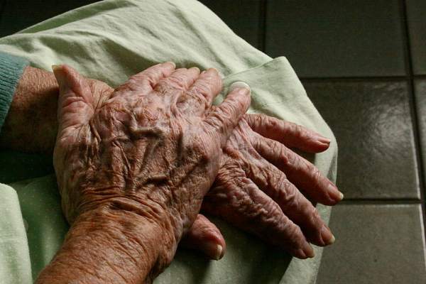
Sun exposure is the largest extrinsic factor that affects the appearance of hands. Resultant changes can include actinic keratosis, seborrheic keratosis, dermatoheliosis, and solar lentigines.
Intrinsic factors that contribute to hand aging include loss of collagen and fatty tissue, resulting in epidermal and dermal atrophy and loss of elasticity. Reduced hydration follows as well. Though hyaluronic acid is less than 1% of dermal dry weight, the loss of glycosaminoglycan-proteoglycan complexes means dermal tissue is much less effective at binding and retaining water, said Dr. Agha.
The consequences are wrinkles and prominence of bones and tendons, large intermetacarpal space, and prominent reticular veins. For many of her patients, “there will be a discordance of the face and the hands,” Dr. Agha nnoted.
Grading scales are available to evaluate hand aging, better to compare apparent hand age before and after treatment. The Merz Hand Grading Scale is used commonly and ranges from 0-4, with increasing scores for more wrinkles, bone and tendon prominence, and photodamage.
Where protuberant veins are a concern, sclerotherapy of the veins on the dorsum of the hand will reduce their prominence. Sclerotherapy options for the hand, where typical vessel size ranges from 1mm to 6 mm, are 0.5% sodium tetradecyl sulfate or polidocanol 1.5% or 3%. Post-sclerotherapy compression is recommended. An option of foam sclerotherapy, mixing the sclerosing agent with carbon dioxide or room air, can have greater efficacy because it prolongs contact between the sclerosant and the vein wall, but it theoretically carries some of the systemic risks associated with air emboli.
Adverse events from sclerotherapy can include telangiectatic matting, or microscopic revascularization; Dr. Agha said the reported incidence neared 15% in one study. Some pain, edema, and ecchymosis may occur; rare radial nerve neuropraxia has been reported. Other rare complications can include ulceration and hyperpigmentation.
Many energy-based, mechanical, and chemical rejuvenation techniques that are used on the face can also be used on the hands, though intensity or duration of treatment has to be modulated in some cases.
Superficial chemical peels only penetrate the epidermis; examples include 70% glycolic acid, salicylic acid, 50% resorcinol, Jessner’s solution, and trichloroacetic acid 10%-20%. Trichloroacetic acid at 30% or greater is an example of a medium-depth peel that extends to the papillary dermis. Deep peels that extend to the midreticular dermis, such as phenol, are not for use on the hands.
Even with light to medium peels, though, caution should be used. Reported complications can include maceration, desquamation, and delayed healing; also, practitioners should take care not to create a line of demarcation between the hand and forearm. “Be conservative and perform these gradually and serially over several weeks,” said Dr. Agha.
A wide range of laser and light therapies can be useful. Q-switched lasers are useful for solar lentigines and other photodamage; intense pulsed light, photodynamic therapy, Nd:YAG lasers, fractionated lasers, and the 2940 erbium:YAG laser are all options, depending on patient preference and what options are available.
“With light therapy, start at lower fluences,” said Dr. Agha. “Limit the number of passes, and the density, if you’re using a fractionated laser.” Complications can include erythema, hypo- or hyper-pigmentation, scarring, textural changes, crusting, and prolonged bleeding. Prompt local wound care can help mitigate long-term effects if any complications are seen post-treatment.
“Strict sun protection, whether with physical block or sunscreen, is a must after hand resurfacing,” whether the procedures are chemical or energy-based, said Dr. Agha.
Another option for hand rejuvenation is to use dermal fillers to plump the dorsum of the hand and obscure some of the exposed hand anatomy. Autologous fat can be used, but more demand is being seen for calcium hydroxylapatite microsphere (Radiesse) fillers, said Dr. Agha. “It is highly biocompatible; therefore, allergic reactions are extremely rare,” she said.
Calcium hydroxylapatite was approved by the Food and Drug Administration for hand rejuvenation in 2015. The particle size of 25-50 micrometers stimulates collagen formation and fibrosis when injected, and the product’s effects can last up to 18 months. In clinical trials, the product produced significant improvement in hand appearance without serious side effects in followup through 1 year.
Dr. Agha reviewed recommended administration of calcium hydroxylapatite, which is meant to be injected in bolus fashion midway between the dorsal crease of the wrist and the metacarpophalangeal joints, in a zone between the second and the fifth metacarpal bones. Dr. Agha says that in her practice, she has the patient make a tight fist, and then she performs vigorous massage of the hand immediately to distribute the product.
This is followed by application of a soapy cleanser, icing, and hand elevation for the first 24 hours post-injection. It’s important that patients be advised to expect moderate edema for up to 10 days, and that they avoid vigorous manual activities in the first week, said Dr. Agha.
Results from clinical trials indicate that between 70% and 80% of patients can expect an improvement of at least one point on a clinician-rated hand appearance scale, while over 98% of patients consider their hands subjectively improved at 1 month after treatment, and 86% of patients still feeling their hands are improved at 1 year post-treatment with calcium hydroxylapatite.
Dr. Agha reported being on the advisory boards of Taro Pharmaceutical Industries and Aqua Pharmaceutical.
On Twitter @karioakes
HOLLYWOOD, FLA. – As more patients seek facial rejuvenation, they find themselves concerned with the mismatch between their refreshed facial appearance and the weathered, aged appearance of their hands. As more resurfacing treatments and fillers are being used beyond the face, options for hand rejuvenation expand.
Dr. Rania Agha, a dermatologist in private practice in Oakbrook Terrace, Ill., discussed a comprehensive approach to hand rejuvenation that addresses photoaging, vein prominence, and atrophy.
Sun exposure is the largest extrinsic factor that affects the appearance of hands. Resultant changes can include actinic keratosis, seborrheic keratosis, dermatoheliosis, and solar lentigines.
Intrinsic factors that contribute to hand aging include loss of collagen and fatty tissue, resulting in epidermal and dermal atrophy and loss of elasticity. Reduced hydration follows as well. Though hyaluronic acid is less than 1% of dermal dry weight, the loss of glycosaminoglycan-proteoglycan complexes means dermal tissue is much less effective at binding and retaining water, said Dr. Agha.
The consequences are wrinkles and prominence of bones and tendons, large intermetacarpal space, and prominent reticular veins. For many of her patients, “there will be a discordance of the face and the hands,” Dr. Agha nnoted.
Grading scales are available to evaluate hand aging, better to compare apparent hand age before and after treatment. The Merz Hand Grading Scale is used commonly and ranges from 0-4, with increasing scores for more wrinkles, bone and tendon prominence, and photodamage.
Where protuberant veins are a concern, sclerotherapy of the veins on the dorsum of the hand will reduce their prominence. Sclerotherapy options for the hand, where typical vessel size ranges from 1mm to 6 mm, are 0.5% sodium tetradecyl sulfate or polidocanol 1.5% or 3%. Post-sclerotherapy compression is recommended. An option of foam sclerotherapy, mixing the sclerosing agent with carbon dioxide or room air, can have greater efficacy because it prolongs contact between the sclerosant and the vein wall, but it theoretically carries some of the systemic risks associated with air emboli.
Adverse events from sclerotherapy can include telangiectatic matting, or microscopic revascularization; Dr. Agha said the reported incidence neared 15% in one study. Some pain, edema, and ecchymosis may occur; rare radial nerve neuropraxia has been reported. Other rare complications can include ulceration and hyperpigmentation.
Many energy-based, mechanical, and chemical rejuvenation techniques that are used on the face can also be used on the hands, though intensity or duration of treatment has to be modulated in some cases.
Superficial chemical peels only penetrate the epidermis; examples include 70% glycolic acid, salicylic acid, 50% resorcinol, Jessner’s solution, and trichloroacetic acid 10%-20%. Trichloroacetic acid at 30% or greater is an example of a medium-depth peel that extends to the papillary dermis. Deep peels that extend to the midreticular dermis, such as phenol, are not for use on the hands.
Even with light to medium peels, though, caution should be used. Reported complications can include maceration, desquamation, and delayed healing; also, practitioners should take care not to create a line of demarcation between the hand and forearm. “Be conservative and perform these gradually and serially over several weeks,” said Dr. Agha.
A wide range of laser and light therapies can be useful. Q-switched lasers are useful for solar lentigines and other photodamage; intense pulsed light, photodynamic therapy, Nd:YAG lasers, fractionated lasers, and the 2940 erbium:YAG laser are all options, depending on patient preference and what options are available.
“With light therapy, start at lower fluences,” said Dr. Agha. “Limit the number of passes, and the density, if you’re using a fractionated laser.” Complications can include erythema, hypo- or hyper-pigmentation, scarring, textural changes, crusting, and prolonged bleeding. Prompt local wound care can help mitigate long-term effects if any complications are seen post-treatment.
“Strict sun protection, whether with physical block or sunscreen, is a must after hand resurfacing,” whether the procedures are chemical or energy-based, said Dr. Agha.
Another option for hand rejuvenation is to use dermal fillers to plump the dorsum of the hand and obscure some of the exposed hand anatomy. Autologous fat can be used, but more demand is being seen for calcium hydroxylapatite microsphere (Radiesse) fillers, said Dr. Agha. “It is highly biocompatible; therefore, allergic reactions are extremely rare,” she said.
Calcium hydroxylapatite was approved by the Food and Drug Administration for hand rejuvenation in 2015. The particle size of 25-50 micrometers stimulates collagen formation and fibrosis when injected, and the product’s effects can last up to 18 months. In clinical trials, the product produced significant improvement in hand appearance without serious side effects in followup through 1 year.
Dr. Agha reviewed recommended administration of calcium hydroxylapatite, which is meant to be injected in bolus fashion midway between the dorsal crease of the wrist and the metacarpophalangeal joints, in a zone between the second and the fifth metacarpal bones. Dr. Agha says that in her practice, she has the patient make a tight fist, and then she performs vigorous massage of the hand immediately to distribute the product.
This is followed by application of a soapy cleanser, icing, and hand elevation for the first 24 hours post-injection. It’s important that patients be advised to expect moderate edema for up to 10 days, and that they avoid vigorous manual activities in the first week, said Dr. Agha.
Results from clinical trials indicate that between 70% and 80% of patients can expect an improvement of at least one point on a clinician-rated hand appearance scale, while over 98% of patients consider their hands subjectively improved at 1 month after treatment, and 86% of patients still feeling their hands are improved at 1 year post-treatment with calcium hydroxylapatite.
Dr. Agha reported being on the advisory boards of Taro Pharmaceutical Industries and Aqua Pharmaceutical.
On Twitter @karioakes
EXPERT ANALYSIS FROM THE AMERICAN ACADEMY OF COSMETIC SURGERY ANNUAL SCIENTIFIC MEETING
