User login
International Association for the Study of Lung Cancer (IASLC): 15th World Conference on Lung Cancer
Radiological guidance does not improve tracheobronchial stent insertion (copy 1)
SYDNEY, AUSTRALIA – Use of radiological guidance for tracheobronchial stent insertion in advanced lung cancer makes no difference to outcomes such as complication rates, length of stay, or survival compared with visually-guided insertion, according to data from 79 patients.
Researchers from Harefield Hospital, London, examined outcomes from patients with advanced primary lung cancer whose airway obstructions were treated with tracheobronchial stenting; 41 were stented under radiological guidance and 38 with direct vision using bronchoscopy.
No cases of stent migration occurred in either group, and there were no significant differences between length of stay and overall survival, Henrietta Wilson, lead investigator, said at a world conference on lung cancer.
However, the use of radiological guidance required more staff and equipment and, by its very nature, led to more radiation exposure than did visually-guided stent insertion, said Ms. Wilson, a thoracic registrar (medical student) at Harefield Hospital.
"I had noticed that with the use of radiological guidance, it seemed to add an amount of time and organization onto the cases that we were doing, with having to get a larger number of people coming to perform the procedure, with the logistics of getting all of the equipment into theater, and that can sometimes also prolong the time of the general anesthetic while getting all of that set up," she said at the conference, which was sponsored by the International Association for the Study of Lung Cancer.
The average length of stay following the procedure was 2.73 days in the radiologically-guided group and 2.26 days in the visually-guided group (P = .93), with 69% of patients discharged on the same day, or the day after. The overall mean survival was 2.6 months, with 20% of patients alive at one year.
"I think probably those who advocate radiological guidance would feel that you get a better position of the stent and so they may have felt that it would get fewer complications from stent migration or malposition of the stent, or people requiring repeat procedures, but that certainly wasn’t something that we found," she said.
Ms. Wilson noted that tracheobronchial stenting was used generally in urgent cases of acute airway obstruction, either as a palliative procedure in itself or to provide short-term relief for patients awaiting further radiotherapy or chemotherapy.
"Acute airway obstruction can be very distressing, so if we’re able to just improve that then it may only be for weeks or a month at most but we find from a quality of life point of view, that’s a real benefit," she said in an interview.
Radiologically-guided stenting required an x-ray C-arm to enable real-time imaging of the stent placement, while in visually-guided placement, the surgeon would use a bronchoscope to assess the position of the tumor, and then use the guide wire to position the stent, Ms. Wilson noted.
Additional research is underway to examine the impact of guidance methods on the duration of the procedure and anesthetic.
Dr. Eleanor Summerhill, FCCP, comments: This small, single- center, retrospective study found no significant differences in rates of stent complications, hospital length of stay, or survival in patients with airway obstruction caused by primary lung cancers stented via direct visualization compared to under radiological guidance.
As the authors note, there are a number of disadvantages to placement under radiologic guidance. These include the need for additional equipment and staffing, as well as more radiation exposure and time under anesthesia.
This study suggests that these measures may not lead to improved outcomes, and will hopefully lead the way to a subsequent multicenter, randomized control trial.
Dr. Summerhill is the director of the Internal Medicine Residency Program at the Memorial Hospital of Rhode Island and Assistant Professor of Medicine
Alpert Medical School of Brown University.
There were no conflicts of interest declared.
AT THE IASLC WORLD CONFERENCE
Major finding: The average length of stay following tracheobronchial stent insertion was 2.73 days in the radiologically-guided group and 2.26 days in the visually-guided group (P = .93).
Data source: Retrospective study of 79 patients.
Disclosures: No conflicts of interest declared.
Radiological guidance does not improve tracheobronchial stent insertion
SYDNEY, AUSTRALIA – Use of radiological guidance for tracheobronchial stent insertion in advanced lung cancer makes no difference to outcomes such as complication rates, length of stay, or survival compared with visually-guided insertion, according to data from 79 patients.
Researchers from Harefield Hospital, London, examined outcomes from patients with advanced primary lung cancer whose airway obstructions were treated with tracheobronchial stenting; 41 were stented under radiological guidance and 38 with direct vision using bronchoscopy.
No cases of stent migration occurred in either group, and there were no significant differences between length of stay and overall survival, Henrietta Wilson, lead investigator, said at a world conference on lung cancer.
However, the use of radiological guidance required more staff and equipment and, by its very nature, led to more radiation exposure than did visually-guided stent insertion, said Ms. Wilson, a thoracic registrar (medical student) at Harefield Hospital.
"I had noticed that with the use of radiological guidance, it seemed to add an amount of time and organization onto the cases that we were doing, with having to get a larger number of people coming to perform the procedure, with the logistics of getting all of the equipment into theater, and that can sometimes also prolong the time of the general anesthetic while getting all of that set up," she said at the conference, which was sponsored by the International Association for the Study of Lung Cancer.
The average length of stay following the procedure was 2.73 days in the radiologically-guided group and 2.26 days in the visually-guided group (P = .93), with 69% of patients discharged on the same day, or the day after. The overall mean survival was 2.6 months, with 20% of patients alive at one year.
"I think probably those who advocate radiological guidance would feel that you get a better position of the stent and so they may have felt that it would get fewer complications from stent migration or malposition of the stent, or people requiring repeat procedures, but that certainly wasn’t something that we found," she said.
Ms. Wilson noted that tracheobronchial stenting was used generally in urgent cases of acute airway obstruction, either as a palliative procedure in itself or to provide short-term relief for patients awaiting further radiotherapy or chemotherapy.
"Acute airway obstruction can be very distressing, so if we’re able to just improve that then it may only be for weeks or a month at most but we find from a quality of life point of view, that’s a real benefit," she said in an interview.
Radiologically-guided stenting required an x-ray C-arm to enable real-time imaging of the stent placement, while in visually-guided placement, the surgeon would use a bronchoscope to assess the position of the tumor, and then use the guide wire to position the stent, Ms. Wilson noted.
Additional research is underway to examine the impact of guidance methods on the duration of the procedure and anesthetic.
Dr. Eleanor Summerhill, FCCP, comments: This small, single- center, retrospective study found no significant differences in rates of stent complications, hospital length of stay, or survival in patients with airway obstruction caused by primary lung cancers stented via direct visualization compared to under radiological guidance.
As the authors note, there are a number of disadvantages to placement under radiologic guidance. These include the need for additional equipment and staffing, as well as more radiation exposure and time under anesthesia.
This study suggests that these measures may not lead to improved outcomes, and will hopefully lead the way to a subsequent multicenter, randomized control trial.
Dr. Summerhill is the director of the Internal Medicine Residency Program at the Memorial Hospital of Rhode Island and Assistant Professor of Medicine
Alpert Medical School of Brown University.
There were no conflicts of interest declared.
AT THE IASLC WORLD CONFERENCE
Major finding: The average length of stay following tracheobronchial stent insertion was 2.73 days in the radiologically-guided group and 2.26 days in the visually-guided group (P = .93).
Data source: Retrospective study of 79 patients.
Disclosures: No conflicts of interest declared.
Partial pleurectomy improved quality of life but not survival in mesothelioma
SYDNEY, AUSTRALIA – Video-assisted thoracoscopic partial pleurectomy significantly improved quality of life and control of pleural effusion compared with talc pleurodesis in patients with malignant mesothelioma in a randomized, controlled, multicenter trial.
However, pleurectomy did not increase overall survival in the study, based on data presented in a plenary session at a world conference on lung cancer.
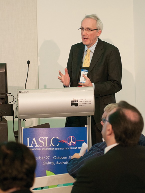
"The problem we now have is, we’ve got a negative result and a positive result because of the primary endpoint, so detractors could say there’s a negative endpoint so there’s no role for surgery," lead researcher Dr. Robert Rintoul said in an interview at the meeting, which was sponsored by the International Association for the Study of Lung Cancer.
The MesoVATS trial is an open-label, parallel-group, multicenter trial that included 175 patients with malignant pleural mesothelioma and pleural effusion. Patients were randomized to either partial pleurectomy (n = 87) or talc pleurodesis with slurry or poudrage (n = 88).
At 1 year, survival rates were 57% in patients who received talc pleurodesis and 52% in those randomized to pleurectomy (hazard ratio, 1.03; 95% confidence interval, 0.76-1.42; P = .83).
In the secondary outcome measures, however, pleural effusion was controlled at 1 month in 37% of patients in the talc group and in 59% of patients in the pleurectomy group (P = .008). At 6 months, pleural effusion was controlled in 57% of talc and 76% of pleurectomy patients (P = .04). Control of pleural effusion did not significantly differ at 9 and 12 months.
The EQ-5D quality-of-life measure was significantly improved with pleurectomy versus talc at 6 months (mean difference, 0.08 [95% CI, 0.003-0.16; P = .042]). Quality of life at 12 months also was significantly better with pleurectomy, but this finding became nonsignificant after adjustment for bias due to missing data.
Median hospital stay was significantly longer in the pleurectomy group (8 vs. 6 days; P < .001), and complications such as prolonged air leak were seen in 26% of those in the surgery arm and 8% of the talc arm (P = .009). The groups did not differ significantly in the incidence of serious adverse events.
Previous, nonrandomized research had suggested that pleurectomy may be associated with increased survival and better control of pleural effusion.
While the study failed to find a significant effect of surgery on the primary endpoint of survival at 12 months, the improvements in quality of life were particularly relevant for patients with malignant mesothelioma, said Dr. Rintoul, consultant respiratory physician at Papworth Hospital, Cambridge, England. "Quality of life in those last 12-13 months of life is very important, and [surgery] is something that is fairly well tolerated and doesn’t require a lot longer hospital stay."
Although the surgical procedure was more expensive than talc pleurodesis, Dr. Rintoul said it was worth giving patients the choice.
No relevant conflicts of interest were declared.
AT THE IASLC WORLD CONFERENCE
Major finding: Pleural effusion was controlled at 1 month in 37% of patients in the talc group and in 59% of patients in the pleurectomy group (P = .008).
Data source: MesoVATS is a randomized, controlled, multicenter trial that included 175 patients.
Disclosures: No relevant financial conflicts of interest were declared.
HPV prevalent in Hispanic nonsmokers with EGFR-positive lung cancer
SYDNEY, AUSTRALIA – Human papillomavirus was prevalent among lung cancer patients in Latin America, and may be a positive prognostic indicator among individuals also carrying the EGFR and KRAS mutations.
Analysis of data from 84 Hispanic patients with EGFR-positive lung adenocarcinoma, and 48 without the EGFR mutation, showed that 39% were positive for human papillomavirus 16 (HPV-16), according to data presented in a poster at a world conference on lung cancer. Overall, 70% of the patients in the study were never-smokers (93 out of 132 individuals – 58.3% of the EGFR-positive individuals and 91.7% of the EGFR-negative individuals).
Progression-free survival also was significantly greater in patients who were EGFR+/HPV+, compared with those who were EGFR+/HPV– (P = .014), as was overall survival (34 months vs. 24 months; P = .0001).
Overall survival was longer for patients who were HPV positive, even in the absence of EGFR mutations (P = .001). HPV status also influenced overall survival in the cohort of patients positive for the KRAS mutation.
"The presence of viral DNA can thus be presumed to be a positive prognostic factor for EGFR- and KRAS-mutated patients, thereby leading to considering infection as a dominant part of carcinogenesis amongst nonsmokers in Latin America," researchers from the CLICaP (Latin American Consortium for the Investigation of Lung Cancer) reported.
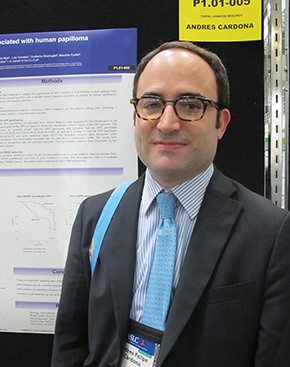
Lead investigator Dr. Andrés Cardona from the Fundación Santa Fe de Bogotá, Colombia, said HPV was known to play a key role in head and neck cancers, and evidence was emerging to support a role in lung cancer, particularly among nonsmokers.
"When you evaluate the perception of the importance of the infection in the patients with lung adenocarcinoma, considering their mutations, EGFR and KRAS, the HPV is an independent predictor of response to tyrosine kinase inhibitors," said Dr. Cardona in an interview.
Human papillomavirus is known to be particularly prevalent in Latin America, as well as in Chinese populations, Dr. Cardona said. The link between HPV and lung adenocarcinoma therefore has implications in terms of future prevention strategies.
"Maybe if this is part of the carcinogenesis of lung cancer in nonsmokers in Latin America as well as China, we can use the vaccine against HPV as a priority for our health system programs to avoid at least a small percentage of lung cancer and head and neck cancers," he said.
The conference was sponsored by the International Association for the Study of Lung Cancer. There were no conflicts of interest declared.
AT THE IASLC WORLD CONFERENCE
Major finding: Progression-free survival was significantly greater in patients who were EGFR-positive/HPV-positive, compared with those who were EGFR-positive/HPV-negative (P = .014), as was overall survival (34 months vs. 24 months; P = .0001).
Data source: Retrospective study of data from 132 patients.
Disclosures: There were no conflicts of interest declared.
Inspiratory muscle training improved subjective breathlessness in lung cancer
SYDNEY, AUSTRALIA – Inspiratory muscle training using a pressure threshold device improved lung cancer patients’ self-reported measures of breathlessness, but did not affect objective measurements of lung function, a pilot trial found.
The study in 46 patients with mostly advanced but stable lung cancer showed statistically and clinically significant differences in ability to cope with breathlessness (P = .02); satisfaction with breathlessness management (P = .024); fatigue (P = .007); emotional function (P = .006); breathlessness mastery (P = .031); anxiety (P = .027); and depression (P = .048).
In the randomized controlled trial, the difference in modified Borg (mBorg) score between the intervention and control groups at 3 months was 0.80, which was borderline clinically significant but not statistically significant. There were no differences in forced expiratory volume in 1 second (FEV1) and forced vital capacity (FVC) levels between the intervention and control groups, according to data presented at a world conference on lung cancer.
Lead investigator Dr. Alex Molasiotis said management of breathlessness in lung cancer is problematic.
"We have very few pharmacological interventions to offer ... so this is one of the fields where we need new evidence, new information, and new interventions," said Dr. Molasiotis, professor and head of the School of Nursing at Hong Kong Polytechnic University.
Inspiratory muscle training has been shown to be effective in some lung diseases such as chronic obstructive pulmonary disease. While the lack of effect on lung function in this pilot study was disappointing, Dr. Molasiotis said the observed improvements in subjective measures were still important.
"Generally we know that FEV1 and FVC don’t really correspond well with subjective measures, and this is something that is clear in the literature," Dr. Molasiotis said in an interview. "[But] it might be that we gave a sense of control to the patients through these techniques; we helped them cope better."
The pressure threshold device increases resistance on inspiration but not on expiration. Patients were instructed to use the device for 30 minutes per day, 5 days a week, for 12 weeks.
While some patients had to break the 30 minutes down into smaller sessions, Dr. Molasiotis said there was unexpectedly high compliance with use of the device.
"This is a population that’s quite ill, but I think it’s also a population that’s quite desperate to have an improvement, so very few of the patients didn’t follow the regime," Dr. Molasiotis said.
The patients were all off first-line treatment to rule out interference from chemotherapy or radiotherapy, and while initially the study investigators set out to recruit nonsmokers only, recruitment difficulties meant they changed the protocol to recruit smokers as well. There were no differences in response between smokers and nonsmokers.
Dr. Molasiotis said inspiratory muscle training is an option that some clinicians might like to consider, but he stressed that it is not appropriate for patients experiencing an acute episode of breathlessness.
The conference was sponsored by the International Association for the Study of Lung Cancer. There were no relevant financial conflicts of interest declared.
AT THE IASLC WORLD CONFERENCE
Major finding: Inspiratory muscle training with a pressure threshold device improved self-reported patient measures of breathlessness and other symptoms such as anxiety and depression, but it did not improve lung function scores.
Data source: Pilot feasibility randomized controlled trial in 46 patients with advanced, stable lung cancer.
Disclosures: There were no relevant financial conflicts of interest declared.
Cetuximab adds no benefit in stage III unresectable lung cancer
SYDNEY, AUSTRALIA – Cetuximab does not improve overall survival when used alongside chemoradiotherapy in patients with unresectable stage III lung cancer, based on data from a randomized, phase III comparison trial.
Cetuximab also appeared to be associated with an increase in therapy-related toxicities, based on data presented during a plenary session of a world conference on lung cancer.
The RTOG 0617 trial is an intergroup trial comparing standard-dose (60 Gy) and high-dose (74 Gy) radiotherapy and standard chemotherapy with and without cetuximab in patients with newly diagnosed, unresectable stage IIIA/IIIB non–small cell lung cancer.
Median survival in patients randomized to cetuximab was 23.1 months, compared with 23.5 months in patients not taking cetuximab; overall survival at 18 months was 60.8% versus 60.2% on the cetuximab and noncetuximab arms, respectively (P = .484; hazard ratio, 0.99), based on data from 465 patients.
There were 167 therapy-related grade 3 nonhematologic adverse events reported in the cetuximab group, compared with 115 in the no-cetuximab group (P less than .0001).
There was no significant interaction between radiation dose and cetuximab, although preliminary analysis did suggest that cetuximab may have more beneficial effect in patients with high-EGFR expression.
Presenting author Dr. Gregory Masters said that cetuximab – a monoclonal antibody used in head and neck, and colon cancer – had shown some survival benefit in previous studies in patients with more advanced lung cancer.
"If a drug shows an effect in stage IV patients, where you may only improve survival by a few months, hopefully you can then translate that into more cures for earlier-stage patients," said Dr. Masters, medical oncologist at the Helen F. Graham Cancer Centre in Newark, Del.
"In fact, we saw more toxicity and we didn’t show a treatment effect, at least in the overall population," Dr. Masters said in an interview at the meeting, which was sponsored by the International Association for the Study of Lung Cancer.
The trial was originally designed to compare high and low-dose radiotherapy; cetuximab was added after other studies suggested promising results in combination with chemotherapy and radiotherapy.
Results from the standard and high-dose radiotherapy analysis of the trial were presented at the annual meeting of the American Society of Clinical Oncology in May of this year and showed no advantage with increased radiation dose.
The study received support from Bristol-Myers Squibb and Eli Lilly, but no other financial conflicts of interest were declared.
AT THE IASLC WORLD CONFERENCE
Major finding: Median survival in patients randomized to cetuximab was 23.1 months, compared with 23.5 months in patients not taking cetuximab; overall survival at 18 months was 60.8% versus 60.2% on the cetuximab and noncetuximab arms, respectively (P = .484; hazard ratio, 0.99).
Data source: Phase III randomized comparison trial in 465 patients with unresectable stage III non–small cell lung cancer.
Disclosures: The study received support from Bristol-Myers Squibb and Eli Lilly, but no other financial conflicts of interest were declared.
One-third of cancer patients over age 70 went untreated, Swedish study finds
SYDNEY – Age should not be a barrier to chemotherapy for lung cancer, even in patients over age 70, based on the results of a retrospective study.
Nearly one-third (32.2%) of 659 patients over age 70 with non–small cell lung cancer received no radiotherapy or chemotherapy, according to data from patients who presented at a single center between 2003 and 2006.
Around one-quarter of the cohort received chemotherapy, 13.9% received surgery, but 10% received second-line chemotherapy, according to data presented at a world conference on lung cancer.
Median survival was 231 days for patients aged 70-75 years, 250 days for those aged 76-80 years, and 213 days for those aged over 80 years.
Median survival was 610 days among patients given second-line chemotherapy, compared with 285 days for those who received only first-line chemotherapy.
Dr. Hirsh Koyi said he and his fellow researchers were surprised to discover how many patients over 70 did not receive any treatment at all. He and his colleagues have been taking steps to address this in their own practice.
There were generally concerns that patients over 70 were more prone to other diseases such as kidney or heart disease. However, the patient population was generally healthy and in good shape, which raised questions about why they did not receive the same treatment as younger patients, said Dr. Koyi, of Gävle (Sweden) Hospital.
Older patients "are undertreated and discriminated against without any reason. We have patients who are older than 80 years and, with surgery, are living still after 10 years," Dr. Koyi said in an interview. No prospective studies specifically address chemotherapy in this age group, he pointed out.
Dr. Koyi said performance status (PS) was a more important consideration in deciding treatment for older patients with lung cancer, with the study finding that median survival for patients with a PS score of 0 was 810 days and was 109 days for those with a PS score of 3.
"If they have a good performance status, age is no problem," Dr. Koyi said. "We have patients who are 50 years old, lying in bed, very sick, with no chemotherapy, but other patients who are 70-80 years have a good performance status and get chemotherapy."
He said patients over 70 years with a PS score of 1-2, with good kidney and heart function, and in otherwise good health, should be considered for chemotherapy.
"We have to encourage our colleagues to look not at the age but at how each patient feels."
The conference was sponsored by the International Association for the Study of Lung Cancer. There were no relevant conflicts of interest declared.
AT THE IASLC WORLD CONFERENCE
Major finding: Nearly one-third (32.2%) of 659 patients over age 70 with non–small cell lung cancer received no radiotherapy or chemotherapy.
Data source: Retrospective study of data from patients who presented at a single center between 2003 and 2006.
Disclosures: The conference was sponsored by the International Association for the Study of Lung Cancer. There were no relevant conflicts of interest declared.
Stereotactic lung radiotherapy of central lung tumors can achieve good overall survival with limited toxicity
SYDNEY – Stereotactic lung radiotherapy for centrally located non–small cell lung carcinomas can result in overall survival outcomes that are similar to those for peripheral tumors, but with an increased risk of local failure and pneumonitis, based on the results of an analysis of a large international data set.
Overall survival at 3 years was similar for 100 centrally located non–small cell lung cancers – defined as being located within 2 cm of the proximal bronchial tree – and for 869 peripheral tumors treated with stereotactic body radiotherapy (50% vs. 51%; P = .70), based on data presented by Dr. Maria Werner-Wasik at a world conference on lung cancer, sponsored by the International Association for the Study of Lung Cancer.
Also, the two groups did not significantly differ in the incidence of chest wall pain and myositis, rib fracture, and dermatitis. Patients with central tumors had a significantly higher incidence of grade 2 or above pneumonitis (8% vs. 1% in those with peripheral tumors; P less than .001). The incidence of grade 3 pneumonitis was similar between the two groups.
Central tumors were associated with a lower rate of cause-specific survival than were peripheral tumors (75% vs. 88%; P less than .001) and higher local failure rates at 3 years (16.2% vs. 5.9%; P less than .001) and 5 years (20.4% vs. 8.3%; P less than .001).
Dr. Werner-Wasik said central tumors had fallen into a "no-fly zone" for stereotactic body radiotherapy since an earlier trial showed unacceptable levels of toxicities from treating centrally located lung tumors. Thereafter, prospective trials examined only peripheral tumors.
"If you have a central tumor you have to do something. You can always treat with standard fractionation, but that’s not the idea. We want to give those patients (with centrally located tumors) the benefit of a short, effective regimen," said Dr. Werner-Wasik, professor and director of clinical research at the department of radiation oncology, Kimmel Cancer Center, Thomas Jefferson University, Philadelphia.
The data from the Elekta Lung Research Groupshowed a higher rate of local failure. Dr. Werner-Wasik said this was still "respectable," especially as the centrally located tumors did receive slightly less radiation than the peripheral tumors did and were significantly larger at baseline (3.1 vs. 2.4 cm; P less than .001).
"The local control was worse for central tumors versus peripheral tumors, presumably because we treated them with lower biologically effective doses," said Dr. Werner-Wasik in an interview. "But also, these tumors were slightly different ... as illustrated by higher values on PET scans."
Results from the upcoming RTOG 0813 study may shed more light on the question of how to deliver stereotactic body radiotherapy to centrally located tumors, she said. "We have to find an effective central dose radiation fractionation and total dose so these patients do not suffer complications and yet are assured local control."
The research was partly supported by a grant from Elekta, makers of stereotactic body radiation technology products. Dr. Werner-Wasik said she had no relevant financial disclosures. The Elekta Lung Research Group includes participants from William Beaumont Hospital in Royal Oak, Mich.; Princess Margaret Cancer Centre, Toronto; Thomas Jefferson University, Philadelphia; Julius Maximilian University of Würzburg (Germany); and the Netherlands Cancer Institute–Antoni van Leeuwenhoek Hospital in Amsterdam.
AT THE IASLC WORLD CONFERENCE
Major finding: Overall survival at 3 years was similar for 100 centrally located non–small cell lung cancers – defined as being located within 2 cm of the proximal bronchial tree – and for 869 peripheral tumors treated with stereotactic body radiotherapy (50% vs. 51%; P = .70).
Data source: A retrospective analysis of data from the Elekta Lung Research Group.
Disclosures: The research is partly funded by a grant from Elekta, makers of stereotactic body radiation technology products. Dr. Werner-Wasik said she had no relevant financial disclosures.
Osteopontin level in early non-small cell lung cancer predicts recurrence
SYDNEY, AUSTRALIA – Elevated plasma levels of osteopontin were predictive of 3-year recurrence rates and second primary cancers in patients with stage 1 non–small-cell lung cancer, based on data from a prospective cohort study presented at a world conference on lung cancer.
Based on preoperative plasma specimens from 137 patients undergoing resection of stage 1 adenocarcinoma of the lung, a preoperative cut-off point of 49.6 ng/mL for plasma osteopontin was predictive of 3-year recurrence. In a multivariate analysis, patients with levels above the cut-off had a fourfold increase in the risk of recurrence (HR 4.2, 95% CI 1.8-10.2, P = .001).
The cohort was followed for at least 3 years, with a median follow-up of 44 months. There was a recurrence in 56 patients (41%) over the study period; 31 patients (22.5%) had recurrences within 3 years and 23 of those patients had high osteopontin levels. Thirteen had systematic progression, ten had local or regional progression, and eight patients had a second primary non–small-cell lung cancer.
After adjustment for variants such as stage, gender, and tumor size, osteopontin levels were not significantly associated with 5-year survival; however, 3-year systemic or local progression was highly predictive of 5-year mortality, reported Dr. Jessica Donington, associate professor of cardiothoracic surgery at NYU Langone Medical Center, New York, and her colleagues.
"We would like to think that this would be something you would use to help guide adjuvant therapy or cancer surveillance or prevention protocols," Dr. Donington said.
Osteopontin is associated with increased inflammation. Previous research had shown that higher levels of osteopontin predicted poor response to chemotherapy in patients with advanced lung cancer. Also, osteopontin levels are known to have prognostic implications in breast and prostate cancers.
Osteopontin levels above the cut-off point also were predictive of second primary lung cancers. The association wasn’t as strong for second cancers as it was for recurrence, but the association was still significant and may help to guide follow-up.
"This might be the group that you’re going to decide to scan every 3 months," Dr. Donington said.
The conference was sponsored by the International Association for the Study of Lung Cancer. The study was supported by an Early Detection Research Network Grant, the Stephen Banner Lung Cancer Foundation, and an IASLC/Lung Cancer Foundation of America grant.
AT THE IASLC WORLD CONFERENCE
Major finding: Preoperative plasma osteopontin levels above 49.6 ng/mL are predictive of recurrence of lung adenocarcinoma in patients with early stage disease.
Data source: Prospective cohort study of 137 patients undergoing resection.
Disclosures: The study was supported by an Early Detection Research Network Grant, the Stephen Banner Lung Cancer Foundation, and an IASLC/Lung Cancer Foundation of America grant.


