User login
Neoadjuvant chemotherapy induces mutational, molecular shifts in ovarian cancers
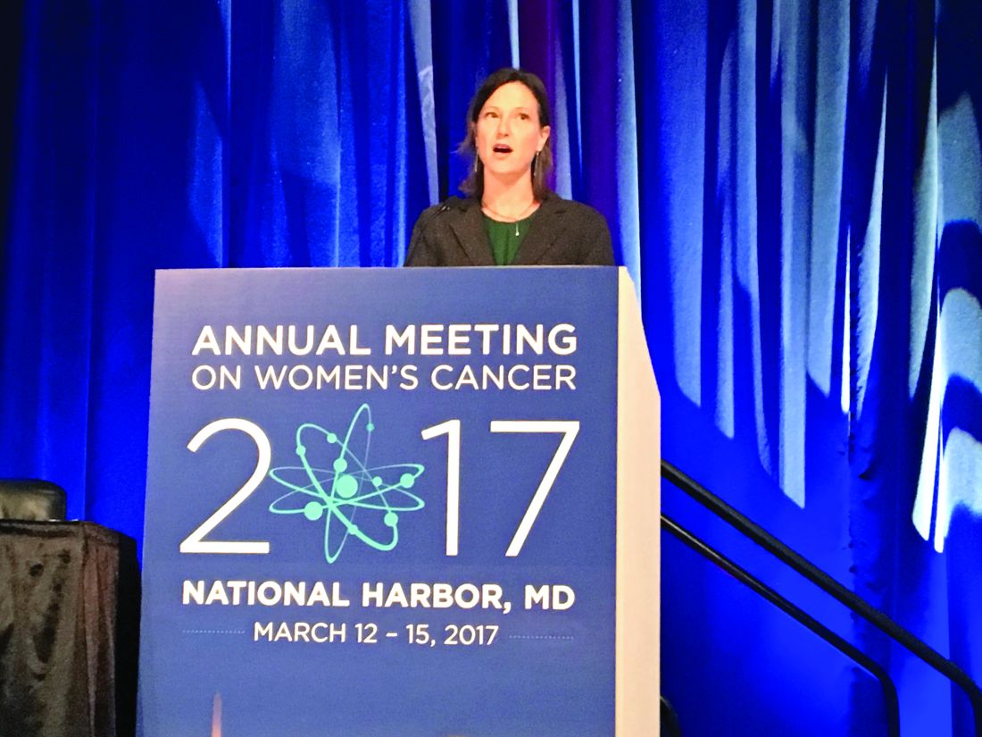
NATIONAL HARBOR, MD. – Treatment of high-grade serous ovarian carcinomas with platinum-based neoadjuvant chemotherapy led to significant changes in the expression of genes encoding “canonical” cell cycle and DNA damage pathways, said Rebecca C. Arend, MD.
Analyses of cell-free (plasma) DNA also revealed mutations that matched those in tumor specimens obtained before and after platinum-based neoadjuvant chemotherapy, Dr. Arend reported at the annual meeting of the Society of Gynecologic Oncology. “Understanding the effect of chemotherapy on gene expression profiles may help guide therapy, and plasma cfDNA could provide a noninvasive approach for monitoring tumor mutations,” said Dr. Arend of the University of Alabama at Birmingham.
High-grade serous ovarian carcinoma is genetically heterogeneous, and chemotherapy further alters gene expression profiles and causes molecular derangement, Dr. Arend noted. “Neoadjuvant chemotherapy provides a unique opportunity to evaluate biospecimens before and afterward,” she added.
Both gene expression and mutational profiles have been used to characterize HGSC, but researchers lack solid methods to evaluate tumor heterogeneity and clonal evolution. To begin filling this gap, Dr. Arend and her associates analyzed plasma and tumor specimens from 19 patients with stage 3 or 4 high-grade serous ovarian carcinoma before and after they underwent three to six cycles of neoadjuvant chemotherapy. Biopsies yielded the baseline tumor specimens, and follow-up specimens were obtained during interval debulking.
The investigators used the NanoString PanCancer 770 gene pathway panel, the Ingenuity Pathway Analysis tool, and nSolver Analysis software to assess changes in gene expression. To quantify mutations, they performed longitudinal next-generation sequencing of 50 genes in tumor and plasma specimens with a 50-gene Ion Torrent panel.
After neoadjuvant chemotherapy, the most up-regulated genes included NR4A1 and NR4A3, which regulate cellular proliferation, differentiation, and apoptosis, and SFRP2, which promotes resistance to chemotherapy by modulating Wnt signaling, Dr. Arend said. The most down-regulated genes included E2F1, which helps mediate the cell cycle and the activity of tumor suppressor genes, and BRCA2, the tumor suppressor gene that encodes a DNA repair protein.
Pathway analysis confirmed that the cell cycle pathway was most up-regulated after neoadjuvant chemotherapy, and that the DNA damage repair pathway was the most downregulated, Dr. Arend reported. Within the DNA damage repair pathway, no gene was significantly up-regulated, while RAD51C, BRCA1, BRCA2, and the FA core complex genes were down-regulated.
Next-generation sequencing of baseline plasma cfDNA identified 57 mutations, of which 6 persisted after neoadjuvant chemotherapy. In contrast, of 38 mutations in tumor at baseline, 33 persisted after chemotherapy.
Only 15 of the 38 mutations in tumor also appeared in cfDNA before treatment. At the time of interval debulking, tumor specimens yielded 36 mutations, of which 11 were detected in cfDNA.
At baseline and after treatment, all patients had TP53 mutations either tumor alone, or in both tumor and plasma. Among four patients whose cancer recurred, three had mutations in cfDNA that were previously detected in tumor. Implicated genes included PIK3CA, TP53, KIT, and KDR.
Overall, the study suggests that gene expression profiling of ovarian HGSC tumor tissue taken at interval debulking could someday help guide treatment decisions after neoadjuvant chemotherapy, Dr. Arend said. “To be able to use cfDNA as liquid biopsy, more studies like this one, which match tumor and cfDNA from multiple time points, are needed,” she added.
Dr. Arend cited no funding sources and reported having no conflicts of interest.
Key clinical point. Neoadjuvant chemotherapy was associated with changes in gene expression and associated pathways in high-grade serous ovarian carcinomas. Some mutations in tumors were also present in plasma cell-free DNA.
Major finding: Pathway analysis confirmed that the cell cycle and apoptosis pathway was the most up-regulated after neoadjuvant chemotherapy, while the DNA damage repair pathway was the most down-regulated. Among 38 baseline mutations in tumor specimens, 15 of 38 appeared in plasma cell-free DNA. At interval debulking, tumor specimens yielded 36 mutations, of which 11 were detected in cfDNA.
Data source: Gene expression profiling, pathway analysis, and next-generation sequencing of 19 patients with high-grade serous ovarian carcinomas.
Disclosures: Dr. Arend cited no funding sources and reported having no conflicts of interest.
VIDEO: When early-stage vulvar cancer recurs, the prognosis is poor
NATIONAL HARBOR, MD – Cancer of the vulva is becoming more common, and as incidence is rising, so are death rates from the relatively rare malignancy. A new examination of women with early-stage vulvar cancer showed that recurrence was common, and carried a poor prognosis.
Cancer in “nearly 25% of women with stage IB squamous cell carcinoma of the vulva will recur within 3 years of initial diagnosis,” said the study’s lead author, Rebecca Stone, MD, at the annual meeting of the Society of Gynecologic Oncology. Although most (85%) of the cancers that recur do so locally, salvage rates for patients with recurrences are poor, she said in a video interview.
Dr. Stone of Johns Hopkins University, Baltimore, said that the study looked at the subset of 59 patients with recurrence of their stage IB vulvar cancer, drawing from a total of 244 stage IB patients for whom complete data were available.
Current National Comprehensive Cancer Network (NCCN) guidelines for the treatment of early-stage vulvar squamous cell carcinoma (meaning T1 or 4 cm or smaller T2 lesions) call for adjuvant radiotherapy for patients with positive margins or unresectable lesions. For patients with negative margins after the primary procedure or re-excision, the guidelines say that radiotherapy decisions should be guided by risk factors including margin width, tumor size, the presence of lymphovascular invasion, and the pattern and depth of invasion.
However, Dr. Stone said, “There’s a knowledge gap in our current management of early-stage disease.” Some unknowns include what the threshold should be for tumor recurrence in terms of depth of invasion and size of tumor, whether perineural invasion is a risk factor, and whether adverse clinical factors are important. Also, she said, it’s not known how risk factors combine to increase recurrence risk.
Tumor size, invasion depth over 6 mm, and patient age were all risk factors identified by the study; perineural and lymphovascular invasion were also potential risk factors for recurrence.
The retrospective study, said Dr. Stone, is the largest multi-institutional cohort study examining stage IB vulvar cancer that has been completed, and it accomplished a detailed examination of which patients have recurrences, factors associated with recurrence, and postrecurrence salvage rates.
Dr. Stone reported no disclosures.
The video associated with this article is no longer available on this site. Please view all of our videos on the MDedge YouTube channel
[email protected]
On Twitter @karioakes
AT THE ANNUAL MEETING ON WOMEN’S CANCER
VIDEO: How to start an oncology sexual health clinic
NATIONAL HARBOR, MD – Start small, but anticipate growth. Engage your administration from the start. Be smart about resources, and consider using advanced practice providers to keep costs down. Above all, keep lines of communication open with physicians and other members of the care team.
In a video interview, Joanne Rash, PA-C, a certified physician assistant at the University of Wisconsin–Madison offers these and other tips. She explains her collaborative work with David Kushner, MD, director of the gynecologic oncology program and professor at the University of Wisconsin School of Medicine and Public Health, and a colleague to develop the Women’s Integrative Sexual Health (WISH) program.
WISH is modeled on the University of Chicago’s Program in Integrative Sex and Medicine for Women and Girls with Cancer (PRISM) and participates in the PRISM registry, which studies ways to prevent and treat sexual problems for women and girls with cancer.
“I think what makes the WISH program unique is that we carve time out,” said Ms. Rash. “Of course, we address some of these issues in my gynecologic oncology practice, but, when we do it in WISH, the format is different,” and there’s just more time for discussion.
Communication is key to the model’s success in safe integration of sexual health into cancer care, she said. “We certainly don’t want to do something that compromises cancer care, and so, it’s important that we have those conversations with that woman’s team. And now we get to be a part of that team, which is a real privilege.”
Ms. Rash reported no conflicts of interest.
The video associated with this article is no longer available on this site. Please view all of our videos on the MDedge YouTube channel
[email protected]
On Twitter @karioakes
AT THE ANNUAL MEETING ON WOMEN’S CANCER
VIDEO: Sex, intimacy part of history in gynecologic oncology care
NATIONAL HARBOR, MD. – Cancer care can feel like a sequence of crises, large and small; a comprehensive cancer care approach can keep the patient at the center of this flurry of events. But understanding a patient’s sexual history and assessing how a gynecologic malignancy has affected that patient’s sexual health can, too often, get lost in the shuffle, said Don Dizon, MD, of Massachusetts General Hospital, Boston.
“I think there are multiple reasons for that, but it has to be the job of the clinician – not necessarily the oncologist-physician – but somebody has to be able to say, ‘What is your sexual history? Can I ask about your sexual history?’ ” he said in a video interview at the annual meeting of the Society of Gynecologic Oncology.
Dr. Dizon, director of the Oncology Sexual Health Clinic at Massachusetts General Hospital*, emphasized the importance of providing sensitive sexual health care while staying true to one’s own framework of care. “I teach clinicians not to run away from their schema,” he said. “Having said that, the job of a clinician is to normalize the history, and to normalize that sex, intimacy, relationships are a part of someone’s history.”
Dr. Dizon sits on the board of the Patty Brisben Foundation and the Young Survival Coalition.
[email protected]
On Twitter @karioakes
*A previous version of this article misstated Dr. Dizon's affiliation.
AT THE ANNUAL MEETING ON WOMEN'S CANCER
Video: Try ‘PLISSIT’ to address postcancer sexual health
NATIONAL HARBOR, MD. – Comprehensive care of patients with gynecologic malignancies should include a sensitive and thorough assessment of sexual health.
In a video interview at the annual meeting of the Society of Gynecologic Oncology, Don Dizon, MD, of Massachusetts General Hospital, Boston, gives a series of practical tips to help physicians take a thorough sexual health history and provide information and guidance for patients and their partners.
Dr. Dizon, professor of gynecologic oncology and director of the oncology sexual health clinic at Brigham and Women’s Hospital, also in Boston, said that he likes to begin with the PLISSIT model, where patients are given permission (P) to talk about sexual problems. Then, the clinician gives the patient limited (LI) scientific or clinical information about the situation, followed by specific suggestions (SS) that might help. Finally, patients may be referred to mental health providers or sex counselors for intensive therapy (IT) if needed.
“It’s also important not to confuse terminology,” said Dr. Dizon. “Intimacy is experienced very differently between men and women. Women experience intimacy through arousal, desire, and, when desire is satisfied, that’s intimacy. Intercourse is not a part of that equation.” For men, he said, intimacy is more often experienced through intercourse. “So the disconnect is greater after cancer is diagnosed,” making it especially important to acknowledge problems sensitively, and to helps patients and partners find a way forward.
“The word I like to use is ‘play,’ ” said Dr. Dizon. When a renegotiation of an intimate relationship is framed in terms of play, the pressure is off, and “men can wrap their hands around that idea,” he said.
Dr. Dizon sits on the board of the Patty Brisben Foundation and the Young Survival Coalition.
The video associated with this article is no longer available on this site. Please view all of our videos on the MDedge YouTube channel
[email protected]
On Twitter @karioakes
AT THE ANNUAL MEETING ON WOMEN’S CANCER
Study supports NCCN recommendations on risk-reducing salpingo-oophorectomy
NATIONAL HARBOR, MD – A large hereditary cancer study supports National Comprehensive Cancer Network guidance to consider risk-reducing salpingo-oophorectomy (RRSO) between ages 45 and 50 years for women with BRIP1, RAD51C, or RAD51D mutations, Lydia Usha, MD, said at the annual meeting of the Society of Gynecologic Oncology.
The average ages for an ovarian cancer diagnosis were 56 years for women with RAD51D mutations, 61 years for RAD51C mutations, and 64 years for BRIP1 mutations, said Dr. Usha of Rush Medical College, Chicago. When appropriate, delaying RRSO “avoids the psychosocial and medical complications of premature menopause,” she said.
Among all women, mutation prevalence was 0.3% for BRIP1, 0.1% for RAD51C and RAD51D, 1.2% for BRCA1, and 1.3% for BRCA2 mutations, Dr. Usha reported. Among 18,719 women who had a personal history of ovarian cancer, the most common mutation was BRCA1 (3.5%), followed by BRCA2 (2.7%). In contrast, the combined prevalence of BRIP1, RAD51C, and RAD51D mutations among cancer patients was only 1.6%.
Cancer prevalence was highest among women who had mutations of RAD51C (22%), followed by RAD51D (19%), BRCA1 and BRIP1 (16% in each case), and BRCA2 (11%). Thus, while BRIP1 and RAD51D mutations were uncommon, their presence signified an ovarian cancer risk that was similar to that with BRCA1 mutations, and a greater risk than with BRCA2, Dr. Usha said.
The average ages for ovarian cancer diagnosis were 64 years for BRIP1, 61 years for RAD51C, 60 years for BRCA2, 56 years for RAD51C, and 54 years for BRCA1. “More than 80% of women with ovarian cancer who had a mutation in BRIP1, RAD51C, or BRCA2 were diagnosed after age 50,” Dr. Usha noted. These findings support considering RRSO closer to age 45 years for RAD51D mutation carriers and closer to age 50 years for women with pathogenic variants of BRIP1, added discussant Kari Ring, MD, of the University of Virginia, Charlottesville.
Mutation type did not significantly correlate with ethnicity or type of ovarian cancer, Dr. Usha noted. “Collectively, these findings may aid clinical decisions about the medical management of women with mutations in these genes,” she said. “Our data may also assist with reproductive decisions, such as age of childbearing.”
Dr. Usha did not report external funding sources, but disclosed travel expenses from Myriad Genetics.
AT THE ANNUAL MEETING ON WOMEN'S CANCER
Key clinical point: A large hereditary cancer study supports National Comprehensive Cancer Network guidance to consider risk-reducing salpingo-oophorectomy between age 45 and 50 years for women with BRIP1, RAD51C, or RAD51D mutations.
Major finding: Average ages for an ovarian cancer diagnosis were 56 years for women with RAD51D mutations, 61 years for RAD51C mutations, and 64 years for BRIP1 mutations.
Data source: Analyses of a 25-gene hereditary panel performed in 345,667 women.
Disclosures: Dr. Usha did not report external funding sources, but disclosed travel expenses from Myriad Genetics.
Complete resection tied to improved survival in low-grade serous ovarian cancer
NATIONAL HARBOR, MD. – Surgical resection to the point of no residual macroscopic disease significantly improved survival among patients with low-grade serous ovarian carcinoma, based on the findings of a large multicenter retrospective cohort study.
Adjuvant platinum-based therapy, however, did not appear to boost survival in the analysis, Tamayaa May, MD, MSc, said at the annual meeting of the Society of Gynecologic Oncology. “Genotyping and targeted sequencing of low-grade serous ovarian carcinoma often identifies actionable mutations, and treatment with MEK-based combination therapy might be a viable therapeutic strategy in patients with KRAS or NRAS mutations,” added Dr. May of Princess Margaret Cancer Center at the University of Toronto. She and her associates plan to examine more subgroups to determine if genomic alterations predict systemic response, she said.
Low-grade (Silverberg grade 1) serous ovarian tumors are slow growing but tend to resist chemotherapy, making optimal debulking a crucial part of treatment. In past studies, debulking that eliminated macroscopic evidence of disease was associated with a median survival time of about 115 months, compared with about 43 months if patients had residual disease, Dr. May noted.
To further explore outcomes after surgical resection, and to help clarify the role of systemic platinum-based therapy in low-grade serous ovarian carcinoma, she and her associates analyzed clinical data from 714 patients with low-grade serous ovarian carcinomas, including 40 from her institution and 674 from the Ovarian Cancer Association Consortium Registry. Most (60%) patients had stage III disease at diagnosis.
Complete data on surgical outcomes were available for 382 patients, of whom 202 (53%) had residual macroscopic disease and 43% did not. Among 439 patients with complete treatment data, 170 (39%) received first-line platinum-based chemotherapy. For the 391 patients with complete data on progression-free survival (PFS), the median follow-up was 4.9 years and median PFS was 3.1 years (95% confidence interval, 2.6-4.5 years). Residual macroscopic disease correlated with shorter PFS (P less than .001), as did higher tumor stage and baseline CA125 (P less than .001), but platinum-based therapy did not (P = .1).
A multivariable analysis of 333 patients confirmed these findings, Dr. May said. Independent correlates of death or disease progression included residual macroscopic disease (hazard ratio, 2.38; 95% CI, 1.68-3.37; P less than .001), older age (HR, 1.15; 95% CI, 1.02-1.30; P = .02), and stage III (HR, 3.28; 95% CI, 1.87-5.76; P less than .001) or stage IV disease (HR, 5.68; 95% CI, 2.73-11.83; P less than .001), compared with stage I disease. In contrast, platinum-based therapy did not correlate with survival (HR, 0.94; 95% CI, 0.69-1.28; P = .69).
The overall survival analysis also linked mortality with higher tumor stage, increased baseline CA125, and residual disease (P less than .001 for each association), but not with platinum-based therapy (P = .2). The multivariable analysis independently tied mortality to older age (HR, 1.25; P less than .001), stage III (HR, 2.31; P = .006) or IV disease (HR, 3.86; P less than .001), and residual disease (HR, 2.53; P less than .001), but not to platinum-based therapy (HR, 1.05; P = .77).
Data consistency and completeness were issues in this study: most notably, 45% of patients had no PFS data, Dr. May commented. Nonetheless, this type of large retrospective multicenter analysis is one of the best ways to study rare tumors, including low-grade serous ovarian carcinoma, she said.
The Ovarian Cancer Research Fund provides financial support for OCAC. Dr. May reported having no conflicts of interest.
AT THE ANNUAL MEETING ON WOMEN’S CANCER
Key clinical point: Unlike platinum-based chemotherapy, resection to the point of no residual disease was associated with improved survival among patients with low-grade serous ovarian carcinoma.
Major finding: Independent correlates of death or disease progression included residual macroscopic disease (hazard ratio, 2.38; P less than .001), older age (HR, 1.15; P = .02), and stage III (HR, 3.28; P less than .001) or stage IV (HR, 5.68; P less than .001) disease, compared with stage I disease. Platinum-based therapy was not associated with improved survival (HR, 0.94; P = 69).
Data source: A retrospective cohort study of 714 patients with low-grade (Silverberg grade 1) serous tumors from the Ovarian Cancer Association Consortium Registry.
Disclosures: The Ovarian Cancer Research Fund provides financial support for OCAC. Dr. May reported having no conflicts of interest.
Adding Pap to plasma testing boosted detection of ovarian tumors
NATIONAL HARBOR, MD. – Combining a liquid-based Pap smear and cell-free DNA plasma testing can boost the sensitivity of screening for primary ovarian tumors, based on the results of a prospective study of 201 patients.
The approach, however, was most sensitive when women already had late-stage disease, Amanda Nickles Fader, MD, said at the annual meeting of the Society of Gynecologic Oncology. Nonetheless, she called combined Pap and plasma testing “a promising step toward a broadly applicable screening methodology for the early detection of ovarian cancer.”
Ovarian cancer is a “legal malignancy,” and detecting it at “earlier, more curable stages is a clinical imperative,” said Dr. Fader of the Johns Hopkins Kelly Gynecologic Oncology Service in Baltimore, Md.
Routine screening with ultrasound or the CA-125 test has not been shown to reduce deaths from ovarian cancer and can lead to unnecessary diagnostic surgeries. Therefore, population-based screening is not recommended by the U.S. Preventive Services Task Force, the American Cancer Society, the American Congress of Obstetricians and Gynecologists, or the National Comprehensive Cancer Network, Dr. Fader noted.
This paradigm, however, is starting to change with the advent of DNA-based tests for ovarian cancer, including the PapGene test and assays for circulating DNA released by apoptotic tumor cells (ctDNA), Dr. Fader said. In a previous pilot study, Pap testing identified oncogenic driver mutations in 41% of primary ovarian tumors. In another study, polymerase chain reaction (PCR) testing detected ctDNA in more than 75% of metastatic solid tumors.
Might combining Pap and plasma testing for ctDNA further boost sensitivity? To test this idea, Dr. Fader and her associates performed liquid-based Pap tests and collected plasma from patients just before they underwent surgery for primary ovarian cancer.
They purified DNA from the Pap tests and used a Safe SeqS assay to amplify for 18 genes with known driver mutations in ovarian cancer: AKT1, APC, BRAF, CDKN2A, CTNNB1, EGFR, FBXW7, FGFR2, KRAS, MAPK1, NRAS, PIK3CA, PIK3R1, POLE, PPP2R1A, PTEN, RN F43, and TP53. Plasma DNA was purified and amplified for 16 genes of interest: AKT1, APC, BRAF, CDKN2A, CTNNB1, EGFR, FBXW7, FGFR2, GNAS, HRAS, KRAS, NRAS, PIK3CA, PPP2R1A, PTEN, and TP53. Tests were considered positive if they identified at least one driver mutation.
By itself, the Pap test identified only 34% of cases of stage I or stage II ovarian cancer and only 37% of cases of stage III/IV cancers, for an overall sensitivity of 36%, Dr. Fader reported. Likewise, ctDNA plasma testing yielded sensitivity values of 34% in stage I/II disease and 54% in stage III/IV disease. But combining these assays yielded sensitivities of 64% for stage I/II ovarian cancer and 97% for stage III/IV cancer, for an overall sensitivity of 76%.
Driver mutations most often involved the TP53 gene, followed by PIK3CA and CDKN2A. Rarer mutations included those of PTEN, KRAS, NRAS, APC, POLE, and PPP2RIA, Dr. Fader said. Study participants were 20-79 years old, with a median age of 61, and about 70% had stage III or stage IV disease. Nearly three-quarters of tumors were serous adenocarcinomas, of which 65% were high grade. Other tumors were mucinous (10%), clear cell (5%), germ cell (3%), carcinosarcomas (2%), or sex cord stromal (1%).
There was less detection of early-stage disease in this study, even though the investigators used a sensitive test that identified relatively rare driver mutations, Dr. Fader said. So far, ctDNA testing seems to be more effective for detecting metastatic ovarian cancer, she said. Future studies should further refine DNA detection by adding Tao Brush testing and should evaluate Pap and plasma testing in a larger cohort of women with early-stage ovarian cancer or precursor lesions, including carriers of BRCA mutations, she added.
Dr. Fader did not acknowledge external funding sources. She reported having no conflicts of interest.
AT THE ANNUAL MEETING ON WOMEN’S CANCER
Key clinical point: Adding a liquid-based Pap smear to cell-free DNA plasma testing increased the sensitivity of screening for primary ovarian cancer.
Major finding: This combined approach detected 64% of stage I/II cases and 97% of stage III/IV cases, for an overall sensitivity of 76%. In the same cohort, Pap or plasma testing alone detected between 34% and 54% of cases.
Data source: A prospective study of 201 patients undergoing surgery for primary ovarian cancer.
Disclosures: Dr. Fader did not acknowledge external funding sources. She reported having no conflicts of interest.
Peptide vaccine shows early promise in ovarian, endometrial cancers
NATIONAL HARBOR, MD. – The folate-binding protein vaccine E39+GM-CSF was well tolerated and exhibited a statistically significant, dose-dependent effect on recurrence and disease-free survival among patients with remitted primary ovarian or endometrial cancer, according to the results of a small prospective controlled phase I/IIa trial.
After a median follow-up of 12 months, cancer recurred in 41% of all vaccine recipients and 55% of controls (P = .41), G. Larry Maxwell, MD, reported at the annual meeting of the Society of Gynecologic Oncology. However, cancer recurred in only 13% of patients who received the highest (1,000 mcg) dose of peptide in the vaccine (P = .01 compared with the control group). A closer look showed that this survival benefit was limited to patients with primary disease, indicating that this vaccine has potential as an adjuvant to standard therapy for primary endometrial or ovarian cancer, he added.
Mortality from these cancers continues to rise in the United States despite conventional treatment with chemotherapy and radiation, noted Dr. Maxwell, who is chairman of the department of obstetrics and gynecology of Inova Fairfax Hospital in Annandale, Va.
“Targeted therapies have been evaluated, but durable response remains limited. Novel agents are needed,” he emphasized. He and his coinvestigators have focused on folate-binding protein, which is overexpressed by 20- to 80-fold in endometrial and ovarian tumors, compared with healthy tissue. To develop the vaccine, they combined E39, an immunogenic peptide of folate receptor 1 that amplifies the lymphocytic tumor response, with the immune adjuvant, granulocyte macrophage-colony stimulating factor (GM-CSF).
The trial included 51 patients, of whom 40 had primary ovarian or endometrial cancer and 11 had recurrent cancer. The 29 patients who were HLA-A2 positive were allocated to the vaccine group, receiving six intradermal inoculations of either 100-mcg, 500-mcg, or 1,000-mcg E39 plus 250-mcg GM-CSF, spaced by 21-28 days. Fifteen of these patients received 1,000-mcg E39, while 14 received 500- or 100-mcg doses. The treatment group also received two booster vaccines spaced 6 months apart. The 22 HLA-A2–negative patients were followed as controls. The intervention and control groups resembled each other clinically and demographically, Dr. Maxwell said.
Estimated rates of 2-year disease-free survival were 77% for patients in the 1,000 mcg–dose group, 44% for controls (P = .05), and 23% for patients who received less than 1,000 mcg vaccine (P = .005). Adverse events mainly included grade 1 or grade 2 myalgias, headaches, or reactions at the vaccination site. Mild adverse events were significantly more common at the 1,000-mcg E39 dose than at lower doses (P = .04). There was one grade 3 toxicity, and no grade 4 or 5 adverse events.
Delayed-type hypersensitivity reactions were more pronounced after vaccination, compared with baseline (5.7 ± 1.5 mm versus 10.3 ± 3.0 mm; P = .06), particularly in the 1,000-mcg group (3.8 ± 2.0 mm vs. 9.5 ± 3.5 mm, P = .03), Dr. Maxwell reported. Among patients whose cancer did not recur, delayed-type hypersensitivity was markedly higher after vaccination than at baseline (P = .06). “Our functional immunologic data show that vaccination is associated with delayed type hypersensitivity, but more important, it is associated with clinical outcome,” Dr. Maxwell said.
Low levels of folate-binding protein expression correlated with better disease-free survival, he also reported. “Possibly, this is because high levels of expression are associated with disease aggressiveness, which may outpace the immune response,” he said.
A phase Ib trial of the E39 folate-binding protein peptide vaccine is underway. Dr. Maxwell did not cite external funding sources and reported having no conflicts of interest.
AT THE ANNUAL MEETING ON WOMEN’S CANCER
Key clinical point: The folate-binding protein vaccine E39+GM-CSF was well tolerated and exhibited a statistically significant, dose-dependent effect on recurrence and disease-free survival among patients with remitted primary ovarian or endometrial cancer.
Major finding: Most adverse events were of grade 1 or grade 2 severity and were local, not systemic. After a median follow-up of 12 months, cancer recurred in 13% of patients who received the highest (1,000 mcg) dose of peptide in the vaccine, versus 55% controls (P = .01 compared with the control group).
Data source: A prospective controlled phase I/IIa trial of 51 patients with primary or recurrent ovarian or endometrial cancer.
Disclosures: Dr. Maxwell did not cite external funding sources and reported having no conflicts of interest.
VIDEO: Pain and impaired QOL persist after open endometrial cancer surgery
NATIONAL HARBOR, MD. – Patient-reported outcomes from a prospective cohort study of minimally invasive versus open surgery for women with endometrial cancer showed that the disability from open surgery persisted for longer than had previously been recognized. Further, for a subset of patients, impairment in sexual functioning was significant, and persistent, regardless of the type of surgery.
At 3 weeks after surgery, patients who had open surgery had greater pain as measured by the Brief Pain Inventory (minimally important difference greater than 1, P = .0004). By 3 months post surgery, responses on the Functional Assessment of Cancer Therapy–General were still significantly lower for the open-surgery group, compared with the minimally invasive group (P = .0011).
Although patients’ pain and overall state of health were better at 3 weeks post surgery, regardless of whether women had open, laparoscopic, or robotic surgery, the reduced overall quality of life experienced by patients who had open surgery persisted.
“What was a bit different from other studies … is that we found that this is maintained even at 3 months, and it was clinically and statistically different,” Sarah Ferguson, MD, said in a video interview at the annual meeting of the Society of Gynecologic Oncology. “So that was really, I think, an interesting finding, that this doesn’t just impact the very short term. Three months is a fairly long time after a primary surgery, and [it’s] important for women to know this.”
The video associated with this article is no longer available on this site. Please view all of our videos on the MDedge YouTube channel
Patients in the eight-center study had histologically confirmed clinical stage I or II endometrial cancer. The open-surgery arm of the study involved 106 patients, and 414 had minimally invasive surgery (168 laparascopic, 246 robotic).
The robotic and laparoscopic arms showed no statistically significant differences for any patient-reported outcome, even after adjusting for potentially confounding variables, said Dr. Ferguson of Princess Margaret Cancer Centre at the University of Toronto. Accordingly, investigators compared both minimally invasive arms grouped together against open surgery.
Overall, about 80% of patients completed the quality-of-life questionnaires. The response rate for the sexual-functioning questionnaires, however, was much lower, ranging from about a quarter to a half of the participants.
When Dr. Ferguson and her colleagues examined the characteristics of the patients who did complete the sexual-functioning questionnaires, they found that these women were more likely to be younger, partnered, premenopausal and sexually active at the time of diagnosis. Both of the surgical groups “met the clinical cutoff for sexual dysfunction” on the Female Sexual Function Index questionnaire, she said.
For the sexual function questionnaires, differences between the open and minimally invasive groups were not significant at any time point throughout the 26 weeks that patients were studied. “Though it’s a small population, I think these results are important,” said Dr. Ferguson. “These variables may be helpful for us to target patients in our practice, or in future studies, who require intervention.”
Though the study was not randomized, Dr. Ferguson said that the baseline characteristics were similar between groups, and the investigators’ intention-to-treat analysis used a statistical model that adjusted for many potential confounding variables.
Dr. Ferguson reported having no conflicts of interest.
[email protected]
On Twitter @karioakes
AT THE ANNUAL MEETING ON WOMEN'S CANCER

