User login
CHMP recommends idelalisib for CLL, FL
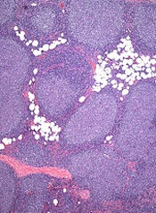
Days after gaining approval for 3 indications in the US, idelalisib (Zydelig) has earned a positive opinion from the European Medicine Agency’s Committee for Medicinal Products for Human Use (CHMP).
The CHMP is recommending the PI3K delta inhibitor for the treatment of chronic lymphocytic leukemia (CLL) and follicular lymphoma (FL).
If approved, the drug would be used as monotherapy for adults with FL that is refractory to 2 prior lines of treatment.
Idelalisib would also be used in combination with rituximab for adults with CLL who have received at least 1 prior therapy or as first-line treatment in CLL patients who have 17p deletion or TP53 mutation and cannot receive chemo-immunotherapy.
The CHMP’s recommendation for idelalisib (150 mg film-coated tablets) will be reviewed by the European Commission, which has the authority to approve medicines for use in the 28 countries of the European Union.
The CHMP’s positive opinion of idelalisib is based on data from 2 clinical trials—Study 116 and Study 101-09.
Study 116: Idelalisib in CLL
This phase 3 trial was stopped early because idelalisib had a significant impact on progression-free survival.
The study included 220 CLL patients who could not receive chemotherapy. Half were randomized to receive idelalisib plus rituximab, and the other half were randomized to rituximab plus placebo.
Patients in the idelalisib arm had a much higher overall response rate than patients in the placebo arm—81% and 13%, respectively (P<0.001). But all responses were partial.
At 24 weeks, the rate of progression-free survival was 93% in the idelalisib arm and 46% in the placebo arm (P<0.001). The median progression-free survival was 5.5 months in the placebo arm and not reached in the idelalisib arm (P<0.001).
At 12 months, the overall survival rate was 92% in the idelalisib arm and 80% in the placebo arm (P=0.02).
Most adverse events, in either treatment arm, were grade 2 or lower. The most common events in the idelalisib arm were pyrexia, fatigue, nausea, chills, and diarrhea. In the placebo arm, the most common events were infusion-related reactions, fatigue, cough, nausea, and dyspnea.
There were more serious adverse events in the idelalisib arm than in the placebo arm—40% and 35%, respectively. The most frequent serious events were pneumonia, pyrexia, and febrile neutropenia (in both treatment arms).
Study 101-09: Idelalisib in FL
In this phase 2 trial, idelalisib was given as a single agent to patients with indolent non-Hodgkin lymphoma who were refractory to rituximab and chemotherapy containing an alkylating agent.
In the 72 patients with FL, the overall response rate was 54%, and the complete response rate was 8%. The median duration of response was not reached (range, 0-14.8 months).
Improvements in patient survival or disease-related symptoms have not been established.
The most common grade 3 or higher adverse events were neutropenia (27%), elevations in aminotransferase levels (13%), diarrhea (13%), and pneumonia (7%).
About idelalisib
Idelalisib is an oral inhibitor of PI3K delta, a protein that plays a role in the activation, proliferation, and viability of B cells. PI3K delta signaling is active in many B-cell leukemias and lymphomas, and, by inhibiting the protein, idelalisib blocks several cellular signaling pathways that drive B-cell viability.
Idelalisib is being developed by Gilead Sciences. On July 23, the drug received US Food and Drug Administration approval for use in combination with rituximab to treat patients with relapsed CLL who cannot receive rituximab alone. The agency also granted idelalisib accelerated approval to treat patients with relapsed FL or small lymphocytic lymphoma who have received at least 2 prior systemic therapies. ![]()
FDA approves idelalisib for CLL, SLL and FL
The US Food and Drug Administration (FDA) has approved the PI3K delta inhibitor idelalisib (Zydelig) for the treatment of chronic lymphocytic leukemia (CLL), small lymphocytic lymphoma (SLL), and follicular lymphoma (FL).
The drug was granted traditional approval for use in combination with rituximab to treat patients with relapsed CLL who cannot receive rituximab alone.
Idelalisib has also received accelerated approval to treat patients with relapsed FL or SLL who have received at least 2 prior systemic therapies.
The FDA’s accelerated approval program allows for approval of a drug to treat a serious or life-threatening disease based on clinical data showing the drug has an effect on a surrogate endpoint that is reasonably likely to predict a clinical benefit to patients.
This program provides earlier patient access to a drug while the developer—in this case, Gilead Sciences—conducts trials confirming the drug’s benefit.
Idelalisib in CLL: Results of a phase 3 study
The approval of idelalisib in CLL is based on results of a phase 3 trial (Study 116), which was stopped early because idelalisib had a significant impact on progression-free survival.
The study included 220 CLL patients who could not receive chemotherapy. Half were randomized to receive idelalisib plus rituximab, and the other half were randomized to rituximab plus placebo.
Patients in the idelalisib arm had a much higher overall response rate than patients in the placebo arm—81% and 13%, respectively (P<0.001). But all responses were partial responses.
At 24 weeks, the rate of progression-free survival was 93% in the idelalisib arm and 46% in the placebo arm (P<0.001). The median progression-free survival was 5.5 months in the placebo arm and not reached in the idelalisib arm (P<0.001).
At 12 months, the overall survival rate was 92% in the idelalisib arm and 80% in the placebo arm (P=0.02).
Most adverse events, in either treatment group, were grade 2 or lower. The most common events in the idelalisib arm were pyrexia, fatigue, nausea, chills, and diarrhea. In the placebo arm, the most common events were infusion-related reactions, fatigue, cough, nausea, and dyspnea.
There were more serious adverse events in the idelalisib arm than in the placebo arm—40% and 35%, respectively. The most frequent serious events were pneumonia, pyrexia, and febrile neutropenia (in both treatment arms).
Idelalisib in FL and SLL: Results of a phase 2 study
Idelalisib’s accelerated approval in FL and SLL is supported by data from a single-arm, phase 2 trial (Study 101-09).
The drug was given as a single agent to patients who were refractory to rituximab and alkylating-agent-containing chemotherapy. Seventy-two patients had FL, and 26 had SLL.
The overall response rate was 54% in FL and 58% in SLL. Eight percent of FL responses were complete, and all responses in SLL patients were partial.
The median duration of response was 11.9 months in SLL patients (range, 0-14.7 months) but was not reached in FL patients (range, 0-14.8 months).
Improvements in patient survival or disease-related symptoms have not been established.
In all patients, the most common grade 3 or higher adverse events were neutropenia (27%), elevations in aminotransferase levels (13%), diarrhea (13%), and pneumonia (7%).
About idelalisib: Dosing, boxed warning and REMS
Idelalisib is an oral inhibitor of PI3K delta, a protein that plays a role in the activation, proliferation, and viability of B cells. PI3K delta signaling is active in many B-cell leukemias and lymphomas, and, by inhibiting the protein, idelalisib blocks several cellular signaling pathways that drive B-cell viability.
The drug is available as 150 mg and 100 mg tablets, administered orally twice-daily, but 150 mg is the recommended starting dose.
Idelalisib has a boxed warning on its label communicating the risks of fatal and serious toxicities, which include hepatic toxicity, severe diarrhea, colitis, pneumonitis, and intestinal perforation.
The drug is being approved with a risk evaluation and mitigation strategy (REMS) comprised of a communication plan to ensure healthcare providers are fully informed about these risks. For more information on this program, visit www.ZydeligREMS.com. ![]()
FDA approves idelalisib for three leukemia and lymphoma indications
The oral kinase inhibitor idelalisib has been approved for treating patients with relapsed chronic lymphocytic leukemia, follicular lymphoma, and small lymphocytic lymphoma, with a boxed warning about fatal and serious toxicities associated with treatment, the Food and Drug Administration announced on July 23.
The three approved indications for idelalisib – administered at a recommended starting dose of 150 mg, twice a day – are for:
• Relapsed chronic lymphocytic leukemia (CLL), in combination with rituximab, in patients for whom rituximab alone would be considered appropriate therapy due to other comorbidities. This is a traditional approval based on progression-free survival (PFS) data.
• Relapsed follicular B-cell non-Hodgkin’s lymphoma (FL) in patients who have received at least two prior systemic therapies.
• Relapsed small lymphocytic lymphoma (SLL) in patients who have received at least two prior systemic therapies.
The FL and SLL indications, based on objective response rates in one study, are accelerated approvals, which provides patients with serious or life-threatening diseases earlier access to a promising drug based on an effect on a surrogate endpoint considered " reasonably likely to predict clinical benefit," according to the FDA. Full approval is contingent on the manufacturer conducting trials confirming clinical benefits; if they do not, the FDA can withdraw the approval.
Idelalisib is being marketed as Zydelig, by Gilead Sciences. There is also a Risk Evaluation and Mitigation Strategy (REMS) in place for the drug, which includes a communication plan to ensure that prescribers are fully informed about treatment-associated risks. The boxed warning lists the risks of fatal and/or serious hepatoxicity (affecting 14% of treated patients); fatal and/or serious and severe diarrhea or colitis (also affecting 14% of treated patients); as well as fatal and serious pneumonitis; and fatal and serious intestinal perforation.
Idelalisib is an oral inhibitor of phosphoinositide 3-kinase (PI3K) delta, "a protein that plays a role in the activation, proliferation, and viability of B cells," according to a Gilead statement announcing the approval, which added that PI3K delta signaling "is active in many B-cell leukemias and lymphomas, and by inhibiting the protein, Zydelig blocks several cellular signaling pathways that drive B-cell viability."
The FDA granted a full approval for the CLL indication, based on a phase III study of 220 patients, which was stopped early in October 2013 at the first-specified interim analysis. Median PFS was not reached among those randomized to on idelalisib plus rituximab, but was at least 10.7 months, and was 5.5 months among those randomized to placebo plus rituximab (N. Engl. J. Med. 2014;370:997-1007). "Results from a second interim analysis continued to show a statistically significant improvement for Zydelig and Rituxan over placebo and Rituxan," the FDA statement said.
The accelerated approvals for relapsed FL and relapsed SLL were based on a single-arm phase II study of 123 patients refractory to rituximab and chemotherapy including alkylating agents, treated with idelalisib. The objective response rates were 54% among those with relapsed FL, and 58% of those with SLL in the study (N. Engl. J. Med. 2014;370:1008-18).
Common adverse events associated with treatment included diarrhea, fever, fatigue, nausea, cough, pneumonia, abdominal pain, chills, and rash; and common lab abnormalities associated with treatment included neutropenia, hypertriglyceridemia, hyperglycemia, and liver enzyme elevations, according to the FDA.
In the FDA statement, Dr. Richard Pazdur, director of the Office of Hematology and Oncology Products in the FDA’s Center for Drug Evaluation and Research, pointed out that in less than a year, "We have seen considerable progress in the availability of treatments for chronic lymphocytic leukemia." The other treatments for CLL are obinutuzumab (Gazyva), approved in November 2013; ibrutinib (Imbruvica), approved in February 2014; and ofatumumab (Arzerra) approved in April 2014.
Prescribing information is available at the FDA website.
Combo appears safe and active in CLL, NHL
Credit: Linda Bartlett
KOHALA COAST, HAWAII—Early results of a small, phase 1 study suggest a novel combination treatment is active and generally well-tolerated in relapsed or refractory patients with chronic lymphocytic leukemia/small lymphocytic lymphoma (CLL/SLL) or non-Hodgkin lymphomas (NHLs).
The treatment consists of ublituximab (TG-1101), a monoclonal antibody that targets a unique epitope on the CD20 antigen, and TGR-1202, a next-generation PI3K delta inhibitor.
The combination appeared to be well tolerated overall, although infusion-related reactions were common, and nearly a quarter of patients experienced grade 3/4 neutropenia.
Four of 5 CLL/SLL patients experienced a partial response (PR), and the remaining patient had stable disease (SD). Among the 10 NHL patients, 1 had progressive disease, 7 had SD, and 2 achieved a PR.
Matthew Lunning, DO, of the University of Nebraska Medical Center in Omaha, and his colleagues presented these results in a poster at the 2014 Pan Pacific Lymphoma Conference.
The study is sponsored by TG Therapeutics, the company developing both drugs.
The researchers presented results from 8 patients with CLL/SLL, 7 patients with diffuse large B-cell lymphoma (DLBCL), 5 with follicular lymphoma (FL), and 1 patient with Richter’s syndrome (RS).
Patients had a median age of 64 years (range, 35-82), and they had received a median of 3 prior therapies (range, 1-9). Fifty-seven percent of patients had received 3 or more prior therapies, and 38% were refractory to their prior therapy.
The patients received escalating doses of TGR-1202, with a fixed dose of ublituximab—900 mg for patients with NHL and 600 mg for patients with CLL.
As of the data cutoff, all 21 patients were evaluable for safety, but only 15 were evaluable for efficacy.
Adverse events
The most common adverse event was infusion-related reactions, which occurred in 48% of patients. All of these events were manageable without dose reductions, and all but 1 event was grade 1 or 2 in severity.
Neutropenia was also common, occurring in 38% of patients. Grade 3/4 neutropenia occurred in 24% of patients. One CLL patient required a dose delay for neutropenia in cycle 1, which met the criteria for a dose-limiting toxicity.
No additional dose-limiting toxicities have been observed to date. Likewise, none of the patients has required dose reductions for either drug, and there were no drug-related AST/ALT elevations.
On the other hand, 1 patient did come off the study due to grade 1 itching that was possibly related to TGR-1202.
Other common adverse events associated with treatment included diarrhea (29%), nausea (29%), hoarseness (10%), muscle aches (10%), and fatigue (10%).
Activity in CLL/SLL
Of the 8 CLL/SLL patients enrolled to date, 5 were evaluable for efficacy. Four patients achieved a PR at the first efficacy assessment. The remaining patient, a CLL patient with both 17p and 11q del, achieved SD with a 44% nodal reduction at the first assessment.
All 5 patients achieved a greater-than-50% reduction in ALC by the first efficacy assessment. One patient achieved complete normalization of ALC (less than 4000/uL), and the other 4 patients achieved at least an 80% reduction by the first efficacy assessment.
The lymphocytosis generally observed in CLL patients treated with TGR-1202, similar to other PI3K delta and BTK inhibitors, appears to be mitigated by the addition of ublituximab.
Activity in NHL
Of the 13 patients in this group, 10 were evaluable for efficacy, including 5 with DLBCL, 4 with FL, and 1 with RS. Results were not as favorable in this group as they were among CLL/SLL patients, but, as the researchers pointed out, these patients were heavily pretreated.
Among the DLBCL patients, 2 achieved PRs with TGR-1202 and ublituximab. Both of these responses occurred at the higher dose of TGR-1202.
Two DLBCL patients had SD, and 1 patient progressed. DLBCL patients had a median of 3 prior treatment lines, and 3 patients had GCB DLBCL, with 1 patient classified as triple-hit lymphoma (overexpression of BCL2, BCL6, and MYC rearrangements).
In the FL group, all 4 patients had SD after treatment and exhibited a reduction in tumor mass at the first assessment. These patients had advanced disease and a median of 6 prior lines of therapy.
The RS patient also had SD following TGR-1202 and ublituximab.
“We have been very impressed with the safety profile and the level of activity observed to date in all patient groups with TGR-1202 in combination with ublituximab, particularly given the advanced stage of disease . . . ,” said Susan O’Brien, MD, a professor at MD Anderson Cancer Center in Houston and study chair for the CLL patient group.
“Of particular interest is the absence of observed elevations in AST/ALT with TGR-1202, which is a known adverse event associated with other PI3K delta inhibitors. We look forward to continuing enrollment at all trial centers of this exciting combination and presenting data on more patients at upcoming medical meetings.” ![]()
Survival differences in blood cancers across Europe

Credit: Rhoda Baer
Differences in treatment access and quality may explain why survival rates vary widely for European patients with hematologic malignancies, researchers have reported in The Lancet Oncology.
“The good news is that 5-year survival for most cancers of the blood has increased over the past 11 years, most likely reflecting the approval of new targeted drugs in the early 2000s . . . ,” said Milena Sant, MD, of the Fondazione IRCCS Istituto Nazionale dei Tumori in Milan, Italy.
“But there continue to be persistent differences between regions. For example, the uptake and use of new technologies and effective treatments has been far slower in eastern Europe than other regions. This might have contributed to the large differences in the management and outcomes of patients.”
Dr Sant and her colleagues uncovered these differences by analyzing data from 30 cancer registries covering all patients diagnosed in 20 European countries.*
The researchers compared changes in 5-year survival for 560,444 adults (aged 15 years and older) who were diagnosed with 11 lymphoid and myeloid cancers between 1997 and 2008, and followed up to the end of 2008.
Some cancers have shown particularly large increases in survival between 1997-1999 and 2006-2008, such as follicular lymphoma (59% to 74%), diffuse large B-cell lymphoma (42% to 55%), chronic myeloid leukemia (32% to 54%), and acute promyelocytic leukemia (50% to 62%).
The greatest improvements in survival have been in northern, central, and eastern Europe, even though adults in eastern Europe (where survival in 1997 was the lowest) continue to have lower survival for most hematologic malignancies than elsewhere.
Survival gains have been lower in southern Europe and the UK. For example, improvements in 5-year chronic myeloid leukemia survival in northern Europe (29% to 60%) and central Europe (34% to 65%) have been persistently higher than in the UK (35% to 56%) and southern Europe (37% to 55%).
Overall, the risk of death within 5 years from diagnosis fell significantly for all malignancies except myelodysplastic syndromes. But not all regions have seen such improvements.
For example, compared with the UK, the excess risk of death was significantly higher in eastern Europe than in other regions for most of the cancers investigated, but significantly lower in northern Europe.
The researchers said the most likely reasons for continuing geographical differences in survival are inequalities in the provision of care and in the availability and use of new treatments.
“We know that rituximab, imatinib, thalidomide, and bortezomib were first made available for general use in Europe in 1997, 2001, 1998, and 2003, respectively,” the researchers wrote.
“The years following general release of these drugs coincided with large increases in survival for chronic myeloid leukemia, diffuse large B-cell lymphoma, and follicular lymphoma, with a smaller but still significant survival increase for multiple myeloma plasmacytoma.”
However, they pointed out that the uptake and use of these drugs has not been uniform across Europe. For example, market uptake of rituximab, imatinib, and bortezomib was lower in eastern Europe than elsewhere and might explain the consistently lower survival in this region.
Writing in a linked comment article, Alastair Munro, MD, of the University of Dundee Medical School in Scotland, questioned whether improvements in survival can be attributed to drugs alone.
He said that better understanding of the conclusions from this study (called EUROCARE-5) requires additional information about changes affecting survival according to disease categories, the distribution of histological subtypes and their relation with the age distribution of the population, the distribution of stages at diagnosis, and the timing of active intervention for indolent tumors. ![]()
*The areas included in the study were northern Europe (Denmark, Iceland, and Norway), the UK (England, Northern Ireland, Scotland, and Wales), central Europe (Austria, France, Germany, Switzerland, and The Netherlands), eastern Europe (Bulgaria, Estonia, Lithuania, Poland, and Slovakia), and southern Europe (Italy, Malta, and Slovenia).
Novel agent shows promising activity in heavily pretreated NHL

©ASCO/Rodney White
CHICAGO—The novel, oral selective inhibitor of nuclear transport known as selinexor (KPT-330) can safely be given as monotherapy to patients with heavily pretreated non-Hodgkin lymphoma (NHL), according to a presentation at the 2014 ASCO Annual Meeting.
“Selinexor has favorable pharmacokinetic and pharmacodynamic characteristics,” said presenter Martin Gutierrez, MD, of the John Theurer Cancer Center at Hackensack University Medical Center in New Jersey.
Selinexor is a slowly reversible, selective inhibitor of nuclear transport that inhibits XPO1, which is elevated in NHL, chronic lymphocytic leukemia (CLL), and other malignances.
“It shows single-agent anti-tumor activity across all NHL types, with durable cancer control of more than 9 months [and] marked activity across germ cell B (GCB), non-germ cell B, and double-hit diffuse large B-cell lymphoma (DLBCL),” Dr Gutierrez said.
He provided an update of the ongoing phase 1 dose-escalation study at the meeting as abstract 8518.
The study now includes 51 patients, half of whom have NHL. Their median age is 60 years.
The patients received selinexor across 8 dose levels, ranging from 3 mg/m2 to 60 mg/m2. Dosing at 60 mg/m2 twice weekly is ongoing, and the maximum-tolerated dose has not been reached.
Among the 43 evaluable patients, the disease control rate was 74%, the overall response rate was 28%, and the complete response rate was 5%.
“All patients who had their disease controlled had a reduction in lymph nodes and some degree of activity across all dose levels,” Dr Gutierrez said. “GCB and non-GCB patients responded similarly.”
The length of response was up to 632 days in the DLBCL group.
Most adverse events were gastrointestinal in nature, and most of them were grade 1 or 2. The most common adverse events were nausea, anorexia, and fatigue.
The investigators found a higher incidence of side effects in the first treatment cycle. The dosing schedule was interrupted and reduced to maintain steady state levels.
“The results suggest that an intermittent dosing schedule optimally induces a steady state with maximal induction of XPO1 mRNA,” Dr Gutierrez said.
There were 3 dose-limiting toxicities, including 1 multiple myeloma patient with grade 4 thrombocytopenia, 1 follicular lymphoma patient with grade 4 thrombocytopenia, and 1 CLL patient with grade 2 fatigue.
ASCO discussant Owen O’Connor, MD, from Columbia University Medical Center in New York, commented, “These clinical data are interesting with a provocative target. I applaud the investigators for doing a trial across a diversity of B- and T-cell lymphomas . . . . The results suggest a potential effect in a rare subset of lymphoma patients that have little treatment options.”
Frontline trials of selinexor are planned, including patients with Richter transformation and follicular lymphoma.
Selinexor recently received orphan drug status from the US Food and Drug Administration for the treatment of DLBCL. ![]()
PET-CT accurately predicts survival of follicular lymphoma patients
CHICAGO – For patients with follicular lymphoma, positron emission tomography–computed tomography performed at the end of induction therapy is strongly predictive of both progression-free and overall survival, a retrospective analysis showed.
The pooled analysis of data on 246 PET-CT scans performed following chemoimmunotherapy in three clinical trials showed that patients with 18-fluorodeoxyglucose (FDG) uptake of 4 or greater on a 5-point scale had a fourfold higher risk for disease progression, compared with patients who became PET negative after induction, reported Dr. Judith Trotman of the University of Sydney (Australia).
At 4.5 years of follow-up, median progression-free survival (PFS) was 16.9 months for patients with a PET uptake of 4 or greater on the 5-point Deauville scale for postinduction response assessment, vs. 74 months for PET-negative patients.
Overall survival at 4.5 years for patients with a higher uptake of FDG PET was 87%, compared with 97% for patients who were PET negative after induction, Dr. Trotman reported at the American Society of Clinical Oncology annual meeting.
"We argue that for the patients who remain PET positive, follicular lymphoma is no longer an indolent histology," Dr. Trotman said.
The study results also showed that conventional CT assessment provides only limited additional value, and that "PET-CT applying the 5-point scale should be the new gold standard for therapeutic response assessment in this lymphoma," she said.
Not so indolent
Although the natural history of follicular lymphoma is a generally indolent course, approximately 15% of patients will die within 5 years of diagnosis, and high-risk scores on the Follicular Lymphoma International Prognostic Index (FLIPI) or its revision (FLIPI2) are not sufficient for predicting which patients are at greatest risk for death, Dr. Trotman said.
Three recent clinical trials reported that positron emission tomography assessment after first-line rituximab-based chemotherapy has good predictive value in patients with high tumor burden follicular lymphoma. Dr. Trotman and his colleagues conducted a pooled analysis of data from the trials with independent review of PET-CT scans to come up with more precise survival estimates and identify the best cutoff for survival using the Deauville scale for response assessment of FDG-avid lymphoma.
The trials were the PRIMA (Primary Rituximab and Maintenance) study of 122 patients, the FOLL05 randomized trial of the Fondazione Italiana Linfomi in 202 patients, and the PET Folliculaire trial in 106 patients.
The Deauville 5-point scale for FDG-avid lymphoma uses the following criteria:
1. No uptake.
2. Uptake less than or equal to mediastinum.
3. Uptake greater than mediastinum but less than or equal to liver.
4. Uptake moderately higher than liver.
5. Uptake markedly higher than liver and/or new lesions.
Dr. Trotman and his colleagues looked at cutoffs of 3 and higher and 4 and higher to see whether they were predictive of prognosis. Reviews of concordance with the trial results, performed by three independent reviewers, showed that a cutoff of 3 or higher had moderate concordance, while a score of 4 or higher had substantial agreement with results.
They then evaluated PET results to see whether they could sharpen the prognostic ability, and found that both cutoffs predicted PFS, but because of the higher concordance score, they chose to focus on the 4+ cutoff.
They found that the hazard ratio (HR) for progression with a score of 4 or greater was 3.9 (P less than .0001), and the hazard ratio for death was 6.7 (P = .0002). Median overall survival in patients with scores of 4 or greater was 79 months, vs. not reached for PET-negative patients.
In multivariate analyses, factors associated with PFS included PET-positive scores of 4 or greater (HR, 3.1; P less than .0001), stable or progressive disease vs. complete responses or complete responses unconfirmed (CR/CRu; HR, 3.7; P = .0013), and partial responses (PRs) vs. CR/CRu (HR, 1.6; P = .04).
Factors associated with OS were PET score and stable/progressive disease vs. CR/CRu (HR, 5.3; P = .05).
"I hope that we can now move on: postinduction PET-CT is a platform for response–adapted therapy, because while achieving PET negativity can better reassure our patients, particularly those otherwise in CRu or PR, the inferior survival of those who remain PET positive compels us to study such PET-response–adapted approaches," Dr. Trotman said.
The invited discussant, Dr. Christopher Flowers of the department of hematology and medical oncology at Emory University, Atlanta, noted that in their analysis, Dr. Trotman and his colleagues included only those scans that were of sufficient quality for central review, and that slightly more than half of all patients had PET scans.
"I think it’s important to try and understand how the PET-available cohort compared to the other clinical trial characteristics of patients to understand whether or not this PET population is a unique population, and [whether] the behavior characteristics may be different from what you might expect from a broader population of patients," he said.
It will be important to see whether the findings will hold up in patients treated with emerging regimens, such as rituximab and bendamustine, and other combinations now in clinical trials, he said.
Dr. Trotman reported having no relevant relationships to disclose. Dr. Flowers disclosed uncompensated consultation from several companies, and receiving research funding from Gilead Sciences, Janssen Pharmaceuticals, Millennium, and Spectrum Pharmaceuticals.
AT THE ASCO ANNUAL MEETING 2014
Key clinical point: For patients with follicular lymphoma, a PET-CT performed at the end of induction therapy is predictive of survival.
Major finding: A PET-CT cutoff score of 4 out of 5 on the Deauville lymphoma-response assessment scale was strongly predictive of both progression-free and overall survival of follicular lymphoma.
Data source: A retrospective analysis of prospectively collected data on 246 patients in three clinical trials.
Disclosures: Dr. Trotman reported having no relevant relationships to disclose. Dr. Flowers disclosed uncompensated consultation from several companies, and receiving research funding from Gilead Sciences, Janssen Pharmaceuticals, Millennium, and Spectrum Pharmaceuticals.
Agricultural chemicals and the risk of NHL

EPA/John Messina
After reviewing nearly 30 years’ worth of data, investigators have compiled a list of agricultural chemicals that appear to increase a person’s risk of developing non-Hodgkin lymphoma (NHL).
Meta-analyses suggested that occupational exposure to phenoxy herbicides, carbamate insecticides, organochlorine insecticides, and organophosphorus insecticides/herbicides can increase the risk of NHL.
The research also revealed associations between certain chemicals and specific NHL subtypes.
Leah Schinasi, PhD, and Maria E. Leon, PhD, of the International Agency for Research on Cancer in Lyon, France, described the analysis and its results in the International Journal of Environmental Research and Public Health.
The investigators reviewed epidemiological research spanning nearly 30 years and identified 44 relevant papers. The papers recounted studies conducted in the US, Canada, Europe, Australia, and New Zealand.
Drs Schinasi and Leon used these data to assess occupational exposure to 80 active ingredients and 21 chemical groups and clarify their role in the development of NHL. Most, but not all, of the studies looked at lifetime exposure to the chemicals in question.
The investigators performed a meta-analysis of the data and found associations between NHL and a range of insecticides and herbicides. But the strongest risk ratios (RRs) were for subtypes of NHL.
There was a positive association between exposure to the organophosphorus herbicide glyphosate and any NHL (RR=1.5), but the link was stronger for B-cell lymphoma in particular (RR=2.0).
Phenoxy herbicide exposure was associated with an increased risk of NHL in general (RR=1.4), B-cell lymphoma (RR=1.8), lymphocytic lymphoma (RR=1.8), and diffuse large B-cell lymphoma (RR=2.0). As for specific phenoxy herbicides, both MCPA (RR=1.5) and 2,4-D (RR=1.4) were associated with NHL.
Carbamate insecticides, as a group, appeared to confer an increased risk of NHL (RR=1.7). The individual insecticides carbaryl and carbofuran showed positive associations with NHL as well (RRs of 1.7 and 1.6, respectively).
There was a positive association with NHL for organophosphorus insecticides as a group (RR=1.6), as well as the individual insecticides chlorpyrifos (RR=1.6), diazinon (RR=1.6), dimethoate (RR=1.4), and malathion (RR=1.8).
Lastly, organochlorine insecticides appeared to confer an increased risk of NHL (RR=1.3). DDT was associated with NHL (RR=1.3), B-cell lymphoma (RR=1.4), diffuse large B-cell lymphoma (RR=1.2), and follicular lymphoma (RR=1.5). And lindane was associated with NHL in general (RR=1.6).
The investigators said this analysis represents one of the most comprehensive reviews on the topic of occupational exposure to agricultural chemicals in the scientific literature.
But it also suggests a need to study a wider variety of chemicals in more geographic areas, especially in low- and middle-income countries, as they were missing from the literature. ![]()
Younger men with goiter at higher risk for thyroid cancers
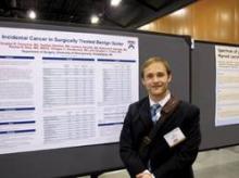
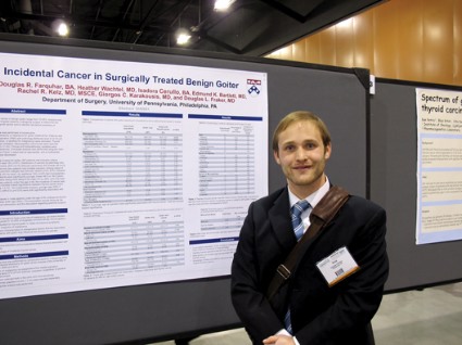
PHOENIX – More than one-fourth of men under age 50 undergoing surgery for benign goiter were found to have thyroid cancers, based on a chart review performed at the University of Pennsylvania.
The overall incidence of thyroid cancers in the patient series was 12%. Among men under age 45, the rate was "surprisingly" 28%, said Douglas R. Farquhar, a medical student at the University of Pennsylvania School of Medicine in Philadelphia.
Although thyroid goiters have traditionally been thought to be associated with a low risk for malignancy, recent studies have suggested otherwise. "In the literature we have seen published rates of up to 35%, which is much higher than we all had anticipated," Mr. Farquhar said at the annual Society of Surgical Oncology Cancer Symposium.
To get a better handle on the preoperative and patient characteristics associated with incident thyroid cancer and characterize the types of thyroid cancer discovered incidentally, Mr. Farquhar and his colleagues reviewed charts on all patients who underwent either total thyroidectomy or thyroid lobectomy for goiter at the center from 2004 through 2012.
Many cases of goiter can be medically managed, but surgery may be indicated in cases of pressure symptoms, cosmesis, or suspicion of malignancy, the investigators noted.
They excluded from their study patients with preoperative fine-needle aspiration pathology findings of Bethesda level III-VI (follicular lesion of undetermined significance, follicular neoplasm, suspicious or positive for malignancy).
Among 418 patients undergoing goiter surgery, 367 had goiter only, and 51 (12%) had an incident thyroid cancer. In all, 38 (75%) had papillary carcinomas, 10 (20%) had follicular carcinomas, 3 (6%) had Hürthle cell carcinomas, and 2 (4%) had thyroid lymphomas (two patients had multiple thyroid cancers, explaining the percentage greater than 100). An additional 67 patients (16%) were found to have micropapillary lesions.
Looking at the population as a whole, the investigators found that patients with thyroid cancer tended to be younger, with a mean age of 49.5 vs. 54.6 years (P = .012). There was a trend toward more cancers among men than women, but it was not significant.
There were no significant differences in any preoperative factors between patients with cancers and those with goiter only, including number of nodules, site of dominant nodule (right, left, or isthmus), thyroid function, thyroid weight, or fine-needle aspiration results (percentage deemed benign or nondiagnostic).
In a multivariate analysis, male sex was associated with a more than twofold risk for thyroid cancer (odds ratio, 2.39; 95% confidence interval, 1.152-4.978). There were also trends toward a lower risk of cancer with each additional decade of life, and a higher risk among patients who had undergone thyroid lobectomy, but these were nonsignificant associations.
"Knowledge of these associations may prove to be useful for both patient counseling and surgical decision making," Mr. Farquhar said.
The study was internally funded. The senior author was Dr. Douglas L. Fraker, chief of the division of endocrine and oncologic surgery at the University of Pennsylvania.
Mr. Farquhar and his coauthors reported having no relevant financial disclosures.
AT SSO 2014
Key clinical point: Men under age 50 undergoing surgery for benign goiter are at elevated risk for thyroid cancer.
Major finding: Among men under 45 undergoing goiter surgery, the rate of incidentally discovered thyroid cancers was 28%.
Data source: A case series of 418 consecutive patients undergoing surgery for goiter.
Disclosures: The study was internally funded. Mr. Farquhar and his coauthors reported having no relevant financial disclosures.
EC approves SC formulation of rituximab
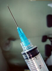
The European Commission (EC) has approved a subcutaneous (SC) formulation of rituximab (MabThera) to treat patients with follicular lymphoma or diffuse large B-cell lymphoma.
This formulation allows for 5-minute administration, a significant decrease over the 2.5-hour infusion time required to administer intravenous (IV) rituximab.
The drug’s maker, Roche, plans to begin launching SC rituximab in a number of European markets this year.
The EC’s approval of this formulation was primarily based on data from the SABRINA trial, which was recently published in The Lancet Oncology and funded by Roche.
In this phase 3 trial, researchers compared 3-week cycles of fixed-dose, SC rituximab to IV rituximab. They enrolled 127 patients with previously untreated, grade 1-3a, CD20-positive follicular lymphoma.
Patients were randomized to receive IV rituximab (375 mg/m2) or SC rituximab (1400 mg). After randomization, they received 1 induction dose of IV rituximab in cycle 1 and then their allocated treatment for cycles 2 through 8. Patients with a complete or partial response continued their treatment as maintenance every 8 weeks.
The study’s primary endpoint was the ratio of observed rituximab serum trough concentrations (Ctrough) between the 2 groups at cycle 7.
Pharmacokinetic data were available for 75% of patients (48/64) in the IV arm and 86% of the patients (54/63) in the SC arm.
An analysis of these data suggested SC rituximab was non-inferior to the IV formulation. The geometric mean Ctrough was 83.13 μg/mL in the IV arm and 134.58 μg/mL in the SC arm (ratio, 1.62).
The rate of adverse events was similar between the 2 arms, occurring in 88% (57/65) of patients in the IV arm and 92% (57/62) of patients in the SC arm. Grade 3 or higher adverse events occurred in 46% (n=30) and 47% (n=29) of patients, respectively.
The most common grade 3 or higher adverse event in both arms was neutropenia. It occurred in 22% (n=14) of patients in the IV arm and 26% (n=16) in the SC arm.
Adverse events related to administration were mostly grade 1-2. And they occurred more often in the SC arm than in the IV arm, in 50% (n=31) and 32% (n=21) of patients, respectively.
The researchers said these results suggest the SC formulation of rituximab is non-inferior to the IV formulation and poses no new safety concerns. ![]()

The European Commission (EC) has approved a subcutaneous (SC) formulation of rituximab (MabThera) to treat patients with follicular lymphoma or diffuse large B-cell lymphoma.
This formulation allows for 5-minute administration, a significant decrease over the 2.5-hour infusion time required to administer intravenous (IV) rituximab.
The drug’s maker, Roche, plans to begin launching SC rituximab in a number of European markets this year.
The EC’s approval of this formulation was primarily based on data from the SABRINA trial, which was recently published in The Lancet Oncology and funded by Roche.
In this phase 3 trial, researchers compared 3-week cycles of fixed-dose, SC rituximab to IV rituximab. They enrolled 127 patients with previously untreated, grade 1-3a, CD20-positive follicular lymphoma.
Patients were randomized to receive IV rituximab (375 mg/m2) or SC rituximab (1400 mg). After randomization, they received 1 induction dose of IV rituximab in cycle 1 and then their allocated treatment for cycles 2 through 8. Patients with a complete or partial response continued their treatment as maintenance every 8 weeks.
The study’s primary endpoint was the ratio of observed rituximab serum trough concentrations (Ctrough) between the 2 groups at cycle 7.
Pharmacokinetic data were available for 75% of patients (48/64) in the IV arm and 86% of the patients (54/63) in the SC arm.
An analysis of these data suggested SC rituximab was non-inferior to the IV formulation. The geometric mean Ctrough was 83.13 μg/mL in the IV arm and 134.58 μg/mL in the SC arm (ratio, 1.62).
The rate of adverse events was similar between the 2 arms, occurring in 88% (57/65) of patients in the IV arm and 92% (57/62) of patients in the SC arm. Grade 3 or higher adverse events occurred in 46% (n=30) and 47% (n=29) of patients, respectively.
The most common grade 3 or higher adverse event in both arms was neutropenia. It occurred in 22% (n=14) of patients in the IV arm and 26% (n=16) in the SC arm.
Adverse events related to administration were mostly grade 1-2. And they occurred more often in the SC arm than in the IV arm, in 50% (n=31) and 32% (n=21) of patients, respectively.
The researchers said these results suggest the SC formulation of rituximab is non-inferior to the IV formulation and poses no new safety concerns.
