User login
TNFRII may play key role in CTCL, speaker says
SAN FRANCISCO—Researchers have found evidence to suggest that tumor necrosis factor receptor II (TNFRII) may be an important driver of cutaneous T-cell lymphomas (CTCLs).
The team discovered that a mutation in this receptor—TNFRII T377I—is present in patients with mycosis fungoides (MF) and those with Sézary syndrome (SS).
And previous research showed that the region encoding TNFRII on chromosome 1 is sometimes amplified in MF and SS patients.
So if, as these factors suggest, TNFRII does play a key role in CTCL, a number of currently available drugs—including proteasome inhibitors and MEK inhibitors—may be effective treatment options.
Alexander Ungewickell, MD, PhD, of Stanford University in California, discussed this possibility and the research supporting it at the 6th Annual T-cell Lymphoma Forum.
A novel mutation
Dr Ungewickell and his colleagues began this research by conducting transcriptome sequencing on samples from 3 patients with SS (Lee et al, Blood 2012). This revealed about 500 genes that were upregulated and about 500 that were downregulated in SS cells.
And pathway enrichment analysis showed that molecular mechanisms of cancer were the most significantly altered pathways. But the researchers also observed PI3 kinase signaling, T-cell receptor signaling, regulation of IL-2, and CD8 signaling.
To better understand the basis for these transcriptional changes, the team performed whole-exome sequencing in 11 CTCL-normal pairs. They uncovered an average of 46 mutations per exome, as well as pathways similar to those observed in the transcriptional analysis.
The researchers then used this information to generate a 245-gene capture reagent. And they used that to perform ultra-deep targeted resequencing on 83 samples from CTCL patients.
“Two things that stood out right away were that TNFRSF1B and KRAS had recurrent point mutations that suggested an activating phenotype,” Dr Ungewickell said. “It’s already known that KRAS is mutated in many human cancers, including CTCL. TNFRSF1B encodes TNFRII and was not previously associated with any malignancies.”
“We also found a smattering of other genes that were mutated, [but] we were most interested in the TNFRII mutation because of the novelty of the finding and also the potential for therapeutic intervention.”
Driving disease
Dr Ungewickell noted that TNFRII is expressed in CD4 and CD8 T lymphocytes but relatively few other cell types. TNFRII is activated by membrane-bound TNFα, which mediates the signal through TRAF proteins and CIAP proteins to activate the NF-κB-inducing kinase (NIK).
This activates the I kappa B kinase (IKK) complex to phosphorylate p100. When phosphorylated, it is processed in the proteasome and translocates to the nucleus. There, it interacts with RelB to mediate transcription that tends to cause T-cell activation and proliferation.
TNFRII also binds to TRAF2 and induces its degradation. The recurrent mutation the researchers identified in TNFRII (T377I) is in the TRAF2 regulatory domain in an evolutionarily conserved residue.
The ultra-deep targeted resequencing of 83 CTCL samples showed 4 mutations at that locus, all of which were acquired in the lymphoma.
This suggests TNFRII is important in CTCL. And the researchers hypothesized that, if that’s the case, TNFRII might be overexpressed in SS cells. So they looked at their transcriptome data and found TNFRII to be overexpressed in all 3 patients.
“Interestingly, the region that encodes TNFRII on chromosome 1 is also amplified in 1 of the 4 commonly used CTCL cell lines, suggesting that amplification may be another way of activating this pathway,” Dr Ungewickell said.
“And we were very interested by a study published by van Doorn et al a few years ago [Blood 2009], which showed that that region of chromosome 1 p36 is, in fact, amplified in 45% of cases of MF and 15% of cases of Sézary syndrome.”
“So we are currently doing FISH studies to confirm that this receptor is actually amplified in as many as half of cases of MF, suggesting that maybe, between mutation and amplification, this is an important driver of CTCL.”
Therapeutic possibilities
The researchers also thought that, if TNFRII is an important driver of CTCL, there would be some kind of transcriptional mark on the lymphoma cells. So they performed gene set enrichment analyses on 24 CTCL samples that had undergone 3-seq.
By comparing tumors expressing high levels of TNFRII and those expressing low levels of TNFRII, the team identified an expression signature that corresponds to the receptor’s known effects on RNA levels in T cells.
When they searched publicly available datasets, the researchers found this signature in 63 cases of MF (Shin et al, Blood 2007). And results of control experiments suggested the signature is specific to CTCL.
“If TNFRII is more active [in CTCL] and the mutation that we found is a hyperactivating mutation, we would expect this pathway to show increased activity downstream; namely, you would expect more processing of p100 to p52,” Dr Ungewickell said.
To investigate this possibility, the researchers generated Jurkat cells expressing empty vector, wild-type TNFRII, or mutant TNFRII and looked at NF-κB processing. They did see an increase in processing with the mutant receptor, compared to the wild-type receptor or empty vector.
“We also found, somewhat surprisingly, increases in phospho-ERK with the mutant receptor, as well as phospho-MEK,” Dr Ungewickell said.
“And to our knowledge, the RAS/MAP kinase pathway has not previously been linked to TNFRII signaling, suggesting that there is some kind of direct or indirect cross-talk between these pathways. We think it’s very interesting, since there are KRAS mutations that activate the RAS/MAP kinase pathway in a subset of these cases, suggesting some kind of synergy.”
Introducing the mutant receptor into primary CD4+ T cells had an effect similar to that observed in the Jurkat cells. The researchers did western blotting for NF-kB processing, and they saw an increase in p100 to p52 processing.
“This is a preliminary experiment, but we’re actually quite excited about this, since Jurkat cells have many abnormalities, due to the fact that they’re a leukemia line, and primary T cells will have the rest of the genome intact,” Dr Ungewickell said.
Now, he and his colleagues are conducting several studies to identify the changes that occur in primary T cells when mutant TNFRII is expressed. They also want to see if they can recapitulate CTCL and identify the transcriptional signature they previously found in patient biopsies and cells.
Lastly, the researchers are performing functional assays to evaluate proliferation, apoptosis, and pharmacological information, with the goal of identifying therapies that might be effective in patients with TNFRII mutation or amplification.
“Patients who have increased TNFRII signaling might respond to proteasome inhibitors, since p100 and p52 processing requires the proteasome,” Dr Ungewickell said. “And given that cross-talk with the RAS/MAP kinase signaling, as well as the KRAS mutations, we also think . . . that MEK inhibitors might be effective in the treatment of CTCL.” ![]()
Inhibitor appears active in relapsed/refractory TCLs
SAN FRANCISCO—Preliminary results of a phase 1 trial suggest the PI3K-delta/gamma inhibitor IPI-145 is active in patients with relapsed or refractory T-cell lymphomas.
Among 26 evaluable patients, 9 experienced partial responses to treatment with IPI-145, and 1 achieved a complete response, for an overall response rate (ORR) of 38%.
The drug also appeared to be well-tolerated, although 30% of patients did experience treatment-related severe adverse events.
Steven Horwitz, MD, of Memorial Sloan-Kettering Cancer Center in New York, and his colleagues presented these results in a poster at the 6th Annual T-cell Lymphoma Forum, which took place January 23-25.
The study was sponsored by Infinity Pharmaceuticals, Inc., the company developing IPI-145.
Patient and treatment characteristics
The trial included 30 patients with peripheral T-cell lymphoma (PTCL) or cutaneous T-cell lymphoma (CTCL). Of the 17 CTCL patients, 16 had mycosis fungoides or Sezary syndrome, and 1 had primary cutaneous anaplastic large-cell lymphoma (ALCL).
Of the 13 patients with PTCL, 3 had angioimmunoblastic T-cell lymphoma (AITL), 3 had subcutaneous panniculitis-like T-cell lymphoma (SPTCL), 3 had PTCL-not otherwise specified, 2 had ALCL, 1 had enteropathy-associated T-cell lymphoma (EATL), and 1 had NK T-cell lymphoma (NKTL).
The patients had advanced disease, with a median of 5 prior systemic therapies (range, 1-11) and a median of 1 month from their last therapy to the first dose on study (range, 0.2-12).
Patients received IPI-145 in escalating doses, from 25 mg to 100 mg twice daily (n=10) and in an expansion cohort at 75 mg twice daily (n=20). All 30 patients were evaluable for the safety analysis, but only 26 were evaluable for clinical activity.
Response by disease type
The ORR for all 26 patients was 38% (1 complete and 9 partial responses).
Among the 11 evaluable PTCL patients, the ORR was 55%. One patient had a complete response, and 5 had partial responses.
Of the 15 evaluable CTCL patients, 4 had partial responses, for an ORR of 27%. In addition, 7 CTCL patients had stable disease.
The median time to response was 1.9 months (range, 1.5-2.7) for patients with PTCL and 2.4 months (range, 1.7-3.8) for patients with CTCL.
Four patients with PTCL and 3 patients with CTCL remain on treatment.
Adverse events
IPI-145 was generally well-tolerated, according to the researchers.
The most common adverse events of any grade were an increases in ALT/AST (47%), fatigue (37%), pyrexia (33%), diarrhea (30%), cough (27%), headache (27%), nausea (27%), rash (23%), increases in alkaline phosphatase (20%), increases in blood creatinine (17%), and weight loss (17%).
Grade 3 side effects included increased ALT/AST (33%), rash (13%), and fatigue (10%). One patient (3%) had grade 4 ALT/AST increases.
Forty percent of patients had severe adverse events, and 30% were treatment-related. Among CTCL patients, the severe events included ALT/AST increases (n=1), pneumonitis (n=1), HSV pneumonitis (n=1), lung infection (n=1), pyrexia (n=1), and staphylococcal sepsis (n=1).
Among PTCL patients, severe events included diarrhea (n=2), pneumonia (n=2), vomiting (n=2), cellulitis (n=1), colitis (n=1), dehydration (n=1), hypotension (n=1), pneumonia cytomegaloviral (n=1), pyrexia (n=1), and rash (macular papular; n=1).
Six CTCL patients and 3 PTCL patients discontinued treatment due to adverse events.
Pharmacodynamics
The data showed that treatment with IPI-145 led to decreases in serum levels of cytokines and chemokines known to play important roles in lymphocyte trafficking and function.
The researchers said this further supports the rationale that inhibiting PI3K-delta and PI3K-gamma has the potential to provide a therapeutic benefit for T-cell lymphomas and other hematologic malignancies.
For more details on this research, see the poster on Infinity’s website: http://www.infi.com/product-candidates-publications.asp. ![]()
Physical activity may cut death risk in male cancer survivors

Credit: Jason E. Miller
Physical activity may reduce the risk of mortality in male cancer survivors, according to research published in the Journal of Physical Activity & Health.
In a study of more than 1000 male cancer survivors, participants who were most active—expending more than 12,600 kilojoules per week in physical activity—cut their risk of death roughly in half.
This was in comparison to the least active cancer survivors—those who burned fewer than 2100 kilojoules per week.
Kathleen Y. Wolin, PhD, of Loyola University Chicago Stritch School of Medicine, and her colleagues conducted this research using data from the Harvard Alumni Health Study, an ongoing study of men who entered Harvard as undergraduates between 1916 and 1950.
The researchers looked at 1021 men, with an average age of 71, who had been diagnosed with cancers.
In 1988, the men completed questionnaires reporting their physical activities, including walking, stair-climbing, and participation in sports and recreational activities. Their physical activities were updated in 1993, and researchers followed the men until 2008.
In all, 777 of the men died—337 from cancer, 190 from cardiovascular disease, 228 from other causes, and 22 from unknown causes.
Compared with men who expended fewer than 2100 kilojoules per week in physical activity, men who expended more than 12,600 kilojoules per week were 48% less likely to die of any cause during the follow-up period. (Expending 12,600 kilojoules can be achieved with about 6 to 8 hours of moderate-intensity physical activity.)
This finding was adjusted for age, smoking habits, body mass index, early parental mortality, and dietary variables.
When the researchers tried to adjust for cancer severity and treatment, they were only able to collect data for 70 men. But the results were not very different from the prior analysis. The most active men were 49% less likely to die of any cause than the least active men.
The team also decided to analyze men who were diagnosed with cancer at least 5 years before baseline (n=421). And among these men, the most active were 52% less likely than the least active to die.
Similarly, among men diagnosed at least 10 years before baseline (n=262), the most active cancer survivors were 63% less likely to die of any cause than the least active survivors.
The researchers also obtained similar results when they assessed mortality from cancer and cardiovascular disease. The most physically active cancer survivors were 38% less likely to die of cancer and 49% less likely to die of cardiovascular disease during follow-up. ![]()
A new prognostic model for PTCL?
SAN FRANCISCO—Unlike its predecessors, a new prognostic model suggests race and histology are important predictors of outcome in patients with peripheral T-cell lymphoma (PTCL).
Researchers analyzed nearly 9000 patients diagnosed with PTCL in the US and found evidence to suggest that patient age and race, as well as histology and disease stage can be used to predict overall survival (OS).
The group’s findings also suggested the use of radiation is associated with improved OS. And later diagnosis may be associated with favorable outcome.
Adam M. Petrich, MD, of Northwestern University in Chicago, presented this research at the 6th Annual T-cell Lymphoma Forum, which took place January 23-25. Dr Petrich was 1 of 2 speakers to receive a Young Investigator Award for his presentation.
Dr Petrich noted that at least 5 models have been used to predict outcomes in patients with newly diagnosed PTCL—the International Prognostic Index (IPI), the Prognostic Index for PTCL (PIT), the International PTCL Project model (IPTCLP), the modified Prognostic Index for T-cell Lymphoma (mPIT), and the Extranodal Natural Killer/T Cell Lymphoma (ENKTL) model.
But these models have limitations, including small patient numbers and issues with applicability. So Dr Petrich and his colleagues wanted to take a closer look at prognostic factors in PTCL, to determine which factors from previously published models remain relevant.
Patient characteristics
The researchers used data from the SEER database, which included 20 state and local registries and captured 28% of the US population. There were 8802 cases of PTCL diagnosed between 2000 and 2010.
Patients ranged in age from 20 to 85 years, and 59% were male. With regard to race, 77% of patients were white, 13% were black, 9% were classified as “other,” and 1% were of unknown race.
Forty-eight percent of patients had stage I-II disease, 31% had stage III-IV, and the stage was unknown for 21% of patients. Extranodal disease was absent in 60% of patients, present in 26%, and unknown in 14%.
Histologies were as follows:
- 38.1% of patients had PTCL-not otherwise specified (PTCL-NOS)
- 24.2% had anaplastic large-cell lymphoma (ALCL)
- 12.7% had angioimmunoblastic T-cell lymphoma (AITL)
- 9.3% had adult T-cell leukemia/lymphoma (ATLL)
- 6.9% had extranodal NK/T-cell lymphoma (ENKTL)
- 3.2% had T-cell-prolymphocytic leukemia(T-PLL)
- 2.5% had T-cell large granular lymphocytic leukemia (T-LGL)
- 1.2% had subcutaneous panniculitis-like T-cell lymphoma (SCPTCL)
- 1.1% had enteropathy-associated T-cell lymphoma (EATL)
- 0.6% had hepatosplenic T-cell lymphoma (HSTL).
Prognostic factors revealed
The researchers performed univariate and multivariate analyses to determine the importance of the aforementioned factors on OS.
“We decided, since we have a large number of patients, to use a very stringent P value,” Dr Petrich said. “Only those that are less than 0.0001 were considered significant.”
In univariate analysis, age, race, disease stage, extranodal disease, use of radiation, and histology all significantly impacted OS. But in multivariate analysis, only race, age, histology, and disease stage retained significance.
“Extranodal disease is associated with protection from overall survival, but that P value did not reach significance (P=0.009),” Dr Petrich said.
Likewise, patients diagnosed from 2009 to 2010 had better OS than patients diagnosed from 2000 to 2008, but this did not meet the significance criterion (P=0.0002).
On the other hand, OS was significantly worse for black patients compared to white patients. And compared to patients aged 20-24, those 55 years of age and older had a significantly increased risk of death.
Compared to patients with PTCL-NOS, those with EATL, ENKTL, and T-LGL all had significantly worse OS. And patients with advanced-stage disease had significantly worse OS.
The researchers also looked at the use of radiation and found that it had a significant impact on survival.
“Of course, this isn’t a pre-treatment variable, but we did add it as sort of an exploratory analysis,” Dr Petrich said. “And regardless of disease stage, [radiation] seems to be associated with improved survival. But when we include it in a multivariate analysis, it’s also highly associated with protection from 5-year mortality (P<0.0001).”
Creating, validating the prognostic model
Dr Petrich and his colleagues created a prognostic model based on some of the aforementioned factors. They assigned points according to hazard ratios (HRs).
Patients received 1 point for each of the following factors: age 55 or older (HR 1.51), black race (HR 1.43), distant-stage disease (HR 1.79), PTCL-NOS (reference), AITL (HR 1.19), ALCL (HR 0.88), and ATLL (HR 1.34).
Patients received 2 points for each of the following histologies: HSTL (HR 1.76), EATL (HR 2.32), ENKTL (HR 1.50), and T-PLL (HR couldn’t be calculated). And they received 0 points for SCPTL (HR 0.71) and T-LGL (HR 0.43).
The researchers then applied the model to the population of 8802 patients and evaluated survival. The median OS was more than 120 months for patients with a score of 0-1, 36 months for those with a score of 2, 14 months for those with a score of 3, and 9 months for those with score of 4 or 5.
“We have good discrimination of outcome based on this scoring system, with patients with the most favorable prognosis having median survival that’s out over 10 years,” Dr Petrich said.
The researchers also obtained good discrimination when they tested the model in a validation cohort of 112 patients, Dr Petrich said. He noted, however, that validating the model with a larger patient population would provide better results.
He also pointed out that this study had its limitations, such as missing data and a lack of uniformity with regard to treatment. But the research does reveal factors that are likely important prognostic indicators in PTCL. ![]()
Ibrutinib trial stopped early

Credit: Steven Harbour
The phase 3 RESONATE study has been stopped early due to positive results in patients receiving ibrutinib.
In this trial, researchers compared the BTK inhibitor ibrutinib to the CD20-directed antibody ofatumumab in patients with relapsed or refractory chronic lymphocytic leukemia (CLL) or small lymphocytic lymphoma (SLL).
The study has ended early because 2 key endpoints were met—namely, ibrutinib significantly improved progression-free and overall survival rates.
The RESONATE study enrolled 391 patients with relapsed or refractory CLL or SLL with measurable nodal disease who were not eligible for treatment with purine-analog-based therapy. Patients had received at least 1 prior therapy.
The researchers randomized patients to receive 420 mg of ibrutinib orally once daily or intravenous doses of ofatumumab over the course of 24 weeks until disease progression or unacceptable toxicity.
At the planned interim analysis, ibrutinib had significantly improved progression-free survival (the primary endpoint) and overall survival (a secondary endpoint).
And the safety profile of ibrutinib was acceptable, according to the companies developing the drug (Pharmacyclics, Inc. and Janssen Research and Development, LLC).
Based on these results, an independent data monitoring committee recommended that patients in the ofatumumab arm be given access to ibrutinib.
Pharmacyclics has informed the US Food and Drug Administration of this recommendation, and Janssen has informed the European Medicines Agency. Both companies are in talks with the health authorities to define the next regulatory steps.
Pharmacyclics has said detailed data from the RESONATE study will be presented at an upcoming oncology conference.
Ibrutinib was recently approved by the US Food and Drug Administration to treat mantle cell lymphoma. ![]()
Brentuximab vedotin approved in Japan

Credit: Linda Bartlett
The Japanese Ministry of Health, Labour and Welfare has approved brentuximab vedotin (Adcetris) for the treatment of patients with relapsed or refractory, CD30+ Hodgkin lymphoma (HL) or anaplastic large-cell lymphoma (ALCL).
The approval was based on a phase 1/2 trial in Japanese patients with relapsed or refractory, CD30+ HL or systemic ALCL, as well as data from two phase 2 trials—one of 102 HL patients and one of 58 patients with ALCL.
Brentuximab vedotin is an antibody-drug conjugate consisting of an anti-CD30 monoclonal antibody attached by a protease-cleavable linker to a microtubule disrupting agent, monomethyl auristatin E.
The conjugate employs a linker system designed to be stable in the bloodstream but release monomethyl auristatin E upon internalization into CD30-expressing tumor cells.
Brentuximab vedotin was approved by the US Food and Drug Administration (FDA) in August 2011 and gained conditional approval from Health Canada in February 2013 for the following indications:
- To treat HL patients who had failed autologous stem cell transplant (auto-SCT) or were not eligible for auto-SCT and had failed at least 2 prior multi-agent chemotherapy regimens
- To treat patients with systemic ALCL after they failed at least 1 multi-agent chemotherapy regimen.
The drug received conditional marketing authorization by the European Commission in October 2012 to treat:
- Adult patients with relapsed or refractory, systemic ALCL
- Adults with relapsed or refractory, CD30-positive HL who had undergone auto-SCT or received 2 prior therapies when auto-SCT or multi-agent chemotherapy were not appropriate.
The FDA has granted brentuximab vedotin orphan designation to treat mycosis fungoides. And trials have suggested the drug is active in diffuse large B-cell lymphoma, as well as leukemias and multiple myeloma.
However, brentuximab vedotin also made the FDA watch list due to adverse events associated with the drug’s use. The FDA added a boxed warning to the drug’s label in January 2012, after 3 cases of progressive multifocal leukoencephalopathy were reported in patients receiving brentuximab vedotin. ![]()
ACA exchanges limiting for patients with blood cancers, report suggests

Credit: CDC
A new report suggests that many health plans in the insurance exchanges mandated by the Affordable Care Act (ACA) will impose high out-of-pocket costs for patients with hematologic malignancies and provide limited access to specialty treatment centers.
Furthermore, although the plans analyzed appear to provide adequate coverage of hematology/oncology drugs, most require prior authorization.
In other words, the insurer must be notified and may not approve the purchase of a drug based on medical evidence or other criteria.
This report, “2014 Individual Exchange Policies in Four States: An Early Look for Patients with Blood Cancers,” was commissioned by the Leukemia & Lymphoma Society and prepared by Milliman, Inc.
It provides a look at the 2014 individual benefit designs, coverage benefits, and premiums for policies sold on 4 state health insurance exchanges—California, New York, Florida, and Texas—with a focus on items of interest for patients with hematologic malignancies.
“[W]hile many new rules under ACA make obtaining insurance easier for people with blood cancers, such as prohibiting companies from turning away patients with pre-existing conditions and eliminating lifetime coverage limitations, the Milliman report identifies several areas of concern that we want cancer patients to be aware of and policymakers to address,” said Mark Velleca, MD, PhD, chief policy and advocacy officer of the Leukemia & Lymphoma Society.
Premium costs
To compare monthly premium rates, the report’s authors captured rates for a 50-year-old non-smoker with an annual income of $90,000 residing in Houston, Los Angeles, Miami, or New York City.
They found considerable variation according to plan type and location, but overall, plans were cheapest in Houston. Monthly premiums for Houston ranged from $234 to $520. The range was $274 to $566 for Los Angeles, $277 to $635 for Miami, and $307 to $896 for New York.
The ranges reflect the costs according to plan type. Each insurer offers 4 different health plans: Platinum (about 10% cost-sharing), Gold (roughly 20%), Silver (roughly 30%), and Bronze (roughly 40%).
Cost-sharing
The authors noted that the lower-tier Bronze and Silver plans require significant cost-sharing for patients. The report revealed high deductibles in the health plans, sometimes nearly as high as the out-of-pocket ceiling.
Deductibles for the Silver and Bronze plans are often at least $2000 and $4000, respectively, for individuals. The maximum out-of-pocket limits set for 2014 are $6350 for an individual policy and $12,799 for a family policy.
Some insurers offer plans in some states with lower out-of-pocket limits. However, the out-of-pocket limit does not apply to non-covered drugs or treatment centers.
Drug coverage
When analyzing drug coverage, the authors decided to look at 3 drugs used to treat chronic myeloid leukemia—imatinib (Gleevec), nilotinib (Tasigna), and dasatinib (Sprycel)—and 5 drugs used to treat multiple myeloma—thalidomide (Thalomid), lenalidomide (Revlimid), pomalidomide (Pomalyst), cyclophosphamide (Cytoxan), and melphalan (Alkeran).
Most of the insurers require prior authorization for these drugs, but most of them cover all 3 chronic myeloid leukemia drugs and a majority of the myeloma drugs. Pomalyst and Cytoxan are often not covered, although most insurers do cover generic cyclophosphamide.
Network adequacy
Most of the insurers studied do not cover all NCI-designated cancer and transplant centers, and a few do not cover any of these centers. The authors said this could discourage patient enrollment in these plans or mean that a patient’s recommended treatment is not covered.
And since it is unlikely that any out-of-network expenses will count toward a patient’s out-of-pocket maximum, cancer patients could accumulate thousands of dollars of medical expenses and never reach their out-of-pocket maximum.
The authors did note, however, that satisfactory cancer care can be provided outside of NCI-designated cancer and transplant centers.
For more details, see the full report. ![]()
Credit: CDC
A new report suggests that many health plans in the insurance exchanges mandated by the Affordable Care Act (ACA) will impose high out-of-pocket costs for patients with hematologic malignancies and provide limited access to specialty treatment centers.
Furthermore, although the plans analyzed appear to provide adequate coverage of hematology/oncology drugs, most require prior authorization.
In other words, the insurer must be notified and may not approve the purchase of a drug based on medical evidence or other criteria.
This report, “2014 Individual Exchange Policies in Four States: An Early Look for Patients with Blood Cancers,” was commissioned by the Leukemia & Lymphoma Society and prepared by Milliman, Inc.
It provides a look at the 2014 individual benefit designs, coverage benefits, and premiums for policies sold on 4 state health insurance exchanges—California, New York, Florida, and Texas—with a focus on items of interest for patients with hematologic malignancies.
“[W]hile many new rules under ACA make obtaining insurance easier for people with blood cancers, such as prohibiting companies from turning away patients with pre-existing conditions and eliminating lifetime coverage limitations, the Milliman report identifies several areas of concern that we want cancer patients to be aware of and policymakers to address,” said Mark Velleca, MD, PhD, chief policy and advocacy officer of the Leukemia & Lymphoma Society.
Premium costs
To compare monthly premium rates, the report’s authors captured rates for a 50-year-old non-smoker with an annual income of $90,000 residing in Houston, Los Angeles, Miami, or New York City.
They found considerable variation according to plan type and location, but overall, plans were cheapest in Houston. Monthly premiums for Houston ranged from $234 to $520. The range was $274 to $566 for Los Angeles, $277 to $635 for Miami, and $307 to $896 for New York.
The ranges reflect the costs according to plan type. Each insurer offers 4 different health plans: Platinum (about 10% cost-sharing), Gold (roughly 20%), Silver (roughly 30%), and Bronze (roughly 40%).
Cost-sharing
The authors noted that the lower-tier Bronze and Silver plans require significant cost-sharing for patients. The report revealed high deductibles in the health plans, sometimes nearly as high as the out-of-pocket ceiling.
Deductibles for the Silver and Bronze plans are often at least $2000 and $4000, respectively, for individuals. The maximum out-of-pocket limits set for 2014 are $6350 for an individual policy and $12,799 for a family policy.
Some insurers offer plans in some states with lower out-of-pocket limits. However, the out-of-pocket limit does not apply to non-covered drugs or treatment centers.
Drug coverage
When analyzing drug coverage, the authors decided to look at 3 drugs used to treat chronic myeloid leukemia—imatinib (Gleevec), nilotinib (Tasigna), and dasatinib (Sprycel)—and 5 drugs used to treat multiple myeloma—thalidomide (Thalomid), lenalidomide (Revlimid), pomalidomide (Pomalyst), cyclophosphamide (Cytoxan), and melphalan (Alkeran).
Most of the insurers require prior authorization for these drugs, but most of them cover all 3 chronic myeloid leukemia drugs and a majority of the myeloma drugs. Pomalyst and Cytoxan are often not covered, although most insurers do cover generic cyclophosphamide.
Network adequacy
Most of the insurers studied do not cover all NCI-designated cancer and transplant centers, and a few do not cover any of these centers. The authors said this could discourage patient enrollment in these plans or mean that a patient’s recommended treatment is not covered.
And since it is unlikely that any out-of-network expenses will count toward a patient’s out-of-pocket maximum, cancer patients could accumulate thousands of dollars of medical expenses and never reach their out-of-pocket maximum.
The authors did note, however, that satisfactory cancer care can be provided outside of NCI-designated cancer and transplant centers.
For more details, see the full report.
CTLs prove effective against EBV lymphomas
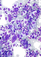
Cytotoxic T lymphocytes (CTLs) targeting Epstein-Barr virus (EBV) proteins appear to be a promising treatment option for patients with aggressive lymphomas.
Researchers tested the autologous CTLs in a cohort of 50 patients with Hodgkin or non-Hodgkin lymphoma.
The treatment produced responses in about 62% of patients with relapsed or refractory disease.
And it sustained remissions in roughly 93% of patients who were at a high risk of relapse.
Catherine Bollard, MD, of the Children’s National Medical Center in Washington, DC, and her colleagues reported these results in the Journal of Clinical Oncology.
The investigators noted that about 40% of lymphoma patients have tumor cells expressing the type II latency EBV antigens latent membrane protein 1 (LMP1) and LMP2. But T cells specific for these antigens are present in low numbers and may not “recognize” the tumors they should attack.
So Dr Bollard and her colleagues decided to test the effects of infusing LMP-directed CTLs into 50 patients with EBV-positive lymphomas.
The researchers used adenoviral vector-transduced dendritic cells and EBV-transformed B–lymphoblastoid cell lines as antigen-presenting cells to activate and expand LMP-specific T cells.
For some patients, the team used an adenoviral vector encoding the LMP2 antigen alone (n=17). And for others, they used a vector encoding both LMP1 and LMP2 (n=33).
Twenty-nine of the patients were in remission when they received CTL infusions, but they were at a high risk of relapse. The remaining 21 patients had relapsed or refractory disease at the time of CTL infusion.
Twenty-seven of the 29 patients who received CTLs as an adjuvant treatment remained in remission from their disease at 3.1 years after treatment.
However, the 2-year event-free survival rate was 82% for this group of patients. None of them died of lymphoma, but 9 died from complications associated with the chemotherapy and radiation they had received.
“That’s why this research is important,” Dr Bollard said. “Patients with lymphomas traditionally have a good cure rate with chemotherapy and radiation. What kills them is the side effects of those treatments—second cancers, lung, and heart disease.”
Of the 21 patients with relapsed or refractory disease, 13 responded to CTL infusions. And 11 patients achieved a complete response.
In this group, the 2-year event-free survival rate was about 50%, regardless of whether patients received CTLs directed against LMP1/2 or LMP2 alone.
The investigators found that responses were associated with effector and central memory LMP1-specific CTLs but not with the patient’s type of lymphoma or lymphopenic status.
Even those patients with limited in vivo expansion of LMP-directed CTLs achieved complete responses. And this effect was associated with epitope spreading.
“This is a targeted therapeutic approach that we hope can be used early in the disease to treat relapse,” Dr Bollard said. “We saw good outcomes here. Eventually, it could be a front-line therapy.”
The researchers noted that the difficulty of tailoring CTLs for each patient has been cited as a barrier to this type of treatment. But currently available treatments can be expensive and induce severe side effects that require hospitalization.
“Although we spend some time making the cells, patients go home with few side effects and few associated hospital costs,” said study author Cliona Rooney, PhD, of the Baylor College of Medicine in Houston, Texas. “It can be less costly than chemotherapy.”
In this study, the investigators did not see any toxicities attributable to CTL infusion. One patient did have CNS deterioration 2 weeks after infusion, but this was attributed to disease progression.
And another patient developed respiratory complications about 4 weeks after a second CTL infusion. But this was attributed to an intercurrent infection, and the patient made a complete recovery.
Histone discovery may have implications for blood cancers

Credit: Eric Smith
Investigators have uncovered an unanticipated mechanism underlying trimethylation of a histone that activates gene expression.
And this finding could have implications for the treatment of leukemias and lymphomas.
Ali Shilatifard, PhD, of the Stowers Institute for Medical Research in Kansas City, Missouri, and his colleagues described the discovery in Genes & Development.
Histones, which come in 4 subtypes—H2A, H2B, H3, and H4—can either coil DNA into inaccessible, silent regions or untwist it to allow gene expression. And small chemical flags, such as methyl groups, affect whether histones silence or activate genes.
Among activator histones is a form of H3 decorated at a precise location with 3 methyl groups, known as H3K4me3.
Previous research showed that the presence of H2B exhibiting a single ubiquitin molecule stimulated the methylase that modifies H3K4, thereby increasing H3K4me3 levels.
But how the methylase’s activity was directed toward the appropriate targets was unclear.
Now, Dr Shilatifard and his colleagues have discovered a mechanism underlying H3K4 trimethylation. Their research explains why H3K4me3 is deposited adjacent to a target gene promoter rather than haphazardly across the entire gene.
The team said this finding is significant because mutations in the human gene encoding the methylase responsible for H3K4me3 are associated with leukemias, lymphomas, and other malignancies.
The methylase in question, named SET1 in yeast and MLL in mammals, is part of a protein aggregate called COMPASS (COMplex of Proteins ASsociated with Set1). Dr Shilatifard was the first to define the role of COMPASS in chromatin modification.
“Over a decade ago, our lab used yeast to show that COMPASS was an H3 methylase,” he said. “Since these fundamental systems are highly conserved from yeast to Drosophila to humans, we took advantage of the awesome power of yeast genetics to identify what regulates H3K4 methylation activity.”
Part of his group’s latest paper addresses SET1/MLL regulation by different proteins within yeast COMPASS.
The investigators knew that if more than half of SET1’s front end was removed, levels of DNA-bound trimethylated H3K4 in cells harboring the remaining “stub” were equal to those in cells containing the full-length protein when analyzed in bulk.
This finding led some researchers to presume that the entire front end of SET1/MLL, as well as factors that interact with it, must not be needed to regulate H3K4me3 activity.
But Dr Shilatifard and his colleagues found evidence suggesting this presumption is incorrect.
The team first employed biochemical methods to capture every piece of DNA bound to H3K4me3 in the genome of yeast harboring either full-length SET1 or the stub missing the front end. They then sequenced all of those DNA fragments and mapped their position in the yeast genome.
Results showed that even though H3K4me3 levels in bulk were equivalent in normal and mutant cells, H3K4me3 was differentially distributed throughout the genome.
In normal cells, H3K4me3 complexes sat primarily on DNA promoter regions. By contrast, the DNA of cells harboring the stub exhibited DNA-binding H3K4me3 complexes in the middle of or between genes.
The work shows that COMPASS factors that bind to the SET1/MLL front end limit H3K4me3 deposition to the correct genomic sites (the promoter regions), while factors that bind the SET1/MLL stub increase the protein’s half-life.
The investigators also discovered how H2B ubiquitin modification machineries stimulate the entire process.
The team said understanding COMPASS regulation is essential, as genes encoding factors in the complex are mutant in numerous cancers.
Inhibitor attacks PEL in vitro and in vivo

Credit: Aaron Logan
A small molecule targeting sphingosine kinase (SPHK) demonstrates considerable activity against virus-associated lymphoma, according to preclinical research published in Molecular Cancer Therapeutics.
The research focused on primary effusion lymphoma (PEL), a variant of diffuse large B-cell lymphoma etiologically linked to Kaposi’s sarcoma-associated herpesvirus (KSHV).
Investigators found that SPHK generates biologically active sphingolipids that keep PEL cells alive.
“It is still early in our understanding of how these special lipids contribute to viral cancers, but this is a major potential advance,” said senior study author Christopher Parsons, MD, of Louisiana State University Health Sciences Center in New Orleans.
“There are no therapies available to fight viral tumors by selectively blocking these pathways, all while not harming normal, uninfected cells.”
So the researchers decided to test ABC294640, a novel small molecule that selectively targets SPHK, in cells from PEL patients and mouse models of PEL.
ABC294640 induced dose-dependent apoptosis in a number of KSHV+ PEL cell lines: KSHV+/EBV- BCBL-1 cells, KSHV+/EBV+ BC-1 cells, KSHV+/EBV- BC-3 cells, and KSHV+/EBV- BCP-1 cells. But the drug showed little to no activity in KSHV-/EBV- BL-41 cells.
Further analyses revealed that ABC294640 induces apoptosis through suppression of KSHV-associated signal transduction.
The investigators observed dose-dependent suppression of ERK, Akt, and NF-kB p65 phosphorylation, as well as cleavage of caspase-3 and caspase-9, in BCBL-1 cells exposed to the drug. But the same effects did not occur in drug-resistant BL-41 cells.
Overexpression of either ERK or p65 in BCBL-1 cells partially suppressed apoptosis induced by ABC294640. And when the researchers used RNAi to target SPHK2, PEL cells exhibited reduced activation of ERK, Akt, and p65 phosphorylation, as well as a 5- to 6-fold increase in apoptosis.
Additionally, the team found that ABC294640 suppressed PEL progression and induced regression of PEL tumors in vivo.
The investigators injected BCL-1 cells into NOD/SCID mice and observed PEL expansion within 3 to 4 weeks. However, when they administered ABC294640 within 24 hours of PEL cell injections, they observed significant reductions in tumor expansion.
The researchers also evaluated ABC294640 activity after PEL tumors had been established. And treated mice showed significant tumor regression compared to untreated mice.
The investigators said these results suggest ABC294640 should be evaluated in clinical trials of KSHV-associated lymphoma.
“Our research thus far indicates that this molecule is safe, with the potential to stand alone as a single, orally administered drug,” Dr Parsons said.