User login
Influenza Vaccine in Pregnancy Seems to Benefit Baby, Too
Major Finding: Receipt of any flu vaccination was significantly associated with higher infant birth weight (3,178 gram vs. 2,903 grams) and longer gestational age (38.3 weeks vs. 36.8 weeks; both P less than .0001).
Data Source: Preliminary analysis of 1,641 women delivering at Duke University Hospital during the 2009-2010 influenza season.
Disclosures: The study was funded by the 2010 American College of Obstetricians and Gynecologists/Merck & Co. Research Award on Immunization. Dr. Fortner and her colleagues reported no relevant financial disclosures.
CHICAGO – Influenza vaccination appears to improve neonatal outcomes, but coverage remains inadequate among pregnant women.
Among 1,641 evaluable women delivering at Duke University Medical Center, Durham, N.C., during the 2009-2010 influenza season, receipt of any flu vaccination was significantly associated with higher infant birth weight (3,178 g vs. 2,903 g) and longer gestational age (38.3 weeks vs. 36.8 weeks; both P values less than .0001).
Women who received at least one flu vaccine also were significantly less likely to require an antepartum visit or hospital admission than were those who did not (39% vs. 44%; P = .005).
“This information supports prior accumulating data that receipt of a flu vaccine improves not only maternal outcomes, but also birth outcomes,” Dr. Kimberly Fortner said at the meeting.
In all, 44% of women in the preliminary analysis received both vaccines in compliance with recommendations, far higher than historical influenza vaccination rates of 12%-34% and comparable to other reports from the season. Another 7% elected no vaccine at all, and 24% of the population had no documented receipt of vaccine in obstetrical records or other electronic medical records.
Uptake of seasonal influenza vaccine was 58% vs. 55% for the 2009 H1N1 influenza vaccine, which is unique among published prior literature. Even though rates were nearly equal, 24% of women elected to receive only one of the two recommended vaccines, resulting in inadequate coverage, said Dr. Fortner of the Translational Medicine Institute at Duke.
The researchers hypothesized that pregnant women may have had inappropriate or inadequate vaccination during the 2009-2010 flu season due to issues of vaccine distribution, sensationalism of the H1N1 influenza pandemic, and recommendations by the Centers for Disease Control and Prevention Advisory Committee on Immunization Practices that pregnant women receive both the seasonal and H1N1 influenza monovalent vaccines.
The earlier women went in for prenatal care, however, the more likely they were to receive a vaccine, she said.
Mean gestational age at first prenatal visit was significantly lower at 14.8 weeks among women who received the vaccine, compared with 18.6 weeks for women who did not receive any vaccine and 21.2 weeks for those with unknown vaccine status (P less than .0001).
Black women and those with public insurance or no insurance were significantly less likely to receive any vaccine.
In multivariate analysis that adjusted for maternal age, black race, less than a high school education, Medicaid or no insurance, and medical comorbidities, receipt of any influenza vaccine during that season was significantly associated with an estimated 133.7-g increase in birth weight (P = .0003).
The vaccine gives pregnant women a greater chance of having a heavier baby who is born closer to term.
Source ©Availablelight/istockphoto.com
From the Annual Meeting of the Infectious Diseases Society for Obstetrics and Gynecology
Pregnancy Weight Gain: Why the Disconnect?
Major Finding: Only 29% of women were counseled about gaining a specific amount or range of weight during pregnancy, and 12% were counseled correctly about how much to gain.
Data Source: A cross-sectional survey of 310 pregnant women with a live, singleton gestation, who visited prenatal clinics.
Disclosures: Dr. McDonald reported that she had no relevant financial disclosures.
VANCOUVER, B.C. – When it comes to counseling women about weight gain during pregnancy, there is plenty of room for improvement, new data suggest.
In a survey of more than 300 pregnant women, fewer than a third reported being counseled on the topic, researchers reported at the meeting. And even fewer, merely an eighth, were counseled correctly about how much weight to gain.
In likely related findings, three-fourths of women who were overweight or obese before conceiving planned to gain more weight than was recommended for them in guidelines.
“A lack of reported counseling has been associated in the literature with inappropriate weight gain, both excessive and inadequate,” said lead investigator Dr. Sarah McDonald, an ob.gyn at McMaster University in Hamilton, Ont. “So these findings were very concerning for us.”
She noted that most women who were approached agreed to participate in the survey and were comfortable about discussing weight. Therefore, “it appeared unlikely that the lack of reported counseling was due to patient-driven factors, apart from possibly forgetting.”
Interestingly, a staggered companion survey of the providers had dramatically different findings, showing high reported rates of counseling. “It was like I was surveying people on a different planet,” she commented. “We think we are doing very well,” yet there is an obvious discrepancy that is as yet unexplained.
Citing the obesity epidemic, Dr. McDonald endorsed repeated counseling of women about weight, both before and during pregnancy.
“Obviously, an optimal BMI [body mass index] prepregnancy is ideal, but that's not the situation where most of us come into contact with our patients; it's when they are already pregnant. Then, I think talking about optimal gestational weight gain to not compound the problems of overweight and obesity is important,” she said. “But given the size of the [obesity] epidemic, [the approach has] got to be multipronged.”
In 2009, the U.S. Institute of Medicine released new recommendations regarding gestational weight gain, tailored to prepregnancy BMI, that have been adopted by Canada and other countries.
“However, previous studies done in the era of the 1990 guidelines have shown that only about 30%-40% of pregnant women gained the appropriate amount of weight during pregnancy,” Dr. McDonald noted. “And we were curious what was going on in the era of the new guidelines.”
The investigators surveyed 310 women (94% of those approached) who made at least one visit to representative Hamilton prenatal clinics, other than for pregnancy diagnosis, and currently had a live, singleton gestation. The women's mean age was 30 years, and the median gestational age was 33.0 weeks. Fully 74% were white, and for 43%, the birth would be their first. They had a mean prepregnancy BMI of 25.1 kg/m
“Interestingly enough, 84% of the women reported that they were either comfortable or very comfortable talking about weight-related issues with their care provider, despite the fact that the mean BMI [in this study] is already in the overweight category prepregnancy,” Dr. McDonald observed.
Only 29% of the women reported that their provider counseled them to gain a specific amount or range of weight, and for just 12% overall, that amount or range was correct according to the new guidelines. Only about a quarter of women reported being told that there were risks associated with gaining too much or too little weight during pregnancy.
The median number of prenatal visits before the survey was 10 for the study population, she pointed out, and “so there were multiple opportunities for discussion about weight gain.”
“We wondered, are clinicians just too busy to be talking about weight and weight-related matters, and nutrition, and preventive-type medicine?” said Dr. McDonald. Yet nearly all of the women (97%) reported being counseled to take a vitamin.
When asked how much weight they planned to gain during pregnancy, only 12%-54% of women, depending on prepregnancy BMI category, cited an amount within the guideline-recommended range for them. In particular, in a finding that she described as “alarming,” 75% of overweight and obese women were planning to gain more weight than was recommended for them.
The proportion of women counseled about weight gain differed by the type of provider that had provided the majority of a woman's pregnancy care; it was 40% for midwives, 24% for obstetricians, 23% for general practitioners, and 28% for other providers. The proportion that was correctly counseled showed a similar pattern, but the differences were not significant.
Clinicians' survey responses were so different, 'it was like I was surveying people on a different planet.'
Source DR. McDONALD
From the Annual Meeting of the Society of Obstetricians and Gynaecologists of Canada
Pediatricians Push Tdap More Than Ob.Gyns.
CHICAGO – Pregnant women were significantly more likely to receive information on pertussis vaccination from their pediatrician than from their obstetrician in a survey of 314 women.
“Multiple opportunities exist for education of obstetricians and gynecologists to improve Tdap vaccination rates in the United States,” Dr. Rachel Gutkin said.
She reported on 314 pregnant women presenting to an academic perinatal center between March and June 2011 who answered an anonymous, multiple-choice questionnaire regarding their knowledge and opinions on vaccination in general, and Tdap (tetanus toxoid, reduced diphtheria toxoid, and acellular pertussis adsorbed) specifically. Overall, 218 (69%) of the women had heard about the Tdap booster vaccine, with 76 (24%) women learning about it at their pediatrician's office and 54 (17%) at their obstetrician's office. Just 8% of women learned about Tdap from the Internet, while 19% did so from friends or family, and 17% from TV or radio, said Dr. Gutkin, a resident in the department of obstetrics and gynecology at the University of California, Los Angeles. The remaining 15% learned of it from other sources.
The majority of respondents knew that pertussis is a significant health risk for children (76%) and newborns (86%), and 50% also thought it was a significant health risk for fetuses. When asked whether they would receive a Tdap vaccination during pregnancy to protect their newborn from whooping cough, 11% said they would if their doctor recommended it, 12% said yes if they knew it was safe, and 66% said they would if both conditions were true. Additionally, 11% said they would not receive Tdap during pregnancy under any circumstances, she said.
Women who discussed Tdap with their obstetrician were nearly five times more likely to be vaccinated than those who did not (odds ratio, 4.93). Only 13% of women who did not discuss Tdap with their ob.gyn. were vaccinated.
Although women were significantly more likely to discuss Tdap with their pediatrician, women were nearly three times more likely to be vaccinated if they were counseled by their ob.gyn. versus their pediatrician (OR, 2.9).
The survey was conducted in California, which in 2010 experienced the largest outbreak of pertussis in 65 years, with 9,120 cases reported, including 10 deaths.
Still, almost one-quarter of women were not sure if vaccines in general are safe (22%). Roughly two-thirds were not sure if vaccines are safe in pregnancy (68%) and a full 10% were not sure if vaccines are effective in preventing illness, Dr. Gutkin said.
Women were significantly more likely to have discussed the influenza vaccine with their ob.gyn. than Tdap (47% vs. 19%), and to be vaccinated for influenza than pertussis (42% vs. 18%). Of note, women were three times more likely to receive Tdap if they had received a flu shot (OR, 3.68). “Discussion with their ob.gyn. was a significant factor in the acceptance of Tdap and influenza vaccines during pregnancy,” she said.
'Discussion with their ob.gyn. was a significant factor in the acceptance of Tdap … vaccines during pregnancy.'
Source DR. GUTKIN
From the Annual Meeting of the Infectious Diseases Society for Obstetrics and Gynecology
Does the use of multiple maneuvers in the management of shoulder dystocia increase the risk of neonatal injury?
- Pelvic injury from the McRoberts maneuver?
(Medical Verdicts, August 2011)
Shoulder dystocia occurs in 0.6% to 1.4% of vaginal births and is highly unpredictable. It can cause maternal lacerations, increase the risk of postpartum hemorrhage, and lead to major neonatal injuries such as fractures of the clavicle or humerus, palsies of the brachial plexus, hypoxic-ischemic encephalopathy (HIE), and, even, death. It is also a leading cause of monetary awards in obstetric-related malpractice litigation.
Hoffman and colleagues analyzed 2,018 cases of shoulder dystocia, 101 of which (5.2%) incurred neonatal injury. A total of 3,751 maneuvers were performed, with an average number of maneuvers per case of 1.86. The total number of maneuvers performed in any given case of shoulder dystocia correlated significantly with the rate of neonatal injury.
When five or more maneuvers were used, the rate of neonatal injury exceeded 20%. The authors did not specify whether the neonatal injuries reflected in this figure included bone fracture as well as peripheral nerve injury, or only the latter. They also failed to specify how many brachial plexus injuries resolved spontaneously.
As for which maneuvers were most effective, delivery of the posterior arm and shoulder had a higher rate of success (84%), compared with the McRoberts maneuver, suprapubic pressure, and other maneuvers (24.3% to 72.0% success rate). However, the authors continue to recommend that the McRoberts maneuver and suprapubic pressure be the first maneuvers utilized when shoulder dystocia occurs.
Large study size was a strength
This study, emanating from 12 centers that make up the Consortium on Safe Labor, is probably the largest to date to examine issues related to shoulder dystocia. The sheer magnitude of this study lends great credence to the findings. Another significant strength: Trained obstetric abstractors reviewed the entire medical record of both the mother and newborn.
That said, as with most studies related to shoulder dystocia, there is the possibility of ascertainment bias. Moreover, the study was not randomized. Nor was there uniformity among cases in terms of the maneuvers used or the order in which they were performed, both of which were based largely on provider preference, theoretical models, and expert opinion.
This analysis encompassed the following maneuvers to manage shoulder dystocia:
- McRoberts maneuver
- suprapubic pressure
- Rubin maneuver
- delivery of the posterior arm
- Woods corkscrew maneuver
- Gaskin maneuver (delivery in the maternal knee-chest position)
- Zavanelli maneuver
- fundal pressure.
ACOG recommends the McRoberts maneuver as an initial intervention, followed by suprapubic pressure, when shoulder dystocia occurs.1
The rate of injury was low
At 5.2%, the rate of clinically relevant injury resulting from shoulder dystocia in this study was low. As the authors note, the reported rate of injury in shoulder dystocia cases in general ranges from 4% to as high as 40%.
In this study, if one excludes the 41 cases of bone fracture (either clavicular or humeral) that occurred, which will heal without long-term sequelae, the rate of injury (i.e., neonatal peripheral nerve injury) was just 3%—60 cases of Erb’s palsy and four cases of Klumpke’s palsy among 2,018 cases of shoulder dystocia.
In general, the rate of HIE related to intractable shoulder dystocia is also exceedingly low. In this study, the authors reported only six cases (0.29%). However, the mean head-to-body delivery time in the cases involving HIE was 10.75 minutes, which is troubling. One case of HIE occurred within a reported time frame of 3 minutes.
No real differences between providers were identified
No significant differences in the rate of injury were observed when shoulder dystocia was managed primarily by a resident physician (7.4%), compared with a midwife (2.9%) or attending physician (5.2%). This finding is somewhat confusing because the authors attributed “primary” management to the individual who delivered the newborn and “thus initiated the maneuvers.” I suspect that when a case was managed by a resident, an attending physician was actively involved.
No causal relationship was established
The authors have made a valiant argument that delivery of the posterior arm should be a priority when shoulder dystocia occurs. However, their findings do not confirm a cause-effect relationship between delivery of the posterior arm and resolution of dystocia, for the following reasons:
- Patients were not randomized to a uniform series of maneuvers.
- The order in which the maneuvers were employed could be determined in only 65.7% of cases.
- Management of shoulder dystocia at the study centers was heterogeneous in nature.
For now, the McRoberts maneuver and suprapubic pressure remain first-line maneuvers of choice. This study, along with several other recent investigations, does suggest that delivery of the posterior arm should be considered if these initial maneuvers are unsuccessful.
As simulation is utilized increasingly in resident learning, providers should become more comfortable facilitating delivery of the posterior arm. —Robert B. Gherman, MD
We want to hear from you! Tell us what you think.
Reference
1. American College of Obstetricians and Gynecologists Committee on Practice Bulletins—Gynecology. ACOG Practice Bulletin No. 40: Shoulder dystocia. Obstet Gynecol. 2002;;100(5 Pt 1)::1045-1050.
- Pelvic injury from the McRoberts maneuver?
(Medical Verdicts, August 2011)
Shoulder dystocia occurs in 0.6% to 1.4% of vaginal births and is highly unpredictable. It can cause maternal lacerations, increase the risk of postpartum hemorrhage, and lead to major neonatal injuries such as fractures of the clavicle or humerus, palsies of the brachial plexus, hypoxic-ischemic encephalopathy (HIE), and, even, death. It is also a leading cause of monetary awards in obstetric-related malpractice litigation.
Hoffman and colleagues analyzed 2,018 cases of shoulder dystocia, 101 of which (5.2%) incurred neonatal injury. A total of 3,751 maneuvers were performed, with an average number of maneuvers per case of 1.86. The total number of maneuvers performed in any given case of shoulder dystocia correlated significantly with the rate of neonatal injury.
When five or more maneuvers were used, the rate of neonatal injury exceeded 20%. The authors did not specify whether the neonatal injuries reflected in this figure included bone fracture as well as peripheral nerve injury, or only the latter. They also failed to specify how many brachial plexus injuries resolved spontaneously.
As for which maneuvers were most effective, delivery of the posterior arm and shoulder had a higher rate of success (84%), compared with the McRoberts maneuver, suprapubic pressure, and other maneuvers (24.3% to 72.0% success rate). However, the authors continue to recommend that the McRoberts maneuver and suprapubic pressure be the first maneuvers utilized when shoulder dystocia occurs.
Large study size was a strength
This study, emanating from 12 centers that make up the Consortium on Safe Labor, is probably the largest to date to examine issues related to shoulder dystocia. The sheer magnitude of this study lends great credence to the findings. Another significant strength: Trained obstetric abstractors reviewed the entire medical record of both the mother and newborn.
That said, as with most studies related to shoulder dystocia, there is the possibility of ascertainment bias. Moreover, the study was not randomized. Nor was there uniformity among cases in terms of the maneuvers used or the order in which they were performed, both of which were based largely on provider preference, theoretical models, and expert opinion.
This analysis encompassed the following maneuvers to manage shoulder dystocia:
- McRoberts maneuver
- suprapubic pressure
- Rubin maneuver
- delivery of the posterior arm
- Woods corkscrew maneuver
- Gaskin maneuver (delivery in the maternal knee-chest position)
- Zavanelli maneuver
- fundal pressure.
ACOG recommends the McRoberts maneuver as an initial intervention, followed by suprapubic pressure, when shoulder dystocia occurs.1
The rate of injury was low
At 5.2%, the rate of clinically relevant injury resulting from shoulder dystocia in this study was low. As the authors note, the reported rate of injury in shoulder dystocia cases in general ranges from 4% to as high as 40%.
In this study, if one excludes the 41 cases of bone fracture (either clavicular or humeral) that occurred, which will heal without long-term sequelae, the rate of injury (i.e., neonatal peripheral nerve injury) was just 3%—60 cases of Erb’s palsy and four cases of Klumpke’s palsy among 2,018 cases of shoulder dystocia.
In general, the rate of HIE related to intractable shoulder dystocia is also exceedingly low. In this study, the authors reported only six cases (0.29%). However, the mean head-to-body delivery time in the cases involving HIE was 10.75 minutes, which is troubling. One case of HIE occurred within a reported time frame of 3 minutes.
No real differences between providers were identified
No significant differences in the rate of injury were observed when shoulder dystocia was managed primarily by a resident physician (7.4%), compared with a midwife (2.9%) or attending physician (5.2%). This finding is somewhat confusing because the authors attributed “primary” management to the individual who delivered the newborn and “thus initiated the maneuvers.” I suspect that when a case was managed by a resident, an attending physician was actively involved.
No causal relationship was established
The authors have made a valiant argument that delivery of the posterior arm should be a priority when shoulder dystocia occurs. However, their findings do not confirm a cause-effect relationship between delivery of the posterior arm and resolution of dystocia, for the following reasons:
- Patients were not randomized to a uniform series of maneuvers.
- The order in which the maneuvers were employed could be determined in only 65.7% of cases.
- Management of shoulder dystocia at the study centers was heterogeneous in nature.
For now, the McRoberts maneuver and suprapubic pressure remain first-line maneuvers of choice. This study, along with several other recent investigations, does suggest that delivery of the posterior arm should be considered if these initial maneuvers are unsuccessful.
As simulation is utilized increasingly in resident learning, providers should become more comfortable facilitating delivery of the posterior arm. —Robert B. Gherman, MD
We want to hear from you! Tell us what you think.
Reference
1. American College of Obstetricians and Gynecologists Committee on Practice Bulletins—Gynecology. ACOG Practice Bulletin No. 40: Shoulder dystocia. Obstet Gynecol. 2002;;100(5 Pt 1)::1045-1050.
Reference
1. American College of Obstetricians and Gynecologists Committee on Practice Bulletins—Gynecology. ACOG Practice Bulletin No. 40: Shoulder dystocia. Obstet Gynecol. 2002;;100(5 Pt 1)::1045-1050.
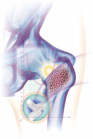
Vitamin D and pregnancy: 9 things you need to know
- How much vitamin D should you recommend to your nonpregnant patients?
Emily D. Szmuilowicz, MD, MS; JoAnn E. Manson, MD, DrPH (July 2011)
With all the publicity surrounding vitamin D lately, it’s no surprise that you have lots of questions. Should you test your patients for deficiency? When? What numbers should you use? And how do you treat a low vitamin D level?
In pregnancy, these issues become critical because there are not one but two patients to consider. Despite the lack of clear guidelines, there is sufficient evidence to suggest that you should at least consider monitoring the vitamin D status of your pregnant patients.
Fetal needs for vitamin D increase during the latter half of pregnancy, when bone growth and ossification are most prominent. Vitamin D travels to the fetus by passive transfer, and the fetus is entirely dependent on maternal stores.1 Therefore, maternal status is a direct reflection of fetal nutritional status.
The vitamin D level in breast milk also correlates with the maternal serum level, and a low vitamin D level in breast milk can exert a harmful effect on a newborn.
In this article, I address nine questions regarding vitamin D and pregnancy:
- Is vitamin D really a vitamin?
- Why do the numbers vary?
- Does the vitamin D level affect pregnancy outcomes?
- Can’t people get enough vitamin D through their diet?
- What level signals deficiency?
- How many women are deficient?
- Should you test all pregnant patients?
- How should you treat vitamin D deficiency in pregnancy?
- Can a person get too much vitamin D?
1. Is vitamin D really a vitamin?
For years, vitamin D was discussed solely in relation to bone metabolism and absorption, and deficiency states were the purview of endocrinologists and gynecologists who treated menopausal patients at risk of osteoporosis. Recent studies demonstrate that vitamin D plays a role in multiple endocrine systems. Indeed, vitamin D may be more correctly considered a hormone because it is a substance produced by one organ (skin) that travels through the bloodstream to target end organs. Vitamin D receptors have been found in bone, breast, brain, colon, muscle, and pancreatic tissues. Not only does vitamin D affect bone metabolism, it also modulates immune responses and even glucose metabolism.2 Vitamin D receptors have also been found in the placenta; their role in that organ remains to be elucidated.
2. Why do the numbers vary?
Some of the confusion surrounding vitamin D concerns the units used to measure and discuss it. Vitamin D can be measured in nanograms per milliliter (ng/mL) or in nanomoles per liter (nmol/L). A measurement of 1 ng/mL equals approximately 2.44 nmol/L. Therefore, deficiency in some articles is described as a vitamin D level below 20 ng/mL and in other articles as a level below 50 nmol/L. As for normal range, it may be listed as a level above 32 ng/mL or as a level above 75 nmol/L.
Compounding the confusion, vitamin D in supplement form can be written in two different measurements—using micrograms or international units. A measurement of 1 μg equals 40 IU, so a supplement of 150 μg/day is the same as one of 6,000 IU/day.
3. Does the vitamin D level affect pregnancy outcomes?
Vitamin D’s role in pregnancy outcomes has yet to be fully described, making it an exciting field to explore. Research into vitamin D and its effects on pregnancy is still in its infancy, but many intriguing associations have been noted. For example, lower levels of vitamin D have been associated with increased rates of cesarean delivery,3 bacterial vaginosis,4 and preeclampsia,5 as well as less efficient glucose metabolism.6
There is biological plausibility for vitamin D to play a role in pregnancy outcomes, given the presence of receptors in gestational tissues. Vitamin D receptors in uterine muscle could affect contractile strength, and vitamin D has been shown to have immunomodulatory effects, thereby potentially protecting the host from infection.
As I mentioned, placental vitamin D receptors and their role need further exploration.
4. Can’t people get enough vitamin D through their diet?
Very few foods contain a large amount of vitamin D, and the few that do (herring, cod liver oil) are not standard fare. Even fortified foods such as milk lack a substantial amount. TABLE 1 lists the amount of vitamin D in various foods.7
TABLE 1
In food, the vitamin D level is generally low
| Source | Amount of vitamin D (IU) |
|---|---|
| Egg yolk | 25 |
| Cereal, fortified with vitamin D, 1 cup | 40–50 |
| Cow’s milk, fortified with vitamin D, 8 oz | 98 |
| Soy milk, fortified with vitamin D, 8 oz | 100 |
| Orange juice, fortified with vitamin D, 8 oz | 100 |
| Quaker Nutrition for Women instant oatmeal, 1 packet | 154 |
| Tuna, canned in oil, 3 oz | 200 |
| Sardines, canned, 3 oz | 231 |
| Mackerel, 3 oz | 306 |
| Most multivitamins | 400 |
| Tri-Vi-Sol infant supplements, 1 drop | 400 |
| Prenatal vitamins | 400 |
| Catfish, 3 oz | 425 |
| Pink salmon, canned, 3 oz | 530 |
| Cod liver oil, 1 tablespoon | 1,360 |
| Herring, 3 oz | 1,383 |
| Over-the-counter vitamin D3 supplements | 2,000 (maximum) |
| Typical prescription of vitamin D2 for deficiency | 50,000 (given weekly until replete) |
5. What level signals deficiency?
Experts disagree about the level of vitamin D that signals deficiency. Many labs report a reference range of 32 to 100 ng/mL as normal. However, in November 2010, the Institute of Medicine (IOM) weighed in on the matter. After examining the data, the IOM suggested that a vitamin D level of 20 ng/mL is sufficient to prevent bone loss and changes seen in rickets and osteoporosis.
This level is hotly contested by experts in other fields, who argue that, although 20 ng/mL may be considered the bare minimum level to prevent negative bone resorption changes, it can hardly be construed as a normal level.
Nor did the IOM recommendation take pregnancy into consideration. Therefore, the IOM made no comment as to whether a level of 20 ng/mL is sufficient for a pregnant woman, given that the fetus will be actively soliciting maternal vitamin D for its own development. Indeed, some researchers have indicated that the actual daily recommended intake for pregnancy and lactation may be as high as 6,000 IU/day.8
6. How many women are deficient?
The rate of deficiency varies, but studies have documented rates as high as 97% in some pregnant populations; the rates vary by race and latitude.9-11
The high prevalence of deficiency in the population is due, in large part, to vitamin D’s mode of production and changes in human lifestyle and culture. Vitamin D is produced primarily through direct exposure of the skin to the sun. Over the past 50 years, as more and more people have come to spend their days in an office or factory instead of on a farm, the opportunity to produce vitamin D has greatly diminished.
Other entities or practices that reduce the production of vitamin D:
- Sunscreen SPF 50 may prevent skin cancer, but it also blocks vitamin D production.
- Fat cells Obese patients produce vitamin D less rapidly than patients of normal weight.
- Melanin Darker-skinned people produce vitamin D at a slower rate than those who have fair skin.
- Cultural practices Some religious and cultural practices mandate full skin coverage in public, particularly for women, leading to minimal sun exposure.
- Age Older people also produce vitamin D more slowly. Among the population of reproductive age, however, the effect of age is minimal.
- Latitude Northern latitudes, with their longer winters and shorter summers, provide less opportunity for sun exposure.
Because vitamin D is, in essence, a “seasonal” vitamin, it makes evolutionary sense that the human body has developed a wide normal range to “store up” vitamin D when sunshine is plentiful and then use its stores during times of scarcity, such as winter. This seasonal variability is another reason why the rate of deficiency can vary, depending on the time and location of study.
Because vitamin D deficiency is clinically silent until severe events such as rickets occur, the best way to check for it is to measure total levels of the two forms of vitamin D found in the body—D2 and D3. The recommended test is total 25-hydroxy vitamin D (25-OHD). Measurement of the activated form of vitamin D—1,25-OHD—will not tell you whether a person’s overall stores are lacking, because the body maintains a normal 1,25-OHD level over a wide range until severe deficiency occurs.
7. Should you test all pregnant patients for deficiency?
ACOG does not recommend that vitamin D be measured routinely in pregnant women.12 In a Committee Opinion published in July 2011, ACOG determined that “there is insufficient evidence to support a recommendation for screening all pregnant women for vitamin D deficiency.”12
Many experts disagree, however, citing the increased rate of rickets being found in the United States.6,8 Pediatricians in the United States have found such a high rate of deficiency in the neonatal population that the American Academy of Pediatricians now recommends that all exclusively breastfed babies be given a supplement of 400 IU of vitamin D daily, beginning in the first few days of life.13
ACOG acknowledged that, for pregnant patients “thought to be at increased risk, measurement of total levels can be considered with “high-risk groups” that have many of the risk factors cited earlier.12
If you want to test your patients, no single plan is recommended. A sample algorithm includes the following steps:
- Measure total 25-OHD at the time of prenatal registration labs
- Select a level of supplementation, based on the findings (see TABLE 2)
- Recheck the 25-OHD level after 3 months. For most patients, this would be around the time of a standard glucose screening test
- Adjust the supplementation level, as needed
- Measure 25-OHD at admission to labor and delivery.
TABLE 2
When (and with how much “D”) to treat pregnant patients
| If the 25-OHD level is… | …then supplement with* |
|---|---|
| <20 ng/mL | 50,000 IU oral vitamin D weekly for 12 weeks |
| 20–32 ng/mL | 2,000–4,000 IU oral vitamin D daily (~15,000–30,000 IU weekly) |
| >32 ng/mL | No action needed |
| *Assuming that the patient will continue taking a prenatal vitamin containing 400 IU/tablet. | |
8. How should you treat vitamin D deficiency in pregnancy?
Here, again, there is a lack of solid evidence. No guidelines exist for pregnant patients. In its Commitee Opinion, ACOG points out that higher-dose regimens have not been studied in pregnancy, but cites studies using up to 4,000 IU daily.12 The question becomes: Can guidelines that have been established for nonpregnant patients be used safely in pregnancy?
Although there is no evidence-based consensus, physiology and previous studies suggest that they can.
In one study, pregnant women were given doses as high as 200,000 IU in the third trimester to treat vitamin D deficiency.14 That investigation produced two key findings:
- There were no signs or symptoms of toxicity in patients or newborns, demonstrating that a single dose of a large amount of vitamin D can be administered safely.
- Despite the treatment, many of the women in this study remained deficient, indicating that continued supplementation would be required beyond the initial dose.
Although the dosage administered in this study seems like a large amount, it should be viewed in context: a Caucasian female can produce 50,000 IU of vitamin D from 30 minutes of sun exposure at midday.14
The IOM acknowledged that it underestimated the amount of vitamin D that can be taken safely and increased its upper limit of normal to 4,000 IU daily. Note that this upper limit is for people who are presumed to have a normal level to begin with. Therefore, it would be expected that a deficiency would require a greater amount for treatment.
As for treatment, both daily and weekly regimens are acceptable. Because vitamin D is fat-soluble, a daily dose of 1,000 IU is equivalent to a weekly dose of 7,000 IU. Many patients prefer the convenience of weekly dosing, which can also improve compliance.
See TABLE 2 for a proposed guideline on how to treat a pregnant patient, based on the 25-OHD level.
9. Can a person get too much vitamin D?
Vitamin D is fat-soluble. Should you worry about toxicity?
Because there is such a wide normal range for vitamin D, a person would have to be taking massive amounts of the nutrient for a substantial time before hypervitaminosis and a potential impact on calcium metabolism occur. Pharmacokinetic data demonstrate that toxicity may not occur until a vitamin D level of 300 ng/mL or higher is reached, which is three times the upper limit of normal for most reference ranges.15 A 2007 review found no cases of toxicity reported in the literature at a total serum level below 200 ng/mL (twice the normal limit) or a dose of less than 30,000 IU/day.16
Last words
Many questions and research opportunities remain regarding optimal vitamin D levels and supplementation in pregnancy, as well as the impact of vitamin D not only on pregnancy-related outcomes but on neonatal and infant health. One thing is certain: No one can argue that a nutritionally deficient state is preferred in pregnancy for maternal or fetal health. As advocates for women’s health, it behooves us to address this situation for the benefit of our patients and their children.
How do you manage the vitamin D requirements of pregnant and nonpregnant patients? Do you agree with the IOM that a vitamin D level of 20 ng/mL is sufficient for most individuals? Do you routinely measure the vitamin D level of your patients? Do you recommend vitamin D supplementation in pregnancy?
To tell us, click here
Study finds vitamin D supplementation in pregnancy to be safe and effective
Daily 4,000-IU vitamin D supplementation from 12 to 16 weeks of gestation is safe and effective in achieving vitamin D sufficiency in pregnant women and their neonates, according to a study published in the July 2011 issue of the Journal of Bone and Mineral Research.
Bruce W. Hollis, PhD, from the Medical University of South Carolina in Charleston, and colleagues assessed the need, safety, and effectiveness of vitamin D supplementation in 350 women with singleton pregnancies at 12 to 16 weeks of gestation. Participants were randomly assigned to receive 400 IU, 2,000 IU, or 4,000 IU vitamin D3 daily until delivery. The outcomes studied included maternal/neonatal circulating serum vitamin D (25-OHD) levels at delivery, achieving 25-OHD of 80 nmol/L or more, and achieving 25-OHD concentration for maximal 1,25-dihydroxycholecalciferol (1,25-OH2D) production.
The investigators found that the percentage of participants who achieved vitamin D sufficiency was significantly different between groups, with the 4,000-IU group having the highest percentage. Within 1 month of delivery, the relative risk (RR) of achieving 25-OHD of 80 nmol/L or more differed significantly between the 2,000-IU versus 400-IU groups and 4,000-IU versus 400-IU groups (RR, 1.52 and 1.60, respectively). There was no significant difference between the 2,000-IU and 4,000-IU groups. Circulatory 25-OHD directly influenced 1,25-OH2D levels throughout pregnancy, with maximal production of 1,25-OH2D in the 4,000-IU group. Vitamin D supplementation was not associated with adverse events, and safety measures were similar between the groups.
“A daily vitamin D dose of 4,000 IU was associated with improved vitamin D status throughout pregnancy, one month prior, and at delivery in both mother and neonate,” the authors write.
One of the study authors disclosed financial ties with the Diasorin Corporation.
Copyright © 2011 HealthDay. All rights reserved.
1. Dror DK, Allen LH. Vitamin D inadequacy in pregnancy: biology outcomes, and interventions. Nutr Rev. 2010;68(8):465-477.
2. Verstuyf A, Carmeliet G, Bouillon R, Mathieu C. Vitamin D: a pleiotropic hormone. Kidney Int. 2010;78(2):140-145.
3. Merewood A, Mehta SD, Chen TC, Bauchner H, Holick MF. Association between vitamin D deficiency and primary cesarean section. J Clin Endocrinol Metab. 2009;94(3):940-945.
4. Bodnar LM, Krohn MA, Simhan HN. Maternal vitamin D deficiency is associated with bacterial vaginosis in the first trimester of pregnancy. J Nutr. 2009;139(6):1157-1161.
5. Robinson CJ, Alanis MC, Wagner CL, Hollis BW, Johnson DD. Plasma 25-hydroxyvitamin D levels in early-onset severe preeclampsia. Am J Obstet Gynecol. 2010;203(4):366.e1-6.
6. Lau SL, Gunton JE, Athayde NP, Byth K, Cheung NW. Serum 25-hydroxyvitamin D and glycated haemoglobin levels in women with gestational diabetes mellitus. Med J Aust. 2011;194(7):334-337.
7. Mulligan ML, Felton SK, Riek AE, Bernal-Mizrachi C. Implications of vitamin D deficiency in pregnancy and lactation. Am J Obstet Gynecol. 2010;202(5):429.e1-9.
8. Hollis BW. Vitamin D requirement during pregnancy and lactation. J Bone Miner Res. 2007;22 (suppl 2):V39-44.
9. Johnson DD, Wagner CL, Hulsey TC, McNeil RB, Ebeling M, Hollis BW. Vitamin D deficiency and insufficiency is common during pregnancy. Am J Perinatol. 2010;28(1):7-12.
10. Bodnar LM, Simhan HN, Powers RW, Frank MP, Cooperstein E, Roberts JM. High prevalence of vitamin D insufficiency in black and white pregnant women residing in the northern United States and their neonates. J Nutr. 2007;137(2):447-452.
11. Ginde AA, Sullivan AF, Mansbach JM, Camargo CA, Jr. Vitamin D insufficiency in pregnant and nonpregnant women of childbearing age in the United States. Am J Obstet Gynecol. 2010;202(5):436.e1-8.
12. ACOG Committee Opinion No. 495: Vitamin D: Screening and supplementation during pregnancy. Obstet Gynecol. 2011;118(1):197-198.
13. Wagner CL, Greer FR. American Academy of Pediatrics Section on Breastfeeding; American Academy of Pediatrics Committee on Nutrition. Prevention of rickets and vitamin D deficiency in infants children, and adolescents. Pediatrics. 2008;122(5):1142-1152.
14. Yu CK, Sykes L, Sethi M, Teoh TG, Robinson S. Vitamin D deficiency and supplementation during pregnancy. Clin Endocrinol (Oxf). 2009;70(5):685-690.
15. Jones G. Pharmacokinetics of vitamin D toxicity. Am J Clin Nutr. 2008;88(2):582S-586S.
16. Hathcock JN, Shao A, Vieth R, Heaney R. Risk assessment for vitamin D. Am J Clin Nutr. 2007;85(1):6-18.
17. Hollis BW. Vitamin D requirement during pregnancy and lactation. J Bone Miner Res. 2007;22 (suppl 2):V39-44.
- How much vitamin D should you recommend to your nonpregnant patients?
Emily D. Szmuilowicz, MD, MS; JoAnn E. Manson, MD, DrPH (July 2011)
With all the publicity surrounding vitamin D lately, it’s no surprise that you have lots of questions. Should you test your patients for deficiency? When? What numbers should you use? And how do you treat a low vitamin D level?
In pregnancy, these issues become critical because there are not one but two patients to consider. Despite the lack of clear guidelines, there is sufficient evidence to suggest that you should at least consider monitoring the vitamin D status of your pregnant patients.
Fetal needs for vitamin D increase during the latter half of pregnancy, when bone growth and ossification are most prominent. Vitamin D travels to the fetus by passive transfer, and the fetus is entirely dependent on maternal stores.1 Therefore, maternal status is a direct reflection of fetal nutritional status.
The vitamin D level in breast milk also correlates with the maternal serum level, and a low vitamin D level in breast milk can exert a harmful effect on a newborn.
In this article, I address nine questions regarding vitamin D and pregnancy:
- Is vitamin D really a vitamin?
- Why do the numbers vary?
- Does the vitamin D level affect pregnancy outcomes?
- Can’t people get enough vitamin D through their diet?
- What level signals deficiency?
- How many women are deficient?
- Should you test all pregnant patients?
- How should you treat vitamin D deficiency in pregnancy?
- Can a person get too much vitamin D?
1. Is vitamin D really a vitamin?
For years, vitamin D was discussed solely in relation to bone metabolism and absorption, and deficiency states were the purview of endocrinologists and gynecologists who treated menopausal patients at risk of osteoporosis. Recent studies demonstrate that vitamin D plays a role in multiple endocrine systems. Indeed, vitamin D may be more correctly considered a hormone because it is a substance produced by one organ (skin) that travels through the bloodstream to target end organs. Vitamin D receptors have been found in bone, breast, brain, colon, muscle, and pancreatic tissues. Not only does vitamin D affect bone metabolism, it also modulates immune responses and even glucose metabolism.2 Vitamin D receptors have also been found in the placenta; their role in that organ remains to be elucidated.
2. Why do the numbers vary?
Some of the confusion surrounding vitamin D concerns the units used to measure and discuss it. Vitamin D can be measured in nanograms per milliliter (ng/mL) or in nanomoles per liter (nmol/L). A measurement of 1 ng/mL equals approximately 2.44 nmol/L. Therefore, deficiency in some articles is described as a vitamin D level below 20 ng/mL and in other articles as a level below 50 nmol/L. As for normal range, it may be listed as a level above 32 ng/mL or as a level above 75 nmol/L.
Compounding the confusion, vitamin D in supplement form can be written in two different measurements—using micrograms or international units. A measurement of 1 μg equals 40 IU, so a supplement of 150 μg/day is the same as one of 6,000 IU/day.
3. Does the vitamin D level affect pregnancy outcomes?
Vitamin D’s role in pregnancy outcomes has yet to be fully described, making it an exciting field to explore. Research into vitamin D and its effects on pregnancy is still in its infancy, but many intriguing associations have been noted. For example, lower levels of vitamin D have been associated with increased rates of cesarean delivery,3 bacterial vaginosis,4 and preeclampsia,5 as well as less efficient glucose metabolism.6
There is biological plausibility for vitamin D to play a role in pregnancy outcomes, given the presence of receptors in gestational tissues. Vitamin D receptors in uterine muscle could affect contractile strength, and vitamin D has been shown to have immunomodulatory effects, thereby potentially protecting the host from infection.
As I mentioned, placental vitamin D receptors and their role need further exploration.
4. Can’t people get enough vitamin D through their diet?
Very few foods contain a large amount of vitamin D, and the few that do (herring, cod liver oil) are not standard fare. Even fortified foods such as milk lack a substantial amount. TABLE 1 lists the amount of vitamin D in various foods.7
TABLE 1
In food, the vitamin D level is generally low
| Source | Amount of vitamin D (IU) |
|---|---|
| Egg yolk | 25 |
| Cereal, fortified with vitamin D, 1 cup | 40–50 |
| Cow’s milk, fortified with vitamin D, 8 oz | 98 |
| Soy milk, fortified with vitamin D, 8 oz | 100 |
| Orange juice, fortified with vitamin D, 8 oz | 100 |
| Quaker Nutrition for Women instant oatmeal, 1 packet | 154 |
| Tuna, canned in oil, 3 oz | 200 |
| Sardines, canned, 3 oz | 231 |
| Mackerel, 3 oz | 306 |
| Most multivitamins | 400 |
| Tri-Vi-Sol infant supplements, 1 drop | 400 |
| Prenatal vitamins | 400 |
| Catfish, 3 oz | 425 |
| Pink salmon, canned, 3 oz | 530 |
| Cod liver oil, 1 tablespoon | 1,360 |
| Herring, 3 oz | 1,383 |
| Over-the-counter vitamin D3 supplements | 2,000 (maximum) |
| Typical prescription of vitamin D2 for deficiency | 50,000 (given weekly until replete) |
5. What level signals deficiency?
Experts disagree about the level of vitamin D that signals deficiency. Many labs report a reference range of 32 to 100 ng/mL as normal. However, in November 2010, the Institute of Medicine (IOM) weighed in on the matter. After examining the data, the IOM suggested that a vitamin D level of 20 ng/mL is sufficient to prevent bone loss and changes seen in rickets and osteoporosis.
This level is hotly contested by experts in other fields, who argue that, although 20 ng/mL may be considered the bare minimum level to prevent negative bone resorption changes, it can hardly be construed as a normal level.
Nor did the IOM recommendation take pregnancy into consideration. Therefore, the IOM made no comment as to whether a level of 20 ng/mL is sufficient for a pregnant woman, given that the fetus will be actively soliciting maternal vitamin D for its own development. Indeed, some researchers have indicated that the actual daily recommended intake for pregnancy and lactation may be as high as 6,000 IU/day.8
6. How many women are deficient?
The rate of deficiency varies, but studies have documented rates as high as 97% in some pregnant populations; the rates vary by race and latitude.9-11
The high prevalence of deficiency in the population is due, in large part, to vitamin D’s mode of production and changes in human lifestyle and culture. Vitamin D is produced primarily through direct exposure of the skin to the sun. Over the past 50 years, as more and more people have come to spend their days in an office or factory instead of on a farm, the opportunity to produce vitamin D has greatly diminished.
Other entities or practices that reduce the production of vitamin D:
- Sunscreen SPF 50 may prevent skin cancer, but it also blocks vitamin D production.
- Fat cells Obese patients produce vitamin D less rapidly than patients of normal weight.
- Melanin Darker-skinned people produce vitamin D at a slower rate than those who have fair skin.
- Cultural practices Some religious and cultural practices mandate full skin coverage in public, particularly for women, leading to minimal sun exposure.
- Age Older people also produce vitamin D more slowly. Among the population of reproductive age, however, the effect of age is minimal.
- Latitude Northern latitudes, with their longer winters and shorter summers, provide less opportunity for sun exposure.
Because vitamin D is, in essence, a “seasonal” vitamin, it makes evolutionary sense that the human body has developed a wide normal range to “store up” vitamin D when sunshine is plentiful and then use its stores during times of scarcity, such as winter. This seasonal variability is another reason why the rate of deficiency can vary, depending on the time and location of study.
Because vitamin D deficiency is clinically silent until severe events such as rickets occur, the best way to check for it is to measure total levels of the two forms of vitamin D found in the body—D2 and D3. The recommended test is total 25-hydroxy vitamin D (25-OHD). Measurement of the activated form of vitamin D—1,25-OHD—will not tell you whether a person’s overall stores are lacking, because the body maintains a normal 1,25-OHD level over a wide range until severe deficiency occurs.
7. Should you test all pregnant patients for deficiency?
ACOG does not recommend that vitamin D be measured routinely in pregnant women.12 In a Committee Opinion published in July 2011, ACOG determined that “there is insufficient evidence to support a recommendation for screening all pregnant women for vitamin D deficiency.”12
Many experts disagree, however, citing the increased rate of rickets being found in the United States.6,8 Pediatricians in the United States have found such a high rate of deficiency in the neonatal population that the American Academy of Pediatricians now recommends that all exclusively breastfed babies be given a supplement of 400 IU of vitamin D daily, beginning in the first few days of life.13
ACOG acknowledged that, for pregnant patients “thought to be at increased risk, measurement of total levels can be considered with “high-risk groups” that have many of the risk factors cited earlier.12
If you want to test your patients, no single plan is recommended. A sample algorithm includes the following steps:
- Measure total 25-OHD at the time of prenatal registration labs
- Select a level of supplementation, based on the findings (see TABLE 2)
- Recheck the 25-OHD level after 3 months. For most patients, this would be around the time of a standard glucose screening test
- Adjust the supplementation level, as needed
- Measure 25-OHD at admission to labor and delivery.
TABLE 2
When (and with how much “D”) to treat pregnant patients
| If the 25-OHD level is… | …then supplement with* |
|---|---|
| <20 ng/mL | 50,000 IU oral vitamin D weekly for 12 weeks |
| 20–32 ng/mL | 2,000–4,000 IU oral vitamin D daily (~15,000–30,000 IU weekly) |
| >32 ng/mL | No action needed |
| *Assuming that the patient will continue taking a prenatal vitamin containing 400 IU/tablet. | |
8. How should you treat vitamin D deficiency in pregnancy?
Here, again, there is a lack of solid evidence. No guidelines exist for pregnant patients. In its Commitee Opinion, ACOG points out that higher-dose regimens have not been studied in pregnancy, but cites studies using up to 4,000 IU daily.12 The question becomes: Can guidelines that have been established for nonpregnant patients be used safely in pregnancy?
Although there is no evidence-based consensus, physiology and previous studies suggest that they can.
In one study, pregnant women were given doses as high as 200,000 IU in the third trimester to treat vitamin D deficiency.14 That investigation produced two key findings:
- There were no signs or symptoms of toxicity in patients or newborns, demonstrating that a single dose of a large amount of vitamin D can be administered safely.
- Despite the treatment, many of the women in this study remained deficient, indicating that continued supplementation would be required beyond the initial dose.
Although the dosage administered in this study seems like a large amount, it should be viewed in context: a Caucasian female can produce 50,000 IU of vitamin D from 30 minutes of sun exposure at midday.14
The IOM acknowledged that it underestimated the amount of vitamin D that can be taken safely and increased its upper limit of normal to 4,000 IU daily. Note that this upper limit is for people who are presumed to have a normal level to begin with. Therefore, it would be expected that a deficiency would require a greater amount for treatment.
As for treatment, both daily and weekly regimens are acceptable. Because vitamin D is fat-soluble, a daily dose of 1,000 IU is equivalent to a weekly dose of 7,000 IU. Many patients prefer the convenience of weekly dosing, which can also improve compliance.
See TABLE 2 for a proposed guideline on how to treat a pregnant patient, based on the 25-OHD level.
9. Can a person get too much vitamin D?
Vitamin D is fat-soluble. Should you worry about toxicity?
Because there is such a wide normal range for vitamin D, a person would have to be taking massive amounts of the nutrient for a substantial time before hypervitaminosis and a potential impact on calcium metabolism occur. Pharmacokinetic data demonstrate that toxicity may not occur until a vitamin D level of 300 ng/mL or higher is reached, which is three times the upper limit of normal for most reference ranges.15 A 2007 review found no cases of toxicity reported in the literature at a total serum level below 200 ng/mL (twice the normal limit) or a dose of less than 30,000 IU/day.16
Last words
Many questions and research opportunities remain regarding optimal vitamin D levels and supplementation in pregnancy, as well as the impact of vitamin D not only on pregnancy-related outcomes but on neonatal and infant health. One thing is certain: No one can argue that a nutritionally deficient state is preferred in pregnancy for maternal or fetal health. As advocates for women’s health, it behooves us to address this situation for the benefit of our patients and their children.
How do you manage the vitamin D requirements of pregnant and nonpregnant patients? Do you agree with the IOM that a vitamin D level of 20 ng/mL is sufficient for most individuals? Do you routinely measure the vitamin D level of your patients? Do you recommend vitamin D supplementation in pregnancy?
To tell us, click here
Study finds vitamin D supplementation in pregnancy to be safe and effective
Daily 4,000-IU vitamin D supplementation from 12 to 16 weeks of gestation is safe and effective in achieving vitamin D sufficiency in pregnant women and their neonates, according to a study published in the July 2011 issue of the Journal of Bone and Mineral Research.
Bruce W. Hollis, PhD, from the Medical University of South Carolina in Charleston, and colleagues assessed the need, safety, and effectiveness of vitamin D supplementation in 350 women with singleton pregnancies at 12 to 16 weeks of gestation. Participants were randomly assigned to receive 400 IU, 2,000 IU, or 4,000 IU vitamin D3 daily until delivery. The outcomes studied included maternal/neonatal circulating serum vitamin D (25-OHD) levels at delivery, achieving 25-OHD of 80 nmol/L or more, and achieving 25-OHD concentration for maximal 1,25-dihydroxycholecalciferol (1,25-OH2D) production.
The investigators found that the percentage of participants who achieved vitamin D sufficiency was significantly different between groups, with the 4,000-IU group having the highest percentage. Within 1 month of delivery, the relative risk (RR) of achieving 25-OHD of 80 nmol/L or more differed significantly between the 2,000-IU versus 400-IU groups and 4,000-IU versus 400-IU groups (RR, 1.52 and 1.60, respectively). There was no significant difference between the 2,000-IU and 4,000-IU groups. Circulatory 25-OHD directly influenced 1,25-OH2D levels throughout pregnancy, with maximal production of 1,25-OH2D in the 4,000-IU group. Vitamin D supplementation was not associated with adverse events, and safety measures were similar between the groups.
“A daily vitamin D dose of 4,000 IU was associated with improved vitamin D status throughout pregnancy, one month prior, and at delivery in both mother and neonate,” the authors write.
One of the study authors disclosed financial ties with the Diasorin Corporation.
Copyright © 2011 HealthDay. All rights reserved.
1. Dror DK, Allen LH. Vitamin D inadequacy in pregnancy: biology outcomes, and interventions. Nutr Rev. 2010;68(8):465-477.
2. Verstuyf A, Carmeliet G, Bouillon R, Mathieu C. Vitamin D: a pleiotropic hormone. Kidney Int. 2010;78(2):140-145.
3. Merewood A, Mehta SD, Chen TC, Bauchner H, Holick MF. Association between vitamin D deficiency and primary cesarean section. J Clin Endocrinol Metab. 2009;94(3):940-945.
4. Bodnar LM, Krohn MA, Simhan HN. Maternal vitamin D deficiency is associated with bacterial vaginosis in the first trimester of pregnancy. J Nutr. 2009;139(6):1157-1161.
5. Robinson CJ, Alanis MC, Wagner CL, Hollis BW, Johnson DD. Plasma 25-hydroxyvitamin D levels in early-onset severe preeclampsia. Am J Obstet Gynecol. 2010;203(4):366.e1-6.
6. Lau SL, Gunton JE, Athayde NP, Byth K, Cheung NW. Serum 25-hydroxyvitamin D and glycated haemoglobin levels in women with gestational diabetes mellitus. Med J Aust. 2011;194(7):334-337.
7. Mulligan ML, Felton SK, Riek AE, Bernal-Mizrachi C. Implications of vitamin D deficiency in pregnancy and lactation. Am J Obstet Gynecol. 2010;202(5):429.e1-9.
8. Hollis BW. Vitamin D requirement during pregnancy and lactation. J Bone Miner Res. 2007;22 (suppl 2):V39-44.
9. Johnson DD, Wagner CL, Hulsey TC, McNeil RB, Ebeling M, Hollis BW. Vitamin D deficiency and insufficiency is common during pregnancy. Am J Perinatol. 2010;28(1):7-12.
10. Bodnar LM, Simhan HN, Powers RW, Frank MP, Cooperstein E, Roberts JM. High prevalence of vitamin D insufficiency in black and white pregnant women residing in the northern United States and their neonates. J Nutr. 2007;137(2):447-452.
11. Ginde AA, Sullivan AF, Mansbach JM, Camargo CA, Jr. Vitamin D insufficiency in pregnant and nonpregnant women of childbearing age in the United States. Am J Obstet Gynecol. 2010;202(5):436.e1-8.
12. ACOG Committee Opinion No. 495: Vitamin D: Screening and supplementation during pregnancy. Obstet Gynecol. 2011;118(1):197-198.
13. Wagner CL, Greer FR. American Academy of Pediatrics Section on Breastfeeding; American Academy of Pediatrics Committee on Nutrition. Prevention of rickets and vitamin D deficiency in infants children, and adolescents. Pediatrics. 2008;122(5):1142-1152.
14. Yu CK, Sykes L, Sethi M, Teoh TG, Robinson S. Vitamin D deficiency and supplementation during pregnancy. Clin Endocrinol (Oxf). 2009;70(5):685-690.
15. Jones G. Pharmacokinetics of vitamin D toxicity. Am J Clin Nutr. 2008;88(2):582S-586S.
16. Hathcock JN, Shao A, Vieth R, Heaney R. Risk assessment for vitamin D. Am J Clin Nutr. 2007;85(1):6-18.
17. Hollis BW. Vitamin D requirement during pregnancy and lactation. J Bone Miner Res. 2007;22 (suppl 2):V39-44.
Shortest Time Between Pregnancies, Most Birth Defects
VANCOUVER, B.C. – The risk of congenital anomalies for a given pregnancy varies according to the time elapsed since the last pregnancy, a retrospective population-based cohort study of more than 46,000 women has shown.
Study results, reported at the meeting, showed that the rate of congenital anomalies was lowest when the interpregnancy interval was 12–17 months and increased with both shorter and longer intervals. The pattern was similar for folate-dependent and folate-independent anomalies individually.
“A J-shaped relationship exists between interpregnancy interval and congenital anomalies,” said principal investigator Dr. Innie Chen, a resident in the department of obstetrics and gynecology at the University of Alberta, Edmonton. “The observation that long intervals were associated with congenital anomalies as well as the preservation of the association for folate-independent anomalies suggests that the mechanism of the observed effect is unlikely to be mediated by folate deficiency alone.
“To date and to our knowledge, this is the most comprehensive data available on this topic. The implications of this study are broad and touch on prenatal risk assessment, prenatal counseling, and future recommendations regarding birth spacing and nutritional supplementation,” she said.
But she also cautioned that it could be problematic to apply the findings to individual women who ask when is the best time to conceive again to minimize risk.
“This is an epidemiological study. Decisions for individuals depend on a lot of things, such as where they are in their life and their career situation,” she explained. “But I think this data adds to the growing literature about the effect of interpregnancy interval and adverse perinatal outcomes, which we see again and again. Compared to 50 or 100 years ago, we have much better contraception, so I think it is within our control.”
A variety of adverse perinatal outcomes – preterm birth, small for gestational age, low birth weight, and perinatal death – have shown a J-shaped association with interpregnancy interval.
“The most-often-cited postulated mechanism for the observed effect is a folate-deficiency hypothesis, which is based on the observation that maternal serum levels are very low in the postpartum period,” Dr. Chen said.
A previous retrospective cohort study found an association between both short and long interpregnancy intervals and major congenital malformations (Contraception 2009;80:512–8). But that study did not evaluate specific types of anomalies.
Dr. Chen and her colleagues began with data from the Alberta Perinatal Health Program Database, which collects information on all hospital and midwife births, and all terminations after 20 weeks' gestation in the Northern part of the province.
They identified women who had a singleton delivery between 1999 and 2007 (the post–folate food fortification era, so that results would be applicable today) and who did not have a miscarriage between their first and second births (so that the interpregnancy interval was more reliable).
They then linked that data with data from other provincial databases to obtain more comprehensive maternal information and ascertain anomalies.
The working data set consisted of 46,559 pregnant women. The interpregnancy interval was 6–59 weeks' gestation for 90% of them.
Most of the women were 20–34 years old (83%) and para 2 (88%) at the time of the second delivery, and most of their infants had a gestational age of at least 37 weeks (93%) and a birth weight of at least 2,500 g (96%).
The rate of congenital anomalies did not vary significantly according to maternal age, maternal weight, smoking in pregnancy, or socioeconomic status, Dr. Chen reported.
For interpregnancy intervals of 59 months or less, there was a J-shaped association between the interval and the rate of congenital anomalies. The rate was lowest, at 1.9%, when the interval was 12–17 months.
It rose to a high of 2.5% when the interval was 0–5 months and 2.4% when the interval was 24–59 months. The corresponding odds ratios were 1.35 and 1.28, respectively.
The pattern was similar for folate-dependent anomalies (neural tube defects, cleft lip and palate, cardiovascular defects, urinary tract anomalies, and limb defects) and for folate-independent anomalies individually.
In addition, an interval of 0–5 months was associated with increased odds of specific anomalies, such as neural tube defects and heart defects, but not significantly so.
“We believe these results to be valid as they are consistent with and corroborate existing studies in the literature,” said Dr. Chen.
“Future directions for research include changing the databases to capture more information on all terminations and folate supplementation, combining the databases for more statistical power, and checking other postulated mechanisms for the observed effect,” she said.
From the Annual Meeting of the Society of Obstetricians and Gynaecologists of Canada
Stillbirth Rates Slightly Higher With ART
Major Finding: The risk of stillbirth among children conceived by assisted reproductive technology is slightly, but significantly higher than that seen among naturally conceived children, accounting for 8 more stillbirths per 1,000 ART pregnancies.
Data Source: A Nordic population registry study of 60,650 ART pregnancies compared with a control group of 360,022 naturally conceived pregnancies.
Disclosures: The study was sponsored by the European IVF Monitoring Group, a working group of the European Society of Human Reproduction and Embryology. Dr. Henningsen said she had no relevant financial disclosures.
While the risk of stillbirth among singleton pregnancies conceived by assisted reproductive technology is marginally higher than is the risk in naturally conceived children, the difference is not large enough to warrant any changes in pregnancy management.
“Although the difference in risk is significant between the two groups, the overall risk is still very low,” Dr. Anna-Karina Henningsen said. “This means that the individual woman need not be concerned about the risk of stillbirth during pregnancy.”
The findings also contribute some data to the argument of whether to induce assisted reproductive technology (ART) pregnancies at term, rather than letting the pregnancy exceed 40 weeks, Dr. Henningsen said during a press briefing.
“In Nordic countries, mothers [can carry up to] 2 weeks past term before they are induced. Obstetricians have debated whether an ART pregnancy should be managed as high risk, with earlier induction. This study tells us that no cases of stillbirth in ART pregnancies can be avoided by inducing birth earlier,” she said.
Dr. Henningsen of the Rigshospitalet, Copenhagen, presented the results of the largest-ever Nordic investigation into this issue. The prospective database study comprised 60,650 singleton pregnancies conceived by ART, comparing them with a control group of 360,022 naturally conceived pregnancies. Information was drawn from population registries in Sweden, Finland, Norway, and Denmark, including ART registries, birth and death registries, and hospital registries. The analysis included singleton pregnancies conceived by in vitro fertilization (IVF), intracytoplasmic sperm injection (ICSI), and frozen embryo transfer.
When defining stillbirth as the birth of a dead infant after 22 weeks' gestation, the study found an overall rate of 0.4% in both groups. However, Dr. Henningsen said, after matching the two groups for maternal parity and the infants' birth year, and controlling for maternal age and the infant's gender, the overall risk was marginally higher (hazard ratio 1.1) in the ART group. This translated into an absolute rate of 8 additional stillbirths per 1,000 among ART pregnancies, compared with the control group.
The researchers also analyzed the stillbirth risk by gestational age. While there was no difference after 40 weeks' gestation, they found a 20% increased risk of stillbirth before 40 weeks among the ART group, compared with the control group. “We believe the difference in risk of stillbirth between ART and naturally conceived children seems to occur before 37 weeks' gestation,” she said. “It is likely that some of this difference in risk is related to factors affecting the subfertile mother or father.” For naturally conceived children, she said, stillbirth is more likely to be related to congenital malformations in the fetus.
In a further analysis of both stillbirth and death within the first year of life, Dr. Henningsen found that children born of ART were 40% more likely to die than were naturally conceived children. After adjusting for confounders, the rate still remained higher at 20%. However, she noted, “This can probably be explained by the higher risk of preterm birth among ART children.”
From the Annual Meeting of the European Society of Human Reproduction and Embryology
Stopping Smoking Anytime Helps Fetal Outcomes
It's never too late for a pregnant woman to stop smoking.
After reviewing the records of more than 50,000 pregnancies, Dr. Nick Macklon concluded that every day a pregnant woman doesn't smoke is a good day for her developing baby. “The more a woman smokes during pregnancy, the worse the effect on the baby,” Dr. Macklon said during a press briefing. “But stopping – even at the time a woman discovers she's pregnant – can completely ameliorate the effects of smoking” on the fetal outcomes of gestational age and birth weight. “For the baby, a mom stopping in the periconceptional phase is as good as her never having smoked at all.”
“We all know that smoking is bad for babies, increasing the rates of stillbirth, neonatal death, congenital malformations, preterm birth, and low birth weight – causing hardship to both parents and child. But it is also a significant public health issue in terms of cost,” said Dr. Macklon of the University of Southampton, England.
Dr. Macklon and his associates reviewed the records of 50,000 women who gave birth at Southampton hospitals from 2002 to 2010. Women were divided into seven groups, depending on how much they smoked: never, stopped in the last year, stopped more than 1 year ago, stopped at confirmation of pregnancy, and current smokers of up to 10 cigarettes each day, 10–20 each day, and more than 20. About 12,000 women decided to stop smoking when they discovered their pregnancy.
For nonsmokers, the mean gestational age at birth was 280 days – significantly longer than for those who smoked up to 10 cigarettes/day (279 days), 10–20/day (277 days), and 20 or more/day (276 days).
The gestational age of infants whose mothers ceased smoking a year or longer before birth was the same as those of never-smoking mothers. The surprise was that the gestational age of infants whose mothers who gave up cigarettes only when they became pregnant was exactly the same as the infants of never-smokers. This relationship remained significant even after the researchers corrected for other factors that affect gestational age, including education and socioeconomic status.
Birth weight also showed a similar relationship with smoking. The infants of current smokers were significantly smaller were than those of nonsmokers, as well as those who had quit a year or more before giving birth. Mothers who smoked up to 10 cigarettes/day had infants with a mean birth weight of 3.25 kg; mothers who smoked 10–20 cigarettes/day had infants weighing a mean 3.2 kg; and the infants of women who smoked more than 20 cigarettes/day weighed in at a mean 3.1 kg. “This effect is quite substantial, with a difference of more than 300 grams,” Dr. Macklon noted. Again, however, mothers who quit smoking as soon as they became pregnant conferred a significant benefit on their infants; these infants weighed a mean 3.4 kg – the same as those of women who had never smoked.
The findings shouldn't be construed as a free license to smoke until conception, he warned. “Many women don't plan their pregnancies and if they come in smoking and pregnant and we tell them it's too late to do anything, this sends a negative, and unnecessary, message. What we can now say is 'If you stop smoking now, you and your baby will get a major health benefit.'”
Smoking directly affects transplacental oxygen and nutrient flow, contributing to low birth weight and premature delivery. But couples who want to conceive should stop smoking for other reasons as well, he advised. “Smoking is a contraceptive. It's been shown to reduce the success of in vitro fertilization by at least 50%. Smoking affects the male partner as well, lowering fertility by impairing the DNA of sperm. Couples who want to conceive quickly and healthily should both stop smoking.” For women who want to quit before or during pregnancy, nicotine replacement therapy is “far less toxic than smoking, and even if we can't get a patient to stop completely I would support its use.” Dr. Macklon said he had no relevant financial disclosures.
From the Annual Meeting of the European Society of Human Reproduction and Embryology
Prenatal Exposure to SSRIs May 'Modestly' Raise Autism Risk
Major Finding: Maternal use of SSRIs during pregnancy was associated with a twofold risk of autism in the exposed offspring, and SSRI use during the first trimester was associated with a threefold risk.
Data Source: A population-based case-control study examining the prenatal exposures to antidepressants of 298 children with ASD and 1,507 unaffected children.
Disclosures: This study was supported by the Kaiser Permanente Community Benefit Clinician Research Fund, and the Centers for Disease Control and Prevention. No relevant financial disclosures were reported.
Prenatal exposure to selective serotonin reuptake inhibitors, particularly during the first trimester, may “modestly” raise the risk of autism spectrum disorders, a preliminary study has shown.
“The fraction of cases of [autism spectrum disorders] that may be attributed to use of antidepressants by the mother during pregnancy is less than 3% in our population, and it is reasonable to conclude that prenatal SSRI exposure is very unlikely to be a major risk factor for ASD,” said Lisa A. Croen, Ph.D., of Kaiser Permanente Northern California, Oakland, and her associates.
“Although these findings indicate that maternal treatment with SSRIs during pregnancy may confer some risk to the fetus with regard to neurodevelopment, this potential risk must be balanced with the risk to the mother or the fetus of untreated mental health disorders,” they noted.
“We recommend that our findings be considered as preliminary and treated with caution, pending results from further studies designed to address the very complex question of whether prenatal exposure to SSRIs may be etiologically linked to later diagnoses of ASD in offspring,” the investigators added.
The study was a population-based case-control assessment of mothers' antidepressant use during pregnancy and the later diagnosis of ASD in their children. The sample was drawn from the Childhood Autism Perinatal Study, and it included 298 children with ASD and 1,507 unaffected controls matched for age, sex, and area of residence within Northern California.
Twenty case mothers (6.7%) and 50 control mothers (3.3%) had at least one prescription for an antidepressant during the year before the birth of the child. Most of these prescriptions – for 5% of the case mothers and 2.3% of the control mothers – were for SSRIs.
After the data were adjusted to account for maternal age, race/ethnicity, and education level and for the child's birth weight and sex, “we found an approximately twofold increased risk of ASD associated with treatment with SSRIs … and an approximately threefold increased risk associated with treatment during the first trimester,” Dr. Croen and her colleagues said (Arch. Gen. Psychiatry 2011 [doi:10.1001/archgenpsychiatry2011.73]).
Mothers of children later diagnosed as having ASD were twice as likely as other mothers to have had at least one antidepressant prescription during the year preceding the birth. In addition, mothers with a prescription for an antidepressant were more than twice as likely as other mothers to have a child who was later diagnosed as having ASD. No such associations were seen among women prescribed any non-SSRI antidepressants, but the number of women in that group was quite small.
The link between SSRIs and ASD risk remained robust in several further statistical analyses. It remained strong when the analysis was restricted to only term births, as well as when it was restricted to only cases in which only one child in the family was affected with ASD (“simplex” cases).
The correlation also remained strong regardless of the indication for which the mother took the drugs. Moreover, ASD risk did not correlate with a history of mental health disorders. These two findings indicate that the SSRIs themselves, not the underlying indications for taking the medications, were the relevant contributing factor, Dr. Croen and her associates said.
However, they cautioned that “despite the significant association, the number of women in this [study] population exposed to SSRIs was modest, and the proportion of children with ASD in this population that can be statistically attributed to SSRI exposure is quite low: 2.1% for exposure during the year before delivery, and 2.3% for exposure during the first trimester.”
There are several biologically plausible explanations for this link between SSRI exposure and ASD. Many studies have implicated serotonin abnormalities and anomalies in serotoninergic pathways in autism.
And several animal studies “suggest the possibility that prenatal exposure to SSRIs may operate directly on the developing brain, perhaps selectively in fetuses with abnormalities in serotonin-related genes,” they said.
“To our knowledge, our study is the first to directly examine antidepressant use during pregnancy as a potential risk factor for childhood ASD. A substantial strength of our study is our reliance on data documented in medical records and thus recorded at the time of diagnosis or treatment, avoiding potential biases associated with the mothers' recall after diagnosis of ASD in the children,” the investigators wrote.
From Archives of General Psychiatry
Is expectant management superior to elective induction of labor in nulliparous women who have an unfavorable cervix?
Over the past 12 years, several studies have demonstrated a higher rate of cesarean delivery among nulliparous women with an unfavorable cervix who undergo induction of labor. However, these studies typically have compared induction of labor with spontaneous labor rather than with its appropriate counterpart—expectant management. In addition, in some cases, the increased rate of cesarean delivery among women who undergo induction of labor may be related to a comorbidity rather than elective induction.
In this retrospective cohort study, Osmundson and colleagues compared elective induction of labor at 39-0/7 to 40-5/7 weeks’ gestation with expectant management beyond 39 weeks. All women in the study were nulliparous, free of comorbidity, and carrying a singleton gestation; they also had an unfavorable cervix, as demonstrated by a modified Bishop score of less than 5.
(According to ACOG, the goal of induction of labor is to achieve vaginal delivery by stimulating uterine contractions before the onset of spontaneous labor.1 Induction is elective when it is not associated with obstetric or medical complications.)
Although the rate of early term (37-0/7 to 38-6/7 weeks) induction increased significantly between 1991 and 2006, especially among non-Hispanic white women,2 there is now strong evidence that early term delivery is associated with significantly higher neonatal, postneonatal, and infant mortality,3 compared with late term delivery (39 to 41 weeks). Therefore, elective induction should not be performed before 39 weeks’ gestation—and it wasn’t in the study by Osmundson and colleagues.
Strengths and weaknesses of the study
This study has a number of strengths:
- the a priori power calculation
- a review of each chart to ensure that no comorbidity was present
- availability of the Bishop score for each case
- documentation of the duration of labor and the time of delivery (i.e., whether it occurred during daytime hours or at night).
However, some weaknesses are also present:
- the retrospective design, with its inherent limitations
- lack of explanation as to why only 102 women met inclusion criteria when the study period was 2 years at a tertiary center (a flow diagram of total deliveries and the reasons for exclusion would have been useful)
- the fact that all inductions were performed using a Foley catheter balloon and oxytocin, thereby limiting appropriate assessment of resource utilization for other techniques, such as prostaglandin administration
- the small sample size, which prevents determination of whether expectant management is linked to uncommon complications such as macrosomia, shoulder dystocia, or meconium-aspiration syndrome.
Until a randomized, controlled trial provides definitive data on the relative outcomes of induction of labor and expectant management among nulliparous women with an unfavorable cervix, these patients may be informed that induction of labor is not associated with an increased rate of cesarean delivery. However, they also should be apprised that they are likely to spend more time in labor and delivery with induction than if they await spontaneous onset of labor.—SUNEET P. CHAUHAN, MD, AND SHILPA BABBAR, MD
We want to hear from you! Tell us what you think.
1. ACOG Practice Bulletin#107: Induction of labor. Obstet Gynecol. 2009;114:386-397.
2. Murthy K, Grobman WA, Lee TA, Holl JL. Trends in induction of labor at early-term gestation. Am J Obstet Gynecol. 2011 Feb 21.
3. Reddy UM, Bettegowda VR, Dias T, Yamada-Kushnir T, Ko CW, Willinger M. Term pregnancy: a period of heterogeneous risk for infant mortality. Obstet Gynecol. 2011;117:1279-1287.
Over the past 12 years, several studies have demonstrated a higher rate of cesarean delivery among nulliparous women with an unfavorable cervix who undergo induction of labor. However, these studies typically have compared induction of labor with spontaneous labor rather than with its appropriate counterpart—expectant management. In addition, in some cases, the increased rate of cesarean delivery among women who undergo induction of labor may be related to a comorbidity rather than elective induction.
In this retrospective cohort study, Osmundson and colleagues compared elective induction of labor at 39-0/7 to 40-5/7 weeks’ gestation with expectant management beyond 39 weeks. All women in the study were nulliparous, free of comorbidity, and carrying a singleton gestation; they also had an unfavorable cervix, as demonstrated by a modified Bishop score of less than 5.
(According to ACOG, the goal of induction of labor is to achieve vaginal delivery by stimulating uterine contractions before the onset of spontaneous labor.1 Induction is elective when it is not associated with obstetric or medical complications.)
Although the rate of early term (37-0/7 to 38-6/7 weeks) induction increased significantly between 1991 and 2006, especially among non-Hispanic white women,2 there is now strong evidence that early term delivery is associated with significantly higher neonatal, postneonatal, and infant mortality,3 compared with late term delivery (39 to 41 weeks). Therefore, elective induction should not be performed before 39 weeks’ gestation—and it wasn’t in the study by Osmundson and colleagues.
Strengths and weaknesses of the study
This study has a number of strengths:
- the a priori power calculation
- a review of each chart to ensure that no comorbidity was present
- availability of the Bishop score for each case
- documentation of the duration of labor and the time of delivery (i.e., whether it occurred during daytime hours or at night).
However, some weaknesses are also present:
- the retrospective design, with its inherent limitations
- lack of explanation as to why only 102 women met inclusion criteria when the study period was 2 years at a tertiary center (a flow diagram of total deliveries and the reasons for exclusion would have been useful)
- the fact that all inductions were performed using a Foley catheter balloon and oxytocin, thereby limiting appropriate assessment of resource utilization for other techniques, such as prostaglandin administration
- the small sample size, which prevents determination of whether expectant management is linked to uncommon complications such as macrosomia, shoulder dystocia, or meconium-aspiration syndrome.
Until a randomized, controlled trial provides definitive data on the relative outcomes of induction of labor and expectant management among nulliparous women with an unfavorable cervix, these patients may be informed that induction of labor is not associated with an increased rate of cesarean delivery. However, they also should be apprised that they are likely to spend more time in labor and delivery with induction than if they await spontaneous onset of labor.—SUNEET P. CHAUHAN, MD, AND SHILPA BABBAR, MD
We want to hear from you! Tell us what you think.
1. ACOG Practice Bulletin#107: Induction of labor. Obstet Gynecol. 2009;114:386-397.
2. Murthy K, Grobman WA, Lee TA, Holl JL. Trends in induction of labor at early-term gestation. Am J Obstet Gynecol. 2011 Feb 21.
3. Reddy UM, Bettegowda VR, Dias T, Yamada-Kushnir T, Ko CW, Willinger M. Term pregnancy: a period of heterogeneous risk for infant mortality. Obstet Gynecol. 2011;117:1279-1287.