User login
Fighting for fresh air: RSV’s connection to environmental pollution
Diffuse Lung Disease and Lung Transplant Network
Occupational and Environmental Health Section
Poor air quality has numerous health hazards for patients with chronic lung disease. Now mounting evidence from pediatric studies suggests a concerning link between air pollution and viral infections, specifically respiratory syncytial virus (RSV).
Multiple studies have shown increased incidence and severity of disease in children with exposure to air pollutants such as particulate matter and nitrogen dioxide.1,2,3 Researchers speculate that these pollutants potentiate viral entry to airway epithelium, increase viral load, and dysregulate the immune response.4 Air pollution, increasingly worsened by climate change, is also associated with acute respiratory infections in adults, though adult research remains sparse.5
The adoption of viral testing during the pandemic has revealed a previously under-recognized prevalence of RSV in adults.
RSV accounts for an estimated 60,000 to 160,000 hospitalizations and 6,000 to 10,000 deaths annually among elderly adults. This newfound awareness coincides with the exciting development of a new RSV vaccine that has shown around 85% efficacy at preventing symptomatic RSV infection in the first year, and new data suggest benefits persisting even into the second year after vaccination.6 With an estimated 60 million adults at high risk for RSV in the US, RSV prevention has become an increasingly important aspect of respiratory care.
While more research is needed to definitively quantify the link between air pollution and RSV in adults, the existing data offer valuable insights for all pulmonologists. These findings suggest a benefit in counseling patients with chronic lung conditions on taking steps to mitigate exposure to air pollutants, either through avoidance of outdoor activities or mask-wearing when air quality levels exceed healthy ranges, as well as promoting RSV vaccination for patients who are at risk.7
References
1. Milani GP, Cafora M, Favero C, et al. PM2.5, PM10 and bronchiolitis severity: a cohort study. Pediatr Allergy Immunol. 2022;33(10). https://doi.org/10.1111/pai.13853
2. Wrotek A, Badyda A, Czechowski PO, Owczarek T, Dąbrowiecki P, Jackowska T. Air pollutants’ concentrations are associated with increased number of RSV hospitalizations in Polish children. J Clin Med. 2021;10(15):3224. https://doi.org/10.3390/jcm10153224
3. Horne BD, Joy EA, Hofmann MG, et al. Short-term elevation of fine particulate matter air pollution and acute lower respiratory infection. Am J Respir Crit Care Med. 2018;198(6):759-766. https://doi.org/10.1164/rccm.201709-1883oc
4. Wrotek A, Jackowska T. Molecular mechanisms of RSV and air pollution interaction: a scoping review. Int J Mol Sci. 2022;23(20):12704. https://doi.org/10.3390/ijms232012704
5. Kirwa K, Eckert CM, Vedal S, Hajat A, Kaufman JD. Ambient air pollution and risk of respiratory infection among adults: evidence from the multiethnic study of atherosclerosis (MESA). BMJ Open Respir Res. 2021;8(1). https://doi.org/10.1136/bmjresp-2020-000866
6. Melgar M, Britton A, Roper LE, et al. Use of respiratory syncytial virus vaccines in older adults: recommendations of the Advisory Committee on Immunization Practices — United States, 2023. MMWR Morb Mortal Wkly Rep. 2023;72(29):793-801. http://dx.doi.org/10.15585/mmwr.mm7229a4
7. Kodros JK, O’Dell K, Samet JM, L’Orange C, Pierce JR, Volckens J. Quantifying the health benefits of face masks and respirators to mitigate exposure to severe air pollution. GeoHealth. 2021;5(9). https://doi.org/10.1029/2021gh000482
A word of caution on e-cigarettes: Retracted paper
Editor’s note: On March 29, 2024, the authors of the study, “Efficacy of Electronic Cigarettes vs Varenicline and Nicotine Chewing Gum as an Aid to Stop Smoking: A Randomized Clinical Trial,” published in JAMA Internal Medicine, issued a formal retraction of their article. The CHEST Physician® Editorial Board apologizes for any confusion this may have caused.
An article in the April issue of the CHEST Physician publication headlined, “E-cigarettes beat nicotine gum for smoking cessation,” was based on an article in JAMA Internal Medicine by Liu Z and colleagues which was subsequently retracted by the author due to coding errors and discrepancies in calculations that cast doubt on the accuracy and reliability of the reported findings.
One should be cautious in evaluating claims of the benefits of electronic cigarettes (e-cigarettes). e-Cigarettes are a highly addictive and largely unregulated product. The fine print in previous clinical trials of e-cigarettes shows greater rates of stopping nicotine products—including e-cigarettes—in the groups assigned to recommendation for nicotine replacement therapy. e-Cigarettes have substantial acute and chronic harms.
Although much of the research to date is from animal models, there is a growing body of evidence in humans that validates the findings from the animal models. In laboratory animal models, e-cigarettes impair airway defenses, contribute to epithelial dysfunction, lead to apoptosis of airway cells, cause emphysematous changes, and lead to increased cancer rates.
Adverse effects on cardiovascular health have also been demonstrated. There is evidence of genotoxicity from e-cigarette exposure, with increased rates of DNA damage and decreased rates of DNA repair. Carcinogenic substances are present in e-cigarettes, and we may not see the carcinogenic effects in humans for several years or even decades. Commonly used flavoring chemicals have substantial pulmonary toxicity. There is evidence that the dual use of e-cigarettes and combustible tobacco can be more harmful than the use of combustible tobacco alone, as the person who smokes is now exposed to additional toxins unique to the e-cigarette.
E-cigarettes can cause severe acute lung disease; 14% of the severe e-cigarette or vaping product use-associated lung injury (EVALI) cases reported use of only nicotine-containing e-cigarette products. There are reports of people who used e-cigarettes who required lung transplant due to complications of their e-cigarette use.
The tobacco industry has a long history of “harm reduction” products that were anything but—from filter cigarettes (the “advanced” Kent Micronite filter contained asbestos) to the so-called low tar and nicotine cigarettes (which were no less harmful). There is a long history of physicians endorsing these products as “must be better.” The growing evidence that e-cigarettes carry distinct health risks of their own should prompt us to consider a broader picture beyond just comparing them with traditional cigarettes to assess their impact on health.
Physicians treating tobacco dependence should recommend US Food and Drug Administration-approved medications for pharmacotherapy. These have a robust evidence base documenting that they help people who smoke to break free of nicotine addiction. The goal of tobacco dependence treatment should be stopping ALL harmful tobacco/nicotine products—including e-cigarettes—not simply changing from one harmful product to another.
References
Liu Z. Notice of retraction: Lin HX et al. Efficacy of electronic cigarettes vs varenicline and nicotine chewing gum as an aid to stop smoking: a randomized clinical trial. JAMA Intern Med. 2024;184(3):291-299. JAMA Intern Med. Preprint. Posted online March 29, 2024. PMID: 38551593. doi: 10.1001/jamainternmed.2024.1125
Farber HJ, Conrado Pacheco Gallego M, Galiatsatos P, Folan P, Lamphere T, Pakhale S. Harms of electronic cigarettes: what the healthcare provider needs to know. Ann Am Thorac Soc. 2021;18(4):567-572. PMID: 33284731. doi: 10.1513/AnnalsATS.202009-1113CME
Proctor RN. Golden Holocaust: Origins of the Cigarette Catastrophe and the Case for Abolition. University of California Press; 2011.
Auer R, Schoeni A, Humair JP, et al. Electronic nicotine-delivery systems for smoking cessation. N Engl J Med. 2024;390(7):601-610. PMID: 38354139. doi: 10.1056/NEJMoa2308815
Hajek P, Phillips-Waller A, Przulj D, et al. A randomized trial of e-cigarettes versus nicotine-replacement therapy. N Engl J Med. 2019;380(7):629-637. Preprint. Posted online January 30, 2019. PMID: 30699054. doi: 10.1056/NEJMoa1808779
Fellow to use diversity scholar mentorship to strengthen care in pediatric-to-adult transitions
During residency training at the Rush University Medical Center in Internal Medicine and Pediatrics, Esha Kapania, MD, quickly became interested in the pulmonary pathologies that span the life of a patient, beginning in childhood and lasting into adulthood.
Now in her first year of fellowship at the University of Louisville and as the recipient of the 2024 Medical Educator Scholar Diversity Fellowship from CHEST and the Association of Pulmonary and Critical Care Medicine Program Directors (APCCMPD), Dr. Kapania will utilize the support of the program to explore this space.
“Recent advancements in pediatric pulmonary medicine have prolonged the expected lifespan of many previously fatal diagnoses, and I have realized that, despite these innovations, there remains very little communication between the adult and pediatric subspecialists,” Dr. Kapania said. “There is minimal education on congenital pulmonary pathology in adult medicine and, perhaps equally as important, negligible instruction on the cultural and social changes that patients experience when they transition from pediatric to adult providers.”
In residency, Dr. Kapania witnessed the success of cystic fibrosis (CF) clinics and hopes to leverage that experience to advance transitional care across disease states. Using the guidelines set to transition patients with CF from pediatric to adult care as a model, Dr. Kapania will focus her time on creating a streamlined process for patients living with severe asthma and patients with neuromuscular diseases who are chronically vented.
“Patients who are chronically vented tend not to have a lot of resources dedicated to them and are a resource- and time-heavy population,” Dr. Kapania said. “Because there is no defined process to transition these patients, we tend to see pediatric providers hold on to these patients for a lot longer than they do with [patients with CF]. A set of evidence-based practices would go a long way in this space.”
Through the APCCMPD and CHEST Medical Educator Scholar Diversity Fellowship, Dr. Kapania will work closely with the program’s selected mentor, Başak Çoruh, MD, FCCP, who is an Associate Professor of Pulmonary, Critical Care, and Sleep Medicine and Director of the Pulmonary and Critical Care Medicine fellowship program at the University of Washington.
“I’m looking forward to working with Dr. Çoruh for career guidance and for support of my area of interest within [pulmonary and critical care medicine],” Dr. Kapania said. “She is an established physician who has a lot of insight to share, and this is a great opportunity to make the best of my fellowship.”
This is the first year for the APCCMPD and CHEST Medical Educator Scholar Diversity Fellowship. To learn more about the scholarship, visit the CHEST website.
Transesophageal ultrasound: The future of ultrasound in the ICU
Thoracic Oncology and Chest Procedures Network
Ultrasound and Chest Imaging Section
Historically, transesophageal ultrasound (TEE) has been regarded as a diagnostic and management tool for structural heart disease in relatively stable patients. However, TEE is more commonly being utilized by intensivists as a first-line tool in the diagnostics and management of patients in the ICU.
TEE, with its unobstructed superior cardiac views, facilitates rapid diagnosis in undifferentiated shock and guides appropriate resuscitation efforts. Studies have shown that TEE alters management strategies in 40% of cases, following transthoracic echocardiography with an extremely low complication rate of 2% to 3% (primarily in the form of self-limited gastrointestinal bleeding).1,2,3,4
TEE also provides ultrasonographic evaluation of the lungs through transesophageal lung ultrasound (TELUS). TELUS allows for visualization of all six traditional lung zones utilized in traditional lung ultrasound.5 Patients with severe acute respiratory distress syndrome may greatly benefit from TEE utilization. TEE enables early detection of right ventricular dysfunction, aids in fluid management, and assesses the severity of lung consolidation, thereby facilitating prompt utilization of prone positioning or adjustments in positive end-expiratory pressure.
Cardiac arrest is another unique opportunity for TEE utilization by providing real-time cardiac visualization during active cardiopulmonary resuscitation. This facilitates optimal chest compression positioning, early recognition of arrhythmia, timely identification of reversible cause, and procedural guidance for ECMO-assisted CPR.6 TEE is an invaluable tool for the modern-day intensivist, providing rapid and accurate assessments, and therefore holds the potential to become standard of care in the ICU.
References
1. Prager R, Bowdridge J, Pratte M, Cheng J, McInnes MD, Arntfield R. Indications, clinical impact, and complications of critical care transesophageal echocardiography: a scoping review. J Intensive Care Med. 2023;38(3):245-272. Preprint. Posted online July 19, 2022. PMID: 35854414; PMCID: PMC9806486. doi: 10.1177/08850666221115348
2. Hüttemann E, Schelenz C, Kara F, Chatzinikolaou K, Reinhart K. The use and safety of transoesophageal echocardiography in the general ICU – a minireview. Acta Anaesthesiol Scand. 2004;48(7):827-36. PMID: 15242426. doi: 10.1111/j.0001-5172.2004.00423.x
3. Mayo PH, Narasimhan M, Koenig S. Critical care transesophageal echocardiography. Chest. 2015;148(5):1323-1332. PMID: 26204465. doi: 10.1378/chest.15-0260
4. Prager R, Ainsworth C, Arntfield R. Critical care transesophageal echocardiography for the resuscitation of shock: an important diagnostic skill for the modern intensivist. Chest. 2023;163(2):268-269. PMID: 36759112. doi: 10.1016/j.chest.2022.09.001
5. Cavayas YA, Girard M, Desjardins G, Denault AY. Transesophageal lung ultrasonography: a novel technique for investigating hypoxemia. Can J Anaesth. 2016;63(11):1266-76. Preprint. Posted online July 29, 2016. PMID: 27473720. doi: 10.1007/s12630-016-0702-2
6. Teran F, Prats MI, Nelson BP, et al. Focused transesophageal echocardiography during cardiac arrest resuscitation: JACC review wopic of the Week. J Am Coll Cardiol. 2020;76(6):745-754. PMID: 32762909. doi: 10.1016/j.jacc.2020.05.074
The pendulum swings in favor of corticosteroids
Critical Care Network
Sepsis/Shock Section
The pendulum swings in favor of corticosteroids and endorses the colloquialism among intensivists that no patient shall die without steroids, especially as it relates to sepsis and septic shock.
In 2018, we saw divergence among randomized controlled trials in the use of glucocorticoids for adults with septic shock such that hydrocortisone without the use of fludrocortisone showed no 90-day mortality benefit; however, hydrocortisone with fludrocortisone showed a 90-day mortality benefit.1,2 The Surviving Sepsis Guidelines in 2021 favored using low-dose corticosteroids in those with persistent vasopressor requirements in whom other core interventions had been instituted.
In 2023, a patient-level meta-analysis of low-dose hydrocortisone in adults with septic shock included seven trials and failed to demonstrate a mortality benefit by relative risk in those who received hydrocortisone compared with placebo. Separately, a network meta-analysis with hydrocortisone plus enteral fludrocortisone was associated with a 90-day all-cause mortality. Of the secondary outcomes, these results offered a possible association of hydrocortisone with a decreased risk of ICU mortality and with increased vasopressor-free days.3
The 2024 Society of Critical Care Medicine recently shared an update of focused guidelines on the use of corticosteroids in sepsis, acute respiratory distress syndrome, and community-acquired pneumonia. These included a conditional recommendation to administer corticosteroids for patients with septic shock but recommended against high-dose/short-duration administration of corticosteroids in these patients. These guidelines were supported by data from 46 randomized controlled trials, which showed that corticosteroid use may reduce hospital/long-term mortality and ICU/short-term mortality, as well as result in higher rates of shock reversal and reduced organ dysfunction.
With the results of these meta-analyses and randomized controlled trials, clinicians should consider low-dose corticosteroids paired with fludrocortisone as a tool in treating patients with septic shock given that the short- and long-term benefits may exceed any risks.
References
1. Venkatesh B, et al. Adjunctive glucocorticoid therapy in patients with septic shock. N Engl J Med. 2018;378:797-808.
2. Annane D, et al. Hydrocortisone plus fludrocortisone for adults with septic shock. N Engl J Med. 2018;378:809-818.
3. Pirracchio R, et al. Patient-level meta-analysis of low-dose hydrocortisone in adults with septic shock. NEJM Evid. 2023;2(6).
Is it time to embrace a multinight sleep study?
Sleep Medicine Network
Respiratory-Related Sleep Disorders Section
Since the 1960s, sleep researchers have been intrigued by the first-night effect (FNE) in polysomnography (PSG) studies. A meta-analysis by Ding and colleagues revealed FNE’s impact on sleep metrics, like total sleep time and REM sleep, without affecting the apnea-hypopnea index, highlighting PSG’s limitations in simulating natural sleep patterns.1
Lechat and colleagues conducted a study using a home-based sleep analyzer on more than 67,000 individuals, averaging 170 nights each.2 This study found that single-night studies could lead to a 20% misdiagnosis rate in OSA, attributed to overlooking real sleep factors such as body posture, environmental effects, alcohol, and medication. Despite this, the wider use of multinight studies for accurate diagnosis is limited by insurance coverage issues.3
The last decade has seen substantial advances in health technology, particularly in consumer wearables capable of detecting various medical conditions. Devices employing techniques like actigraphy and accelerometry have reached a level of performance comparable with US Food and Drug Administration-approved clinical tools. However, these technologies are still in development for the diagnosis and classification of sleep-disordered breathing.
Tech companies are actively innovating sleep sensing technologies, smartwatches, bed sensors, wireless EEG, radiofrequency, and ultrasound sensors. With significant investments in this sector, these technologies could be ready for widespread use in the next 5 to 10 years. Health care professionals should consider data from sleep-tracking wearables when there are inconsistencies between a patient’s sleep study results and symptoms. The insights from these devices could provide crucial diagnostic information, enhancing the accuracy of sleep disorder diagnoses.
References
1. Ding L, Chen B, Dai Y, Li Y. A meta-analysis of the first-night effect in healthy individuals for the full age spectrum. Sleep Med. 2022;89:159-165. Preprint. Posted online December 17, 2021. PMID: 34998093. doi: 10.1016/j.sleep.2021.12.007
2. Lechat B, Naik G, Reynolds A, et al. Multinight prevalence, variability, and diagnostic misclassification of obstructive sleep apnea. Am J Respir Crit Care Med. 2022;205(5):563-569. PMID: 34904935; PMCID: PMC8906484. doi: 10.1164/rccm.202107-1761OC
3. Abreu A, Punjabi NM. How many nights are really needed to diagnose obstructive sleep apnea? Am J Respir Crit Care Med. 2022;206(1):125-126. PMID: 35476613; PMCID: PMC9954337. doi: 10.1164/rccm.202112-2837LE
GI Doc Aims to Lift Barriers to CRC Screening for Black Patients
In gastroenterology, a good bedside manner is a vital attribute. Visiting with an anxious patient before a colonoscopy, Adjoa Anyane-Yeboa, MD, MPH, knew what to say to calm him down.
“I could tell he was really nervous about the procedure, even though he wasn’t letting on,” said Dr. Anyane-Yeboa, a gastroenterologist with Massachusetts General Hospital in Boston. She put him at ease by cracking jokes and making him smile during the consent process. After it was over, he thanked her for making him feel more comfortable.
“I will have it done again, and I’ll come back to you next time,” said the patient.
GI doctors perform colonoscopies all day, every day, “so we sometimes forget how nervous people are. But it’s nice to be able to connect with people and put them at ease,” she said.
Interacting with patients gives her joy. Addressing health disparities is her long-term goal. Dr. Anyane-Yeboa’s research has focused on the barriers to colorectal cancer screening in the Black population, as well as disparities in inflammatory bowel disease (IBD).
“I think there’s a lot that still needs to be done around colorectal cancer screening,” she said.
In an interview, she talks more in depth about her research and her ongoing work to increase public knowledge and awareness about colorectal cancer screening.
Q: Why did you choose GI?
Dr. Anyane-Yeboa: When I got to residency, GI was the rotation that was the most fun. I was the most excited to read about it, the most excited to go to work the next day.
I remember people saying, “You should look at the people who are in the field and look at their personalities, and then think about which personalities match you best.” In residency I considered hematology, cardiology, and GI. The cardiologists were so serious, so intense, talking about research methods all the time. Whereas, the GI folks were joking, laughing, making fart jokes. I felt like these were my people, lighthearted and easy-going. And I genuinely enjoyed going to work every day and learning about the disorders of the GI tract. I still do to this day.
Q: Let’s discuss your research with IBD in Black populations and colorectal cancer screening.
Dr. Anyane-Yeboa: My two main areas of work are in IBD and minority populations, predominantly Black populations, and in colorectal cancer screening in minority populations, and again, mostly in historically marginalized populations.
With colon cancer, we know that there are disparities with incidence in mortality. Black individuals have had the second highest incidence in mortality from colorectal cancer. For me, being a Black female physician and seeing people who look like me, time and time again, being diagnosed with colorectal cancer and dying is really what drives me, because in GI, colon cancer screening is our bread and butter.
Some of the work that I’m doing now around colorectal cancer is in predominantly Black community health centers, working on increasing colorectal cancer screening rates in this population, and figuring out what the barriers are to screening and how we can address them, and what are some strategies that will work in a health center setting to get people screened.
Q: One study of yours surveyed unscreened Black individuals age ≥ 45 and found age-specific barriers to CRC screening in this population, as well as a lack of targeted messaging to incentivize screening.
Dr. Anyane-Yeboa: That mixed method study was done in partnership with the National Colorectal Cancer Roundtable and American Cancer Society.
In that study, we found that the most common barrier to screening was self-procrastination or delay of screening, meaning, “I’m going to get screened, just not right now.” It’s not a priority. What was unique about this is we looked at it from age breakdown, so 45-49, 50-54, 55-plus. With the younger 45-49 group, we don’t know as much about how to get them screened. We also saw that healthcare providers weren’t starting conversations about screening with these younger newly eligible patients.
We also described effective messages to get people screened in that paper as well.
Q: What changes would you like to see going forward with screening? What still needs to happen?
Dr. Anyane-Yeboa: In some of the other work that I’ve done, particularly with the health centers and younger populations interviewed in focus groups, I’m seeing that those who are younger don’t really know much about colorectal cancer screening. Those who do know about it have seen commercials about popular stool-based testing brands, and that’s how they’ve learned about screening.
What I would like to see is ways to increase the knowledge and awareness about colorectal cancer screening and colorectal cancer on a broad scale, on a more national, public-facing scale. Because I’m realizing that if they’re healthy young folks who aren’t going to the physician, who don’t have a primary care provider, then they might not even really hear about colorectal cancer screening. We need ways to educate the general public so individuals can advocate for themselves around screening.
I also want to see more providers discussing screening with all patients, starting from those 45-49, and younger if they have a family history. Providers should screen every single patient that they see. We know that every single person should be screened at 45 and older, and not all providers, surprisingly, are discussing it with their patients.
Q: When you’re not being a GI, how do you spend your free weekend afternoons?
Dr. Anyane-Yeboa: Saturday morning is my favorite time of the week. I’m either catching up on my TV shows, or I might be on a walk with my dog, particularly in the afternoon. I live near an arboretum, so I usually walk through there on the weekend afternoons. I also might be trying out a new restaurant with my friends. I love traveling, so I might also be sightseeing in another country.
Lightning Round
Texting or talking?
Texting
Favorite junk food?
Cookies
Cat or dog person?
Both; love cats, have a dog
If you weren’t a gastroenterologist, what would you be?
Fashion boutique owner
Best place you’ve traveled to?
Morocco
How many cups of coffee do you drink per day?
Two
Favorite ice cream?
Don’t eat ice cream, only cookies
Favorite sport?
Tennis
Optimist or pessimist?
Optimist (glass half full)
Impact of the AGA Research Foundation
The AGA Research Foundation, the charitable arm of the American Gastroenterological Association (AGA), plays an important role in medical research by providing grants to young scientists at a critical time in their career. The AGA Research Foundation’s mission is to raise funds to support young researchers in gastroenterology and hepatology.
The research program of the AGA has had an important impact on digestive disease research for the last 30 years. Ninety percent of investigators who received an AGA Research Scholar Award over the past 10 years have stayed in gastroenterology and hepatology research.
AGA grants have led to discoveries, including new approaches to down-regulate intestinal inflammation, a test for genetic predisposition to colon cancer, and autoimmune liver disease treatments. The importance of these awards is evidenced by the fact that virtually every major advance leading to the understanding, prevention, treatment, and cure of digestive diseases has been made in the research laboratory of a talented young investigator.
At a time when funds from the National Institutes of Health and other traditional sources of support are in decline, the AGA Research Foundation is committed and ready to support young investigators and fund discoveries that will continue to improve GI practice and better patient care.
The AGA Research Foundation provides a key source of funding at a critical juncture in a young researcher’s career. By joining AGA members and donors in donating to the AGA Research Foundation, you will ensure that researchers have opportunities to continue their life-saving work.
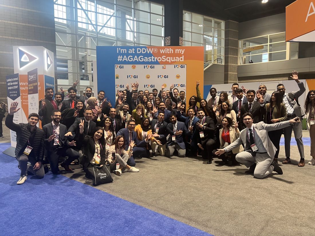
Let’s Mingle at DDW
We are looking forward to seeing you in our hometown for Digestive Disease Week® (DDW) 2024!
As you plan your schedule, here’s a listing of AGA’s free networking events. For more details and featured programming, visit www.gastro.org/DDW.
Meetups at AGA Central (L Street Bridge)
Network with like-minded attendees, build your #AGAGastroSquad and enjoy refreshments at our meetups.
Saturday, May 18
- 3 p.m.: Advocacy champions meetup – A “thank you” for everyone who supported our grassroots advocacy efforts this year!
Sunday, May 19
- 11 a.m.: NPPA meetup
- 1 p.m.: Dietitian meetup
- 3 p.m.: IBD meetup – Happy World IBD Day!
Monday, May 20
- 11 a.m.: Trainee meetup – Mingle with AGA journal editors!
- 1 p.m.: Psychologists meetup
- 3 p.m.: Clinician meetup
Tuesday, May 21
- 11 a.m.: Innovator meetup
RSVP and add to your calendar: www.signupgenius.com/go/10C0E4EA4AE2DA2F5C43-48529281-agacentral#/
Additional events for trainees
We have more opportunities for you to network at DDW! The following events all take place on Sunday, May 19.
- 10 a.m.: Live recording: Small Talk, Big Topics – Mingle with fellow trainees and early career GIs during a live recording of AGA’s podcast. Our hosts will interview fellowship program director Dr. Janice Jou.
[Location: AGA Central (L Street Bridge)]
- 1 p.m.: Meet-the-Experts: AGA Leadership – Held in the DDW Trainee and Early Career Lounge, these sessions are an opportunity for early career attendees to get tips from those further along in their career.
[Location: DDW Trainee and Early Career Lounge]
- 2 p.m.: AGA/DHPA Networking Hour – Join us for an hour of guided networking and 4-way Jeopardy!
[Location: DDW Trainee and Early Career Lounge]
We Have a New Congressional Champion in the Fight Against CRC!
Rep. Yadira Caraveo, MD (D-CO), recently introduced the Colorectal Cancer Early Detection Act along with Reps. Donald Payne Jr. (D-NJ), Haley Stevens (D-MI), and Terri Sewell (D-AL).
The Colorectal Cancer Early Detection Act would award grants to states to promote colorectal cancer prevention and early detection efforts to individuals under age 45.
Grants would be used to:
- Screen increased risk and high-risk individuals under age 45 for colorectal cancer.
- Provide appropriate referrals for medical treatment.
- Develop and carry out a public education and awareness campaign for the detection and control of CRC.
- Improve the education and training of health providers in detecting and controlling CRC.
- Establish mechanisms through which states can monitor the quality of CRC screening procedures.
- Develop strategies to assess family history and genetic predispositions to CRC.
- Design patient and clinician decision support tools for CRC.
- Conduct surveillance to determine other risk factors for CRC in this population.
“Colorectal cancer is the second-leading cause of cancer death in the US and is increasing at an alarming rate in younger people. AGA celebrates Rep. Caraveo’s work to address this trend through education and awareness” said Barbara Jung, MD, AGA President.
We look forward to working with our congressional champions to increase screening rates and reverse the trend of early onset colorectal cancer!

