User login
Updates Presented on Robotic Vena Cava Surgery
Developments in robotic vena cava surgery will be presented Friday afternoon during the session entitled “Endovascular and Open Solutions for Inferior Vena Cava Tumors and Occusions, Vena Cava Filtration Strategies, Pitfalls, and Complications and More About Iliac Vein Stenting.”
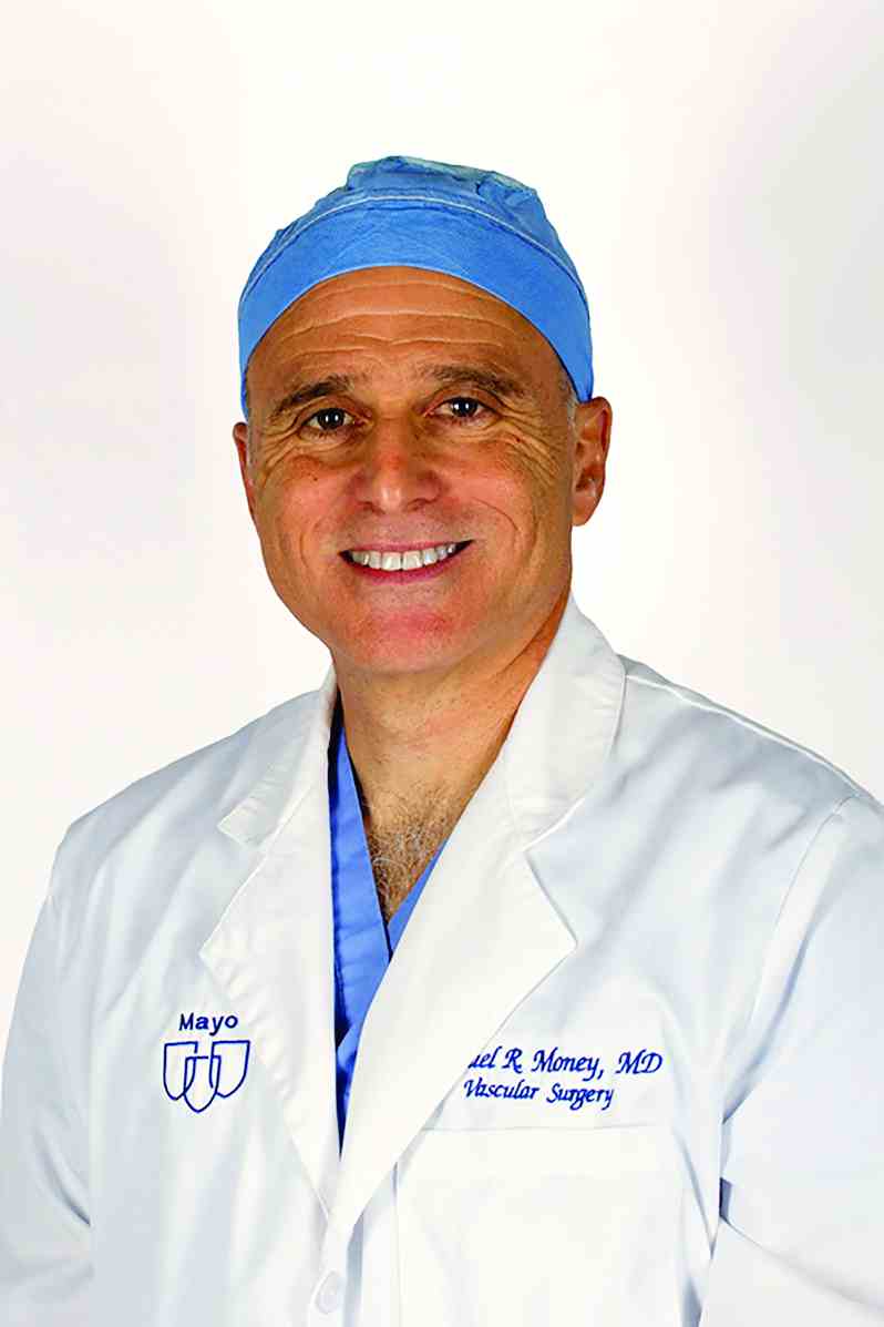
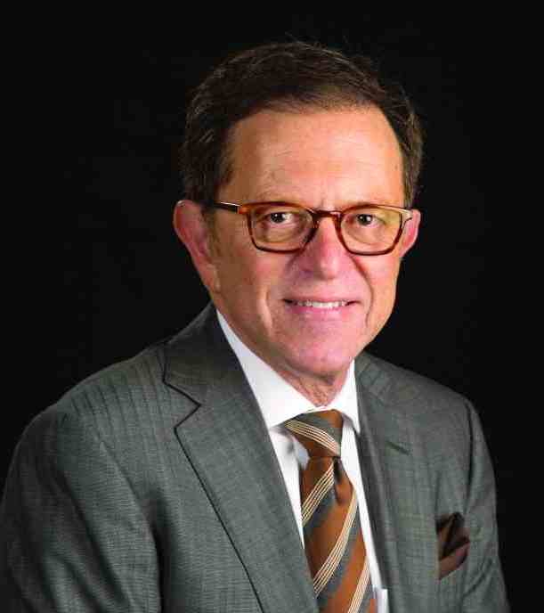
“The selective use of robotic surgery on the vena cava is a tremendous step forward because of its minimal invasiveness and also because of the magnification that the robot gives and the technical advantages the robot gives over open surgery or laparoscopic surgery,” said Dr. Money.
Traditionally, inferior vena cava thrombectomy is done using a large open incision, since it is difficult otherwise to gain access to the vein. The surgery is done most often to address involvement of the inferior vena cava by an adjacent tumor. In addition, surgery may be needed to remove filters inserted to prevent migration of pulmonary emboli when problems develop with the filter.
The open incision approach carries a considerable risk of morbidity with the approach. A minimally invasive treatment option has been needed. Minimally invasive laparoscopy is an option. But drawbacks include the technically challenging nature of the procedure which makes for a steep learning curve.
Within the past decade, the use of robotic surgery has been utilized for interior vena cava thrombectomy. The approach involves multiple but smaller incisions to allow access of robotic tools. The robotic approach provides better visualization with magnification, three-dimensional vision, tremor filtration, and seven degrees of freedom, among other advantages.
“The overall take-home message from my presentation is that robotic vena caval surgery can be done and it should be used. Overall, robotic surgery for the vena cava is a field that is developing. It requires collaboration with other specialties and a realization that minimally invasive procedures are not just endovascular procedures but may be laparoscopic or robotic in nature,” said Dr. Money.
Dr. Money will describe the outcomes of a prospective study undertaken at the Scottsdale Mayo Clinic that evaluated the efficacy of a vena cava surgical system for radical nephrectomy and inferior vena cava tumor thrombectomy and, in a few patients, inferior vena cava filter removal. More recently, the robotic surgery was used to treat compression of the left renal vein between the superior mesenteric artery and aorta.
A focus of the presentation will be the key importance of the collaboration between vascular and robotic surgeons. “As time has developed I have become more aware and more adept at what the robot can do. However, it is not a routine part of my practice. By collaborating with others who use it more frequently, we have developed a practice where vena caval surgery can be done in a minimally invasive fashion with patients going home following a shorter length of stay, with less significant blood loss, and overall an easier recovery,” said Dr. Money.
Session 91:Endovascular and Open Solutions for Inferior Vena Cava Tumors and Occusions, Vena Cava Filtration Strategies, Pitfalls, and Complications and More About Iliac Vein Stenting
Robotic Vena Cava Surgery
Friday, 3:48 p.m. – 3:53 p.m.
Trianon Ballroom, 3rd Floor
Don’t Miss Out On. . .
The Challenging World of the Diagnosis and Treatment of Vascular Malformations: An Orphan Disease that Has Now Come of Age
(Sessions 92-98; Friday, 6:45 a.m. to 1:30 p.m.)
Location: Gramercy Suites East and West, 2nd Floor
Vascular malformations constitute one of the most challenging clinical entities encountered in Vascular Medicine today. They can occur in every anatomy in the body insinuating themselves throughout an organ or tissue. Being totally comprised of vascular structures, any surgical attempt at resection is fraught with potential hemorrhagic complications. Compounding their inherent difficulty in management , they are also very rare entities. The clinical presentations are extremely protean and can range from an asymptomatic birthmark, to fulminant, life-threatening CHF. Attributing any of these extremely varied symptoms that a patient may present with to a vascular malformation can be challenging to the most experienced clinician. Patients typically bounce from clinician to clinician experiencing disappointing outcomes, complications, and recurrence or worsening of their presenting symptoms. Due to their extreme rarity (>1% of the population), it is difficult for clinicians to gain any experience at all in their diagnosis and optimal management and make definitive statements.
The purpose of these Vascular Malformation Sessions is to offer the attendee the current state-of-the-art multi-disciplinary endovascular and surgical approaches to accurately diagnose and optimally treat all types of vascular malformations (AVMs, AVFs, venous malformations, lymphatic malformations, capillary-venous malformations, and mixed lesions) in all the various problematic anatomies in which they occur. Being that there are controversies regarding the various treatment strategies regarding vascular malformations, the attendees will be exposed to those multiple approaches and philosophies regarding the science, the multiple management strategies, the results, the long-term outcomes, and the complications inherent in the various palliative and curative treatments proffered by international experts.
Improving Outcomes in Hemodialysis Access
(Sessions 106-110; Saturday; 7:55 a.m. – 4:00 p.m.) Registration Begins At 6:00 a.m.
Location: Grand Ballroom West, 3rd Floor
The Hemodialysis Access sessions on Saturday (Sessions 106 – 110) will include presentations on planning, optimizing outcomes, political, economic and legal issues, new technologies and an update on clinical issues related to hemodialysis access. Experts will address specific topics including vein preservation and planning for access, use of ultrasound to facilitate cannulation, use of simulators, the role of drug eluting balloons and stents, management of complications including steal syndrome, infections and access hemorrhage, coding for access procedures, pharmacologic and mechanical approaches to improve fistula outcomes, treatment of fistula aneurysms, use of the HeRO graft and many more important topics. The keynote speaker will be Dr. Michael Brescia who will give his unique historical perspective on the development of the AV fistula. This session should be of interest to surgeons, nephrologists, interventionalists, nurses, dialysis technicians and others interested in the care of patients with end stage renal disease.
The Robots Are Coming to Vascular Surgery
Emerging technologies in robotics systems will be the subject of a key session at the 2016 Veithsymposium, entitled “Vascular Robotics and Guidance Systems” and co-chaired by Dr. Kenneth Ouriel, founder of Syntactx, and Dr. Anton N. Sidawy of George Washington University.
According to Dr. Sidawy, the major focus of the session will be the Magellan System, which is a steerable catheter system controlled by a remote physician console. The system has two major components: a robotic arm and the aforementioned remote console. This design is meant to keep the operator away from the table, thereby decreasing exposure to the radiation source.
The promise of such technology is becoming increasingly acknowledged in the field of vascular surgery. The Magellan can be used in a number of endovascular procedures, including – but not necessarily limited to – embolization, endovascular aneurysm repair (EVAR), angioplasty, stenting, crossing total occlusions, and venous procedures. Such applications will be highlighted in-depth during this session, with presentations by some of the most prominent names in the field of vascular robotic surgery.
The session will conclude with a panel discussion involving all the speakers and which will cover the current state and future of robotics in vascular surgery.
Thursday, November 17, 2016
Session 53: VASCULAR ROBOTICS AND GUIDANCE SYSTEMS
Moderators: Kenneth Ouriel, MD, MBA / Anton N. Sidawy, MD, MPH
3:52 PM - 4:52 PM
Lower Extremity Occlusive Disease and Alternative Graft Options
Vascular surgeons spend much of their clinical time taking care of patients with lower extremity occlusive disease, so the “New Devices and Techniques for Treating Lower Extremity Occlusive Disease; Prosthetic Grafts and Heparin Bonding” session on Thursday afternoon will be particularly useful for the practicing surgeon, according to Dr. Russell Samson who will co-moderate the session along with Dr. Ali F. AbuRahma of West Virginia University.
Two recent advances with respect to such alternative graft options, which will be addressed during the session, are heparin-bonded ePTFE and spiral laminar flow grafts.
“Whether these two modalities improve patency rates compared to standard ePTFE grafts remains controversial,” Dr. Samson said.
Heparin-bonded ePTFE will be the topic of a debate presented by Dr. Richard Neville of George Washington University Hospital, and Dr. Jonathan Beard of Sheffield Vascular Institute, in the United Kingdom. These two experts will conduct a debate. Dr. Neville will discuss long-term patency data suggesting that heparin bonding is valuable in PTFE Fempop Bypass Grafts, and Dr. Beard will argue that it does not improve results.
Additionally, Dr. Yann Gouëffic of the University of Nantes, France, will address the cost-efficiency of heparin bonding.
“In an age where surgeons are becoming more cost conscious, new graft configurations, which are often more costly, need to offer significant improvement in long-term outcomes,” Dr. Samson said.
As for spiral laminar flow grafts, Dr. Hosam F. El Sayed of Ohio State University will provide insight into a specific format of PTFE that creates a spiral laminar flow that more accurately represents flow within the arterial system, Dr. Samson noted.
Therefore, Dr. Enrico Ascher of Lutheran Medical Center, who is highly regarded for his experience in performing distal tibial bypass, will provide insight into how to deal with “pipe-like” calcified arteries that some surgeons may consider inoperable, Dr. Samson added.
Further, since many surgeons may prefer an endovascular procedure as a first-line alternative to bypass, this session will also include a talk by Dr. Hiroyoshi Yokoi of Fukuoka Sanno Hospital in Japan on advanced guidewire techniques to open these distal vessels, and another by noted inventor, Dr. Timothy A.M. Chuter of the University of California, San Francisco, who will discuss advantages and early clinical experience with a novel balloon that does not straighten when it inflates.
Another talk during the session, which will be given by Dr. Joseph L. Mills of Baylor College of Medicine, will focus on the impact of foot infections on leg bypass outcomes in CLI patients and what can be done to offset that impact.
“Dr. Mills will remind us that no matter what treatment we choose, the final result can be spoiled by an infection, so managing foot infections in patients requiring surgical bypass becomes a preeminent consideration,” Dr. Samson said.
“The practicing vascular surgeon deals with challenging infrainguinal atherosclerotic conditions on a daily basis. Therefore, information gained from attending this program should enhance their ability to offer long-lasting therapies,” he added.
Session 59
“New Devices and Techniques for Treating Lower Extremity Occlusive Disease; Prosthetic Grafts and Heparin Bonding”
Thursday, 1:00 p.m. – 1:54 p.m.
Grand Ballroom West, 3rd Floor
Highlight on Innovative Devices for Embolectomy, Clot Retrieval, and Embolization
With the development of ever-more-sophisticated endovascular intervention systems, limb ischemia need no longer be an automatic sentence of lifelong pain or amputation.
But the burgeoning supply of these specialized devices brings its own challenge to the area of endovascular intervention. Which device is best for which patient? And which have strong data to back up their claims of effectiveness? Thursday’s session, “New Devices for Embolectomy, Clot Removal, and Embolization,” will offer some guidance.
“The presentations represent significant further advances in methods and technology that can treat patients with severe, limb-threatening, and life-altering vascular diseases,” said Dr. McNamara. “Also, comparisons are made between competing new technologies. This will be very helpful in deciding which method and/or technology to use. And it will, I think, be very helpful in deciding whether to use interventional or open surgical treatments.“These presentations will not only guide the vascular specialist, but help to substantiate that the interventional techniques continue to be more and more effective. This information could, and should, be shared with primary care physicians.”
This point can’t be underestimated, Dr. McNamara noted. Primary care physicians are on the front line of venous disorders, and should know that there are good treatment options for many patients – treatments than can save limbs and vastly improve pain and function.
“An increasing percentage of patients should be referred for consultation and possible treatments for arterial and venous disease. This will result in decreased morbidity and improved quality of life for an increasing number of patients.”
Surgeons should expect to see more and more such patients as the U.S. population continues to age – and effective treatment not only benefits each individual, but society as a whole.
“The disease significantly decreases the quality of life, and reduces the independence of our aging population. This significantly increases the costs, and increases the burden to society and families of the growing percentage of our population in their ‘Golden Years.’”
The session opens with a lecture by Dr. Ali Amin, a vascular surgeon from West Reading, Pa. He will discuss the role of mechanical thrombectomy and thrombolysis in acute limb ischemia, and when open surgery rather than endovascular intervention is indicated
Dr. Jos C. van den Berg of the University of Bern, Switzerland, will then tackle how to utilize suction techniques and devices to deal with the embolic complications of peripheral vascular interventions.
Rotarex and Aspirex, two mechanical debulking devices created by Straub Med, will be the topic of a presentation by Dr. Michael K.W. Lichtenberg chief of the Angiology Clinic and Venous Center at Klinikum Arnsberg, Germany.
There will be three presentations on thrombectomy with the Indigo System by Penumbra. Indigo provides mechanical clot engagement and vacuum extraction and can be used in both arteries and veins.
Dr. James F. Benenati, of Florida International University Herbert Wertheim College of Medicine, and Dr. Richard R. Saxon of San Diego Imaging, will discuss the PRISM Registry data. The registry recently reported that mechanical aspiration with the Indigo device is a safe and effective option for the treatment of peripheral or visceral arterial occlusions either as a frontline therapy or in patients who failed thrombolysis.
Dr. Frank R. Arko of Carolinas Healthcare System, will share his thoughts on why the Indigo system is an excellent choice for clot removal, and how it decreases the need for lytic therapy.
Dr. George L. Adams, MD, Director of Cardiovascular and Peripheral Vascular Research will make the case for why the Indigo system could be a “game-changer” in the treatment of acute limb ischemia and visceral artery thomboses and emboli.
Dr. Andrej Schmidt of the University Clinic of Leipzig, Germany, will discuss the advantages and limitations of several other Penumbra products, including the Ruby Coil for peripheral embolization, and the Penumbra Occlusion Device (POD). He will also touch on the Lantern microcatheter, Penumbra’s newest product, launched in April. The low-profile, high-flow microcatheter can pass through the distal vasculature, but can also be used for high-flow contrast injections.
Finally, Dr. Aaron Fischman of Mount Sinai Medical Center, will speak on the challenge of endovascular rescue procedures for acute visceral ischemia from thromboembolism.
Session 62
“New Devices for Embolectomy, Clot Removal, and Embolization”
4:32 p.m. – 5:30 p.m.
Grand Ballroom West, 3rd Floor
Study of Bioengineered Vessels for Dialysis Vascular Access Promising
The idea of using bioengineered human acellular vessels for dialysis vascular access used to be a pie-in-the-sky notion, but research presented today will demonstrate just how close they are to becoming a realistic option.
Dr. Jeffrey H. Lawson will present results from two Phase II single-arm trials of bioengineered human acellular vessels for dialysis access in 60 patients with end-stage renal disease conducted in six hospitals: three in the United States and three in Poland. His presentation is entitled “Tissue Engineered Blood Vessels for Arterial Bypass and Dialysis Access: Midterm Results n Patients.” To date, the early clinical experience with these vessels “suggests that they are safe to implant as a dialysis access vessel and lower leg bypass graft,” said Dr. Lawson, chief medical officer at Humacyte and professor of surgery and pathology in the department of surgery at Duke University, Durham, N.C. “They enjoy enduring patency, tolerate needed interventions, appear durable and have a low risk of infection.”
The researchers observed that during 82 patient-years of follow-up, only one vessel became infected. The vessels had no dilatation and rarely had postcannulation bleeding. At 6 months, 63% of patients had primary patency, 73% had primary assisted patency, and 97% had secondary patency, with most loss of primary patency because of thrombosis. At 1 year, 28% had primary patency, 38% had primary assisted patency, and 89% had secondary patency. “Following implantation into patients, the human acellular vessel (HAV) appears to completely repopulate with the host’s (recipient’s) own vascular tissue,” Dr. Lawson said. “The vessel is filled with cells that look like vascular smooth muscle cells and the implanted vessel is lined with endothelial cells from the host, suggesting that the manufactured acellular vessel becomes repopulated with the hosts own cells making it part of their own body. I will also be discussing some unpublished work that we have done for lower leg arterial bypass work in 20 patients to date.”
Dr. Lawson acknowledged that the Phase II experience at a few clinical sites is a limitation of the current analysis. “These findings need to be confirmed and validated in a large Phase III clinical trial, which is now underway,” he said.
Session 60
“New Developments in Arterial Grafts; Stents and Stent-Grafts; Concepts and Techniques to Improve Their Use and Results”
Thursday, 1:54 p.m.– 3:24 p.m.
Grand Ballroom East, 3rd Floor
ASVAL Can Save Refluxing Saphenous Veins
Ambulatory selective varicose vein ablation under local anesthesia (ASVAL) can abolish reflux while preserving the great saphenous vein, and benefits can last at least a decade, said Dr. Silvain Chastanet, who helped create the technique and will discuss it Thursday morning.
Historically, a refluxing great saphenous vein was treated by removing it. The ASVAL method preserves the vein by using microphlebotomy to instead remove the varicose reservoir, said Dr. Chastanet of the Riviera Vein Institute in Monaco. “Because each patient is different, this strategy offers tailored, à la carte treatment, as opposed to systematic ablation,” he said. “Doing precise and thorough microphlebectomies is not as simple as one might think, but we will share tips and tricks for performing saphenous sparing venous surgery in an easy and elegant way.”
The ASVAL technique is based on the premise that reflux usually ascends from the tributaries toward the saphenous trunk, rather than following the opposite path, said Dr. Chastanet. Although the ascending pattern is not universal, increasing evidence suggests that it is most common. For example, a study of more than 700 consecutive patients with chronic venous disease showed that patients of CEAP category C4-C6 had significantly greater involvement of the saphenofemoral junction and saphenopopliteal trunk than patients with less advanced venous disease. The authors concluded that reflux usually ascends up from the tributaries and accessory saphenous veins (J Vasc Surg. 2010;51(1):96-103).
Thus, when evaluating a patient with saphenous reflux, surgeons should always ask whether they can spare the great saphenous vein, Dr. Chastanet said. Evidence indicates that if the patient is classified as C2 (varicose veins) and the great saphenous vein is dilated less than 10 mm, ASVAL may be a valid option, especially if reflux is localized above the knee, with the varices localized to the thigh.
Techniques such as ASVAL are a major advance, but questions persist about surgically treating varicose disease, Dr. Chastanet noted. For example, although ascending disease appears to be most common, it can convert to descending disease or patients can have a refluxing saphenous trunk without tributaries. “We still do not know which patients are likely to have descending or ascending disease, and why,” Dr. Chastanet said. Likewise, experts continue to debate whether phlebectomy should be performed together with saphenous ablation. “Considering that some varicosities will disappear after endothermal ablation of the saphenous vein, some surgeons consider concomitant phlebectomy to be overtreatment,” Dr. Chastenet noted. “Conversely, if we consider that the disease begins at the tributary level according to the ascending theory, it seems obvious to focus treatment on the varicose tributaries, which are the main concern for the majority of patients.”
Bringing techniques such as ASVAL into community practice involves practical challenges, too. Microphlebectomy is “clearly” the best cosmetic option, but is not taught during surgical or phlebology training, Dr. Chastanet said. “There is a need to develop new tools for treating the varicose tributaries in an easier and faster way.” Such advances could help transition procedures such as ASVAL to outpatient settings, which is not now the norm in most countries, he noted. “Using local anesthesia, mini-invasive surgical procedures with immediate ambulation will enable us to reach this goal.”
Session 63
“Venous Clinical Examination and Hemodynamics”
Thursday 8:13 a.m. – 8:18 a.m.
Trianon Ballroom, 3rd Floor
Treating Lower Arterial Occlusive Disease
How to proceed when a patient needs a bypass that can’t be done using the saphenous vein, and how to measure arterial flow to the foot are two novel areas that help improve a revascularization algorithm for lower arterial occlusive disease, according to Dr. Kenneth Ouriel, co-moderator of the Wednesday session, “Value of Deep Vein Grafts; New Concepts in Assessing Foot Perfusion and Improving It; More About the Angiosome Controversy; Some Ongoing CLI Trials.”
“A lot of patients don’t have a suitable bypass conduit to bring blood to the lower limb. But when the saphenous vein isn’t available, and you’re concerned an artificial graft won’t be as effective, there is another alternative,” said Dr. Ouriel, president and CEO of the New York–based clinical research firm, Syntactx.
Dr. Ouriel said “it’s also a vein that is not that long, so if you need a long vein for a bypass, from the top of the leg to the ankle, for example, it might not be long enough.”
Clinicians who attend this session also will be able to incorporate novel ways of quantifying perfusion to the foot, according to Dr. Ouriel. “The angiosome isn’t controversial so much as just new. Most of us didn’t learn about it in medical school, but we will explore it in depth during this session.”
The general principle of the angiosome is that arterial flow to the foot is “compartmentalized” into regions supplied by discrete vessels. This leads to seeing perfusion not only as a whole, but also in terms of specific regions, said Dr. Ouriel.
By focusing on the revascularization of the source artery to a specific angiosome, data indicate there are better rates of wound healing and limb salvage, according to Dr. Ouriel.
To round out the session are results and questions posed by a series of “exciting” trials both completed and still recruiting, including the LIBERTY, SPINACH, and BEST trials. The discussion of these trial findings will add nuance to the overall discussion about whether surgical reconstruction or peripheral intervention in patients with critical limb ischemia is appropriate and when to do it.
Session 30:
Value of Deep Vein Grafts; New Concepts in Assessing Foot Perfusion and Improving It; More About the Angiosome Controversy; Some Ongoing CLI Trials
Wednesday, 4:22 p.m. – 5:58 p.m.
Grand Ballroom East, 3rd Floor
Innovations in treating AAA: Innovative Endovascular Approaches
Aortic aneurysms involving aortic branches pose significant challenges for open and endovascular repair. Tuesday morning’s session, “New Developments with Juxta and Pararenal Aortic Aneurysms and Thoracoabdominal Aneurysms,” will be a discussion of surgical experiences with the first fenestrated graft approved in the United States and a review several other approaches to treatment.
The session will provide “an important update about the most recent advances in this field,” Dr. Cao added. “It will be focused on the most recent adjuncts to be used not only in patients with a failed EVAR but also in preventing the failure as well as the indications for using endovascular versus an open approach.”
The session kicks off with presentations on the current status of ZFEN by Dr. Andres Schanzer of UMass Memorial Medical Center and on circumstances in when superior mesenteric artery (SMA) scallops can cause problems during F/EVAR with ZFEN by Dr. Carlos Timaran of the University of Texas Southwestern Medical School.
“The topics covered in this session will aid vascular surgeons in deciding the best approach to treat such patients, by emphasizing the strengths, weaknesses, and limitations of the various options,” he said. “Tips and tricks to address specific circumstances can be invaluable in optimizing patient outcomes.”
The last portion of the session will cover F/EVAR for failed EVAR, including when F/EVAR is indicated, adjuncts to improve outcomes of treatment for challenging AAAs, and branched EVAR (B/EVAR). The session will wrap with a presentation by Dr. Piotr Szopinski of the Medical University of Warsaw on early clinical results from the COLT endograft system for treatment of thoracoabdominal aortic aneurysms (TAAAs).
“Attendees will have the opportunity to compare the different treatment options,” Dr. Torsello said. “We look forward to an open discussion that will lead to practical guidelines for decision making. The audience can develop their own treatment algorithms for patients with hostile or absent aortic aneurysm neck.”
“We currently have multiple modalities to prevent and treat complications,” Dr. Cao said. “Vascular surgeons should be familiar with different techniques and should tailor their approach to each patient according to the morphology of the native aorta and access vessels, as well as the clinical conditions and risk factors for open surgery.”
Session 12:
New Developments with Juxta and Pararenal Aortic Aneurysms and Thoracoabdominal Aneurysms
Tuesday, 10:20 a.m. – 12:00 p.m.
Grand Ballroom West, 3 rd Floor
Session Tackles Merits and Shortcomings of Drug Coated Balloons
Most vascular surgeons have a preferred method for managing femoropopliteal lesions, but drug-coated balloons are emerging as a option for lower extremity lesions.
The Wednesday morning session, “More on Lower Extremity Occlusive Disease: New Developments in Drug Coated Balloons (DCBs); Dealing with Complex Lesions and Trials,” will feature opposing views on how to manage the most severe disease morphology.
In this important session, world renown experts also will discuss the current status and immediate future of drug-coated balloons (DCB) for use on the lower extremity.
“The treatment with DCB is a story of great success. In general, the patency rate is increased and this is with no side effects due to the coating,” said Dr. Gunnar Tepe of Rosenheim Hospital in Rosenheim, Germany. “In the near future treatment with uncoated balloons will be mainly replaced by DCBs.” Dr. Tepe will present a paper on the current status and future prospects of DCBs.
Understanding important technical aspects remains essential to successful outcomes with DCBs, according to Dr. Tepe. “We have to know that treatment with DCBs is not a stand-alone therapy which can be successful in all lesions,” said Dr. Tepe. “If DCBs fail to obtain long-term patency, it is either due to recoil or drug uptake. For recoil, the combination of DCBs and stents is perfect, but for better drug uptake vessel, preparation is needed.”
Dr. Tepe and others will offer a detailed analysis of the potential shortcomings of DCBs, such as in long lesions, and in calcified arteries, and how to overcome them.
“The future of DCBs for use in patients with the potential threat of amputation is under development internationally,” said Dr. Schneider. “Practitioners will leave the session fully updated on the latest data and analysis of the value of DCBs in daily practice, since the most current results from randomized trials, and large registry datasets from the United States and around the world will be presented side by side.”
The presentations include the 3-year results from the international, randomized, controlled IN.PACT SFA Trial that compared the IN.PACT DCB against standard angioplasty. On hand to analyze and discuss the results will be co-principal investigators Dr. Schneider, Dr. Tepe, as well as panelist Dr. John R. Laird of the Vascular Center at the University of California, Davis. Co-moderator and study investigator Dr. Dierk Scheinert, MD, of the Leipzig University Hospital in Germany, will also join the discussion.
“I believe the data will show that DCBs have significantly changed how we practice and that by using them when possible, we can substantially improve what we can offer to patients in terms of long-term durability of the reconstructions we perform for their blocked and damaged arteries,” Dr. Tepe said.
The session will also cover second- and third-generation DCBs that are currently under development.
“As has been the tradition for many years, the VEITH planning committee has been able to bring together the world experts with the most important experience in these topics to educate, debate, inform the broader community,” said Dr. Schneider.
Session 26:
More on Lower Extremity Occlusive Disease: New Developments in Drug Coated Balloons (DCBs); Dealing with Complex Lesions and Trials
Wednesday 10:13 p.m. –12:00 p.m.
Grand Ballroom East, 3rd Floor
