User login
Painful rash on face
A Good-quality patient-oriented evidence
B Inconsistent or limited-quality patient-oriented evidence
C Consensus, usual practice, opinion, disease-oriented evidence, case series
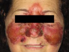
A 68-year-old woman came into the clinic for treatment of a painful, erythematous skin rash over the bridge of her nose. She’d had the rash for 4 days, and it was spreading to the malar area and up around her eyelids and forehead (FIGURE 1). The patient said she felt “out of sorts,” was nauseous, and had a low fever. She said she’d recently had a sore throat. Her past medical history included hypertension.
The patient had elevated and indurated shiny skin plaques involving the nose, cheeks, eyelids, and forehead. She also had some blisters and crusty lesions. On palpation, the skin was hot and tender. The pharynx was unremarkable and the neck supple, with no palpable nodes.
FIGURE 1
Spreading rash
The 68-year-old patient said that she’d recently had a sore throat. She indicated that she felt nauseous and out of sorts.
WHAT IS YOUR DIAGNOSIS?
HOW WOULD YOU TREAT THIS PATIENT?
Diagnosis: Erysipelas
Erysipelas is an acute superficial cellulitis with lymphatic involvement. It is characterized by the abrupt onset of a warm, erythematous rash with a sharply demarcated, indurated, elevated margin. There are no suppurative foci; sometimes, however, there are bullae or vesicles.
In facial “butterfly” erysipelas (which this patient had), the plaques may involve the eyelids, cheeks, nose, and forehead. Upon palpation, the skin is hot and tender. As the process develops, the color becomes a dark, fiery red and vesicles appear at the advancing border and over the surface. Associated regional lymphadenopathy may be present. There is no necrosis.1
Most cases are caused by the Streptococcus species—usually group A (Streptococcus pyogenes)—and groups B, C, and G. (Erysipelas is occasionally caused by Staphylococcus aureus.) After prodromal symptoms that last for 4 to 48 hours and include malaise, chills, fever, anorexia, and vomiting, more red, tender firm spots develop.
Predisposing conditions for erysipelas include disruption to the skin barrier as a result of trauma, lymph stasis (prior radiation, mastectomy, saphenous vein harvest, lymphadenectomy), injection drug use, ulcers, wounds, dermatophytic infections, and edema. Toe intertrigo is perhaps the most common site for pathogen entry. The source of infection in facial erysipelas, however, is often the nasopharynx.2,3
Differential diagnosis includes cellulitis, rosacea
The differential diagnosis for erysipelas include cellulitis, rosacea, lupus erythematous “butterfly rash,” and seborrheic dermatitis.
Cellulitis is associated with skin erythema, edema, and warmth in the absence of underlying suppurative foci. Lymphangitis and inflammation of regional lymph nodes may occur. Vesicles, bullae, and ecchymoses (or petechiae) may also be present.4
Rosacea is a disorder that occurs in middle-aged and older adults, and is more common in women.5 It is characterized by symmetrical vascular dilation on the central face, including the nose, cheek, eyelids, and forehead. Facial erythema and telangiectasias—typically on the cheeks—are also present, as well as late papules and pustules.6 There are no comedones, which you would see with acne vulgaris.6 Rosacea is made worse by heat or sunlight, emotional stressors, and drinking alcohol or eating spicy foods.
Lupus erythematous “butterfly rash” is characterized by a macular confluent erythema over the cheeks and bridge of the nose with fine scaling, erosions, and crusts. It appears in approximately half of patients with systemic lupus erythematosus, usually after sun exposure.7,8
Seborrheic dermatitis is characterized by intermittent, active phases that manifest with burning, scaling, excess oil secretion, and itching. Seborrheic dermatitis usually appears over areas that are rich in sebaceous glands, such as the lateral sides of the nose and the nasolabial folds, eyebrows, glabella, and scalp.9
The exam holds the key to diagnosis
The diagnosis of erysipelas is based on clinical manifestations. Blood cultures, needle aspirations, and punch biopsies are not usually helpful. Blood cultures are positive in less than 5% of cases.4 Punch biopsy is positive in 20% to 30% of cases.4
Needle aspiration and skin biopsies may be considered for a patient with a severe infection that is not responding to treatment, or for a patient who has diabetes, a malignancy, or unusual predisposing associated factors, such as an immersion injury, an animal bite, neutropenia, or immunodeficiency.
Treatment centers on penicillin
With early diagnosis and proper treatment, the prognosis for erysipelas is excellent. Empiric antimicrobial therapy should include activity against beta-hemolytic Streptococcus. Penicillin is the treatment of choice4,10 (strength of recommendation [SOR]: A). Streptococcus strains are susceptible to penicillin and 99.5% are susceptible to clindamycin. A 7% macrolide resistance has been reported in the United States.4
Oral therapy is recommended for mild infections (or for those who have improved after intravenous [IV] antibiotics). Treat with penicillin V 500 mg PO 4 times daily or amoxicillin 500 mg PO 3 times daily. Other choices include clindamycin 300 mg PO for 7 to 10 days; azithromycin 500 mg PO for 1 day, followed by 250 mg PO daily for 4 days; or clarithromycin 250 mg PO twice a day for 7 to 10 days4,10 (SOR: A).
Consider hospitalization for severely ill patients, for those who are unable to tolerate oral medications, and for those who require parenteral antibiotic therapy for systemic symptoms and rapidly progressing erythema4 (SOR: A). Regimens include penicillin G 2 million to 4 million units IV every 4 to 6 hours; cefazolin 0.5 to 1 g IV every 8 hours; cefotaxime 1 to 2 g IV every 8 hours; and ceftriaxone 1 to 2 g IV every 24 hours.
If staphylococcal infection is suspected, a penicillinase-resistant semisynthetic penicillin or a first-generation cephalosporin, such as cefazolin, can be selected. For oral therapy, cephalexin 500 mg 4 times daily can be used4,11 (SOR: A). Macrolides (erythromycin 250 mg PO every 6 hours) are also an option4 (SOR: A). For presumed methicillin-resistant Staphylococcus aureus (MRSA), you can prescribe clindamycin 600 mg IV every 8 hours; vancomycin 15 mg/kg IV every 12 hours; or linezolid 600 mg every 12 hours. The total duration of antibiotic therapy may be extended to up to 14 days, and tailored to clinical improvement.4,10
Steroids can slightly reduce healing time and antibiotic duration in patients with erysipelas. You can prescribe prednisone 30 mg PO and taper over 8 days.4,12
Be sure to treat underlying predisposing conditions such as tinea pedis, venous eczema, and trauma sites. Encourage supportive measures, such as elevating the affected extremity, as well.
Improvement in a few days. Patients with erysipelas typically show improvement in 24 to 48 hours of beginning antibiotic therapy. Don’t be too quick to consider antibiotic failure in patients with worsening erythema after initiating antimicrobial therapy; this may be related to the destruction of pathogens that release enzymes (including deoxyribonuclease B [DNase B] and hyaluronidase) that increase local inflammation.
A good outcome for the patient
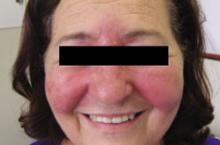
The patient was hospitalized and received cefazolin 1 g IV every 8 hours for 3 da ys. She was discharged on cephalexin 500 mg PO, every 6 hours for 7 days, with excellent results. FIGURE 2 shows the patient 4 days after she finished taking the cephalexin.
FIGURE 2
A few days, a big difference
The patient received cefazolin 1 g IV every 8 hours for 3 days, followed by oral cephalexin 500 mg every 6 hours for 7 days. This photo shows the patient 4 days after she finished taking the cephalexin.
CORRESPONDENCE Felix B. Chang, MD, Leominster Community Health Connections Family Health Center, 14 Manning Ave, Suite 402, Leominster, MA 01453; [email protected].
1. Chang F, Lopes A. Erysipelas. In: Domino FJ, ed. The 5-Minute Clinical Consult 2008. 16th ed. Philadelphia, Pa: Lippincott Williams & Wilkins; 2008:448–449.
2. Jorup-Ronstrom C. Epidemiological, bacteriological and complicating features of erysipelas. Scand J Infect Dis. 1986;18:519-524.
3. Davis L, Cole J. Erysipelas. Dermatology-bacterial infections. Available at: http://www.imedicine.com/printtopic.asp?bookid=2&topic=129. Accessed at January 11, 2010.
4. Stevens DL, Bisno AL, Chambers HF. Infectious Diseases Society of America (ISDA) practice guidelines for diagnosis and management of skin and soft tissue infections. Clin Infect Dis. 2005;41:1373-1406.
5. Wilking J, Dahl M, Detmar M, et al. Standard classification of rosacea: report of the National Rosacea Society Expert Committee on the Classification and Staging of Rosacea. J Am Acad Dermatol. 2002;46:584-587.
6. Crawford GH, Pelle MT, James WD. Rosacea: I. Etiology, pathogenesis, and subtype classification. J Am Acad Dermatol. 2004;51:327-341.
7. Patel P, Werth V. Cutaneous lupus erythematosus: a review. Dermatol Clin. 2002;20:373-385.
8. Madhok R, Wu O. Systemic lupus erythematosus. Am Fam Physician. 2007;76:1351-1353.
9. Gupta AK, Bluhm R. Seborrheic dermatitis. J Eur Acad Dermatol Venereol. 2004;18:13-26.
10. Van Beneden CA, Facklam R, Lynfield R, et al. Erythromycin resistance among invasive group A streptococcal infections, United States, 1999-2001 [abstract 345]. In: Proceedings and Abstract of the 42nd Annual Meeting of the Infectious Diseases Society of America (Boston). Alexandria, Va: Infectious Diseases Society of America; 2004:102.
11. Stulberg DL, Penrod MA, Blatny RA. Common bacterial skin infections. Am Fam Physician. 2002;66:119-124.
12. Bergkvist PI, Sjobeck K. Relapse of erysipelas following treatment with prednisolone or placebo in addition to antibiotics: a 1-year follow up. Scand J Infect Dis. 1998;30:206-207.
CORRESPONDENCE Felix B. Chang, MD, Leominster Community Health Connections Family Health Center, 14 Manning Ave, Suite 402, Leominster, MA 01453; [email protected].
A Good-quality patient-oriented evidence
B Inconsistent or limited-quality patient-oriented evidence
C Consensus, usual practice, opinion, disease-oriented evidence, case series
A 68-year-old woman came into the clinic for treatment of a painful, erythematous skin rash over the bridge of her nose. She’d had the rash for 4 days, and it was spreading to the malar area and up around her eyelids and forehead (FIGURE 1). The patient said she felt “out of sorts,” was nauseous, and had a low fever. She said she’d recently had a sore throat. Her past medical history included hypertension.
The patient had elevated and indurated shiny skin plaques involving the nose, cheeks, eyelids, and forehead. She also had some blisters and crusty lesions. On palpation, the skin was hot and tender. The pharynx was unremarkable and the neck supple, with no palpable nodes.
FIGURE 1
Spreading rash
The 68-year-old patient said that she’d recently had a sore throat. She indicated that she felt nauseous and out of sorts.
WHAT IS YOUR DIAGNOSIS?
HOW WOULD YOU TREAT THIS PATIENT?
Diagnosis: Erysipelas
Erysipelas is an acute superficial cellulitis with lymphatic involvement. It is characterized by the abrupt onset of a warm, erythematous rash with a sharply demarcated, indurated, elevated margin. There are no suppurative foci; sometimes, however, there are bullae or vesicles.
In facial “butterfly” erysipelas (which this patient had), the plaques may involve the eyelids, cheeks, nose, and forehead. Upon palpation, the skin is hot and tender. As the process develops, the color becomes a dark, fiery red and vesicles appear at the advancing border and over the surface. Associated regional lymphadenopathy may be present. There is no necrosis.1
Most cases are caused by the Streptococcus species—usually group A (Streptococcus pyogenes)—and groups B, C, and G. (Erysipelas is occasionally caused by Staphylococcus aureus.) After prodromal symptoms that last for 4 to 48 hours and include malaise, chills, fever, anorexia, and vomiting, more red, tender firm spots develop.
Predisposing conditions for erysipelas include disruption to the skin barrier as a result of trauma, lymph stasis (prior radiation, mastectomy, saphenous vein harvest, lymphadenectomy), injection drug use, ulcers, wounds, dermatophytic infections, and edema. Toe intertrigo is perhaps the most common site for pathogen entry. The source of infection in facial erysipelas, however, is often the nasopharynx.2,3
Differential diagnosis includes cellulitis, rosacea
The differential diagnosis for erysipelas include cellulitis, rosacea, lupus erythematous “butterfly rash,” and seborrheic dermatitis.
Cellulitis is associated with skin erythema, edema, and warmth in the absence of underlying suppurative foci. Lymphangitis and inflammation of regional lymph nodes may occur. Vesicles, bullae, and ecchymoses (or petechiae) may also be present.4
Rosacea is a disorder that occurs in middle-aged and older adults, and is more common in women.5 It is characterized by symmetrical vascular dilation on the central face, including the nose, cheek, eyelids, and forehead. Facial erythema and telangiectasias—typically on the cheeks—are also present, as well as late papules and pustules.6 There are no comedones, which you would see with acne vulgaris.6 Rosacea is made worse by heat or sunlight, emotional stressors, and drinking alcohol or eating spicy foods.
Lupus erythematous “butterfly rash” is characterized by a macular confluent erythema over the cheeks and bridge of the nose with fine scaling, erosions, and crusts. It appears in approximately half of patients with systemic lupus erythematosus, usually after sun exposure.7,8
Seborrheic dermatitis is characterized by intermittent, active phases that manifest with burning, scaling, excess oil secretion, and itching. Seborrheic dermatitis usually appears over areas that are rich in sebaceous glands, such as the lateral sides of the nose and the nasolabial folds, eyebrows, glabella, and scalp.9
The exam holds the key to diagnosis
The diagnosis of erysipelas is based on clinical manifestations. Blood cultures, needle aspirations, and punch biopsies are not usually helpful. Blood cultures are positive in less than 5% of cases.4 Punch biopsy is positive in 20% to 30% of cases.4
Needle aspiration and skin biopsies may be considered for a patient with a severe infection that is not responding to treatment, or for a patient who has diabetes, a malignancy, or unusual predisposing associated factors, such as an immersion injury, an animal bite, neutropenia, or immunodeficiency.
Treatment centers on penicillin
With early diagnosis and proper treatment, the prognosis for erysipelas is excellent. Empiric antimicrobial therapy should include activity against beta-hemolytic Streptococcus. Penicillin is the treatment of choice4,10 (strength of recommendation [SOR]: A). Streptococcus strains are susceptible to penicillin and 99.5% are susceptible to clindamycin. A 7% macrolide resistance has been reported in the United States.4
Oral therapy is recommended for mild infections (or for those who have improved after intravenous [IV] antibiotics). Treat with penicillin V 500 mg PO 4 times daily or amoxicillin 500 mg PO 3 times daily. Other choices include clindamycin 300 mg PO for 7 to 10 days; azithromycin 500 mg PO for 1 day, followed by 250 mg PO daily for 4 days; or clarithromycin 250 mg PO twice a day for 7 to 10 days4,10 (SOR: A).
Consider hospitalization for severely ill patients, for those who are unable to tolerate oral medications, and for those who require parenteral antibiotic therapy for systemic symptoms and rapidly progressing erythema4 (SOR: A). Regimens include penicillin G 2 million to 4 million units IV every 4 to 6 hours; cefazolin 0.5 to 1 g IV every 8 hours; cefotaxime 1 to 2 g IV every 8 hours; and ceftriaxone 1 to 2 g IV every 24 hours.
If staphylococcal infection is suspected, a penicillinase-resistant semisynthetic penicillin or a first-generation cephalosporin, such as cefazolin, can be selected. For oral therapy, cephalexin 500 mg 4 times daily can be used4,11 (SOR: A). Macrolides (erythromycin 250 mg PO every 6 hours) are also an option4 (SOR: A). For presumed methicillin-resistant Staphylococcus aureus (MRSA), you can prescribe clindamycin 600 mg IV every 8 hours; vancomycin 15 mg/kg IV every 12 hours; or linezolid 600 mg every 12 hours. The total duration of antibiotic therapy may be extended to up to 14 days, and tailored to clinical improvement.4,10
Steroids can slightly reduce healing time and antibiotic duration in patients with erysipelas. You can prescribe prednisone 30 mg PO and taper over 8 days.4,12
Be sure to treat underlying predisposing conditions such as tinea pedis, venous eczema, and trauma sites. Encourage supportive measures, such as elevating the affected extremity, as well.
Improvement in a few days. Patients with erysipelas typically show improvement in 24 to 48 hours of beginning antibiotic therapy. Don’t be too quick to consider antibiotic failure in patients with worsening erythema after initiating antimicrobial therapy; this may be related to the destruction of pathogens that release enzymes (including deoxyribonuclease B [DNase B] and hyaluronidase) that increase local inflammation.
A good outcome for the patient
The patient was hospitalized and received cefazolin 1 g IV every 8 hours for 3 da ys. She was discharged on cephalexin 500 mg PO, every 6 hours for 7 days, with excellent results. FIGURE 2 shows the patient 4 days after she finished taking the cephalexin.
FIGURE 2
A few days, a big difference
The patient received cefazolin 1 g IV every 8 hours for 3 days, followed by oral cephalexin 500 mg every 6 hours for 7 days. This photo shows the patient 4 days after she finished taking the cephalexin.
CORRESPONDENCE Felix B. Chang, MD, Leominster Community Health Connections Family Health Center, 14 Manning Ave, Suite 402, Leominster, MA 01453; [email protected].
1. Chang F, Lopes A. Erysipelas. In: Domino FJ, ed. The 5-Minute Clinical Consult 2008. 16th ed. Philadelphia, Pa: Lippincott Williams & Wilkins; 2008:448–449.
2. Jorup-Ronstrom C. Epidemiological, bacteriological and complicating features of erysipelas. Scand J Infect Dis. 1986;18:519-524.
3. Davis L, Cole J. Erysipelas. Dermatology-bacterial infections. Available at: http://www.imedicine.com/printtopic.asp?bookid=2&topic=129. Accessed at January 11, 2010.
4. Stevens DL, Bisno AL, Chambers HF. Infectious Diseases Society of America (ISDA) practice guidelines for diagnosis and management of skin and soft tissue infections. Clin Infect Dis. 2005;41:1373-1406.
5. Wilking J, Dahl M, Detmar M, et al. Standard classification of rosacea: report of the National Rosacea Society Expert Committee on the Classification and Staging of Rosacea. J Am Acad Dermatol. 2002;46:584-587.
6. Crawford GH, Pelle MT, James WD. Rosacea: I. Etiology, pathogenesis, and subtype classification. J Am Acad Dermatol. 2004;51:327-341.
7. Patel P, Werth V. Cutaneous lupus erythematosus: a review. Dermatol Clin. 2002;20:373-385.
8. Madhok R, Wu O. Systemic lupus erythematosus. Am Fam Physician. 2007;76:1351-1353.
9. Gupta AK, Bluhm R. Seborrheic dermatitis. J Eur Acad Dermatol Venereol. 2004;18:13-26.
10. Van Beneden CA, Facklam R, Lynfield R, et al. Erythromycin resistance among invasive group A streptococcal infections, United States, 1999-2001 [abstract 345]. In: Proceedings and Abstract of the 42nd Annual Meeting of the Infectious Diseases Society of America (Boston). Alexandria, Va: Infectious Diseases Society of America; 2004:102.
11. Stulberg DL, Penrod MA, Blatny RA. Common bacterial skin infections. Am Fam Physician. 2002;66:119-124.
12. Bergkvist PI, Sjobeck K. Relapse of erysipelas following treatment with prednisolone or placebo in addition to antibiotics: a 1-year follow up. Scand J Infect Dis. 1998;30:206-207.
CORRESPONDENCE Felix B. Chang, MD, Leominster Community Health Connections Family Health Center, 14 Manning Ave, Suite 402, Leominster, MA 01453; [email protected].

