User login
Smart prescribing strategies improve antibiotic stewardship
“Antibiotic stewardship is never easy, and sometimes it is very difficult to differentiate what is going on with a patient in the clinical setting,” said Valerie M. Vaughn, MD, of the University of Utah, Salt Lake City, at SHM Converge, the annual conference of the Society of Hospital Medicine.
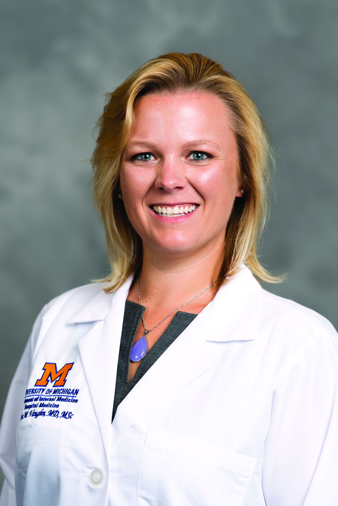
“We know from studies that 20% of hospitalized patients who receive an antibiotic have an adverse drug event from that antibiotic within 30 days,” said Dr. Vaughn.
Dr. Vaughn identified several practical ways in which hospitalists can reduce antibiotic overuse, including in the management of patients hospitalized with COVID-19.
Identify asymptomatic bacteriuria
One key area in which hospitalists can improve antibiotic stewardship is in recognizing asymptomatic bacteriuria and the harms associated with treatment, Dr. Vaughn said. For example, a common scenario for hospitalists might involve and 80-year-old woman with dementia, who can provide little in the way of history, and whose chest x-ray can’t rule out an underlying infection. This patient might have a positive urine culture, but no other signs of a urinary tract infection. “We know that asymptomatic bacteriuria is very common in hospitalized patients,” especially elderly women living in nursing home settings, she noted.
In cases of asymptomatic bacteriuria, data show that antibiotic treatment does not improve outcomes, and in fact may increase the risk of subsequent UTI, said Dr. Vaughn. Elderly patients also are at increased risk for developing antibiotic-related adverse events, especially Clostridioides difficile. Asymptomatic bacteriuria is any bacteria in the urine in the absence of signs or symptoms of a UTI, even if lab tests show pyuria, nitrates, and resistant bacteria. These lab results are often associated with inappropriate antibiotic use. “The laboratory tests can’t distinguish between asymptomatic bacteriuria and a UTI, only the symptoms can,” she emphasized.
Contain treatment of community-acquired pneumonia
Another practical point for reducing antibiotics in the hospital setting is to limit treatment of community-acquired pneumonia (CAP) to 5 days when possible. Duration matters because for many diseases, shorter durations of antibiotic treatments are just as effective as longer durations based on the latest evidence. “This is a change in dogma,” from previous thinking that patients must complete a full course, and that anything less might promote antibiotic resistance, she said.
“In fact, longer antibiotic durations kill off more healthy, normal flora, select for resistant pathogens, increase the risk of C. difficile, and increase the risk of side effects,” she said.
Ultimately, the right treatment duration for pneumonia depends on several factors including patient factors, disease, clinical stability, and rate of improvement. However, a good rule of thumb is that approximately 89% of CAP patients need only 5 days of antibiotics as long as they are afebrile for 48 hours and have 1 or fewer vital sign abnormalities by day 5 of treatment. “We do need to prescribe longer durations for patients with complications,” she emphasized.
Revisit need for antibiotics at discharge
Hospitalists also can practice antibiotic stewardship by considering four points at patient discharge, said Dr. Vaughn.
First, consider whether antibiotics can be stopped. For example, antibiotics are not needed on discharge if infection is no longer the most likely diagnosis, or if the course of antibiotics has been completed, as is often the case for patients hospitalized with CAP, she noted.
Second, if the antibiotics can’t be stopped at the time of discharge, consider whether the preferred agent is being used. Third, be sure the patient is receiving the minimum duration of antibiotics, and fourth, be sure that the dose, indication, and total planned duration with start and stop dates is written in the discharge summary, said Dr. Vaughn. “This helps with communication to our outpatient providers as well as with education to the patients themselves.”
Bacterial coinfections rare in COVID-19
Dr. Vaughn concluded the session with data from a study she conducted with colleagues on the use of empiric antibacterial therapy and community-onset bacterial coinfection in hospitalized COVID-19 patients. The study included 1,667 patients at 32 hospitals in Michigan. The number of patients treated with antibiotics varied widely among hospitals, from 30% to as much as 90%, Dr. Vaughn said.
“What we found was that more than half of hospitalized patients with COVID (57%) received empiric antibiotic therapy in the first few days of hospitalization,” she said.
However, “despite all the antibiotic use, community-onset bacterial coinfections were rare,” and occurred in only 3.5% of the patients, meaning that the number needed to treat with antibiotics to prevent a single case was about 20.
Predictors of community-onset co-infections in the patients included older age, more severe disease, patients coming from nursing homes, and those with lower BMI or kidney disease, said Dr. Vaughn. She and her team also found that procalcitonin’s positive predictive value was 9.3%, but the negative predictive value was 98.3%, so these patients were extremely likely to have no coinfection.
Dr. Vaughn said that in her practice she might order procalcitonin when considering stopping antibiotics in a patient with COVID-19 and make a decision based on the negative predictive value, but she emphasized that she does not use it in the converse situation to rely on a positive value when deciding whether to start antibiotics in these patients.
Dr. Vaughn had no financial conflicts to disclose.
“Antibiotic stewardship is never easy, and sometimes it is very difficult to differentiate what is going on with a patient in the clinical setting,” said Valerie M. Vaughn, MD, of the University of Utah, Salt Lake City, at SHM Converge, the annual conference of the Society of Hospital Medicine.
“We know from studies that 20% of hospitalized patients who receive an antibiotic have an adverse drug event from that antibiotic within 30 days,” said Dr. Vaughn.
Dr. Vaughn identified several practical ways in which hospitalists can reduce antibiotic overuse, including in the management of patients hospitalized with COVID-19.
Identify asymptomatic bacteriuria
One key area in which hospitalists can improve antibiotic stewardship is in recognizing asymptomatic bacteriuria and the harms associated with treatment, Dr. Vaughn said. For example, a common scenario for hospitalists might involve and 80-year-old woman with dementia, who can provide little in the way of history, and whose chest x-ray can’t rule out an underlying infection. This patient might have a positive urine culture, but no other signs of a urinary tract infection. “We know that asymptomatic bacteriuria is very common in hospitalized patients,” especially elderly women living in nursing home settings, she noted.
In cases of asymptomatic bacteriuria, data show that antibiotic treatment does not improve outcomes, and in fact may increase the risk of subsequent UTI, said Dr. Vaughn. Elderly patients also are at increased risk for developing antibiotic-related adverse events, especially Clostridioides difficile. Asymptomatic bacteriuria is any bacteria in the urine in the absence of signs or symptoms of a UTI, even if lab tests show pyuria, nitrates, and resistant bacteria. These lab results are often associated with inappropriate antibiotic use. “The laboratory tests can’t distinguish between asymptomatic bacteriuria and a UTI, only the symptoms can,” she emphasized.
Contain treatment of community-acquired pneumonia
Another practical point for reducing antibiotics in the hospital setting is to limit treatment of community-acquired pneumonia (CAP) to 5 days when possible. Duration matters because for many diseases, shorter durations of antibiotic treatments are just as effective as longer durations based on the latest evidence. “This is a change in dogma,” from previous thinking that patients must complete a full course, and that anything less might promote antibiotic resistance, she said.
“In fact, longer antibiotic durations kill off more healthy, normal flora, select for resistant pathogens, increase the risk of C. difficile, and increase the risk of side effects,” she said.
Ultimately, the right treatment duration for pneumonia depends on several factors including patient factors, disease, clinical stability, and rate of improvement. However, a good rule of thumb is that approximately 89% of CAP patients need only 5 days of antibiotics as long as they are afebrile for 48 hours and have 1 or fewer vital sign abnormalities by day 5 of treatment. “We do need to prescribe longer durations for patients with complications,” she emphasized.
Revisit need for antibiotics at discharge
Hospitalists also can practice antibiotic stewardship by considering four points at patient discharge, said Dr. Vaughn.
First, consider whether antibiotics can be stopped. For example, antibiotics are not needed on discharge if infection is no longer the most likely diagnosis, or if the course of antibiotics has been completed, as is often the case for patients hospitalized with CAP, she noted.
Second, if the antibiotics can’t be stopped at the time of discharge, consider whether the preferred agent is being used. Third, be sure the patient is receiving the minimum duration of antibiotics, and fourth, be sure that the dose, indication, and total planned duration with start and stop dates is written in the discharge summary, said Dr. Vaughn. “This helps with communication to our outpatient providers as well as with education to the patients themselves.”
Bacterial coinfections rare in COVID-19
Dr. Vaughn concluded the session with data from a study she conducted with colleagues on the use of empiric antibacterial therapy and community-onset bacterial coinfection in hospitalized COVID-19 patients. The study included 1,667 patients at 32 hospitals in Michigan. The number of patients treated with antibiotics varied widely among hospitals, from 30% to as much as 90%, Dr. Vaughn said.
“What we found was that more than half of hospitalized patients with COVID (57%) received empiric antibiotic therapy in the first few days of hospitalization,” she said.
However, “despite all the antibiotic use, community-onset bacterial coinfections were rare,” and occurred in only 3.5% of the patients, meaning that the number needed to treat with antibiotics to prevent a single case was about 20.
Predictors of community-onset co-infections in the patients included older age, more severe disease, patients coming from nursing homes, and those with lower BMI or kidney disease, said Dr. Vaughn. She and her team also found that procalcitonin’s positive predictive value was 9.3%, but the negative predictive value was 98.3%, so these patients were extremely likely to have no coinfection.
Dr. Vaughn said that in her practice she might order procalcitonin when considering stopping antibiotics in a patient with COVID-19 and make a decision based on the negative predictive value, but she emphasized that she does not use it in the converse situation to rely on a positive value when deciding whether to start antibiotics in these patients.
Dr. Vaughn had no financial conflicts to disclose.
FROM SHM CONVERGE 2021
Keep antibiotics unchanged in breakthrough UTIs
Changing the continuous antibiotic prophylactic agent had no significant effect on the risk of a second infection in children with breakthrough urinary tract infections (UTIs), based on data from 62 children treated at a single center.
Continuous antibiotic prophylaxis (CAP) is often used for UTI prevention in children with febrile UTIs or anomalies that predispose them to UTIs, such as vesicoureteral reflux (VUR) or bladder and bowel dysfunction, said Lane M. Shish, MPH, of the University of Washington, Bothell, and colleagues in a poster (#1245) presented at the Pediatric Academic Societies annual meeting.
CAP, once initiated, is used until a planned endpoint or a breakthrough UTI, at which point alternative treatments usually include surgical intervention or a CAP agent change, the researchers said. However, changing the CAP agent is based on consensus without evidence of benefit, they noted.
To evaluate the potential effect of switching or maintaining CAP in cases of breakthrough UTIs, the researchers conducted a retrospective cohort study of all patients younger than 18 years on CAP for UTI prevention enrolled in a pediatric urology registry between January 2013 and August 2020.
All patients experienced a breakthrough UTI while on CAP; CAP was changed for 24 patients and left unchanged for 38 patients.
The primary outcome of second-breakthrough infections occurred in 12 of the changed CAP group and 22 of the unchanged group, with a relative risk of 0.86. The percentage of second breakthrough UTIs resistant to the current CAP was not significantly different between the changed and unchanged CAP groups (75% vs. 77%; P = 0.88).
The researchers also identified a rate ratio of 0.67 for a second breakthrough UTI in the changed CAP group, and found that approximately one-third of these patients (33.3%) developed antibiotic resistance to their initial antibiotic agent and the changed antibiotic agent.
The study findings were limited by several factors, including the retrospective design and small sample size, the researchers noted.
However, the results suggest that changing the CAP after an initial breakthrough UTI in children did not increase the risk of a second breakthrough UTI, and that CAP changing did introduce a risk of developing a second UTI with increased CAP resistance, the researchers noted. The results support leaving a child’s CAP unchanged after an initial breakthrough UTI, although additional research is needed to verify the findings, including studies involving a larger cohort with a multi-institutional prospective evaluation, they concluded.
Manage UTIs to reduce recurrence and resistance
“As we know, avoiding recurrent UTIs is important in preserving renal function in pediatric patients,” said Tim Joos, MD, a Seattle-based clinician with a combination internal medicine/pediatrics practice, in an interview.
“Avoiding recurrent UTIs is also important to avoid the development and spread of multidrug-resistant organisms,” he said.
Dr. Joos said he was surprised by some of the study findings. “I was surprised that, over the course of this 7-year retrospective review, overall only approximately 50% of patients with a first breakthrough UTI on CAP developed a second breakthrough UTI,” he noted. “Also, the relative risk of a second UTI was not significantly affected by whether the CAP antibiotic was changed after the first infection,” he said. “It would be interesting to see whether these results hold up in a randomized, prospective study,” he added.
The study received no outside funding. The researchers had no financial conflicts to disclose. Dr. Joos had no financial conflicts to disclose, but serves as a member of the Pediatric News Editorial Advisory Board.
FROM PAS 2021
Palliative care in the pandemic: How one hospital met the challenge
Clarissa Johnston, MD, said during a virtual presentation at the annual meeting of the Society of General Internal Medicine.
Dr. Johnston, of the University of Texas at Austin, and colleagues experienced an extreme COVID-19 surge when they reopened after initial closure in the first weeks of the pandemic.
“Our hospital and clinics are the health care safety net in Austin, and we serve a predominantly uninsured and Hispanic population that experienced a greater burden of COVID-19 than other populations in our area,” she said in the presentation.
The rapid onset and spread of COVID-19 locally required physicians and staff to innovate quickly, and “we developed and implemented collaborative and novel partnerships between generalists and palliative care specialists to help ensure that our core humanizing values were not lost in the pandemic,” Dr. Johnston emphasized.
Collaboration between internal medicine and palliative care involved developing relationship-centered communication for families and health care workers, as well as engaging medical students in a Transitions of Care elective, Dr. Johnston said.
The early weeks of the pandemic impacted families with the no visitor policy and the loss of death rituals, she said. Health care providers suffered, too, as nurses experienced an overload of work, fears for their own health and safety, and feelings of disconnect from their patients. Physicians dealt with the challenges of a unique illness, and their own fears and uncertainty, Dr. Johnston said.
Meeting communication challenges
One of the strategies used to bridge the communication gap caused by the lack of visitors and family contact was the adoption of the Meet My Loved One program, adapted from a similar program at the University of Alabama, said Dr. Johnston. Meet My Loved One was a collaborative effort focused on ICU patients, Dr. Johnston said. Members of the primary care team, including medical students in the Transitions of Care elective, called family members of ICU patients to collect personal details and humanizing information about the patient, such as preferred name, favorite foods, favorite activities, and some personal history (i.e. played basketball when he was young), and this information was collated, summarized, and posted on the door of the patient’s room.
Advanced care conversations
Advanced care planning (ACP) benefits include not only the promotion of patient-centered care, but also decreases in ICU admissions, length of stay, and cost. Dr. Johnston and colleagues developed a multipronged curriculum that trainees could use to have ACP conversations with clinic patients who would be considered high risk if they developed COVID-19 infections, Dr. Johnston explained. As part of the elective, medical students were trained to have ACP conversations with patients via telehealth; students practiced role-playing conversations with each other via Zoom and met virtually as a group to review the conversations, she said.
Maintaining Humanity
“COVID-19 has changed the way we interact with patients and families,” Dr. Johnston said in an interview. The inability to rely on face-to-face discussions means that “we really need to think carefully how we maintain humanity and the human touch,” she said.
Challenges in providing palliative care during the pandemic include “maintaining humanity, remembering that there is a person behind the prone, paralyzed patient, with family members who love them, and are desperate to be with them but unable,” Dr. Johnston said.
“The Meet My Loved One program helped, as well as multidisciplinary rounds, chaplain services, and frequent check ins with the bedside nurses,” she said.
“I tried hard to call families every day to start to build that trust and rapport that was lost by all the distancing and lack of visits. I didn’t realize how much the day in and day out care of ICU patients is witnessed by families when they are in the room,” she noted. “During COVID-19, it was so much harder to build trust, especially when you add in the inequities and structural racism problems in our health care system,” she said.
“Why would a family member believe and trust some random doctor calling them on the phone? Were we really trying our hardest? Families didn’t have a way to assess that, at least not like they do when they are at bedside and see how hard everyone works,” Dr. Johnston said. “Video visits helped but were not the same.”
Some key lessons about palliative care Dr. Johnson said she learned from the pandemic were how important it is to remember the patient and family, “how we need to work to build trust,” and that clinicians should be mindful that video visits don’t work for everyone, and to “ask, ask, ask about what you don’t know, including death rituals.”
Additional research needs in palliative care in the wake of COVID-19 include more information on what works and what doesn’t work, from the patient and family perspective, said Dr. Johnston. Communication strategies are important, and “we need to address how we can better communicate around serious illness and end-of-life issues with Black and Brown communities,” she said.
Challenges of COVID care
One of the main challenges to providing palliative care in the early days of the pandemic was navigating the constantly evolving science of COVID-19, Aziz Ansari, DO, of Loyola University Chicago, Maywood, Ill., said in an interview.
“It was, and remains, very hard to prognosticate on how a patient will do having respiratory failure with COVID,” said Dr. Ansari, who was the leader of the Palliative Care interest group at the SGIM meeting.
“So, the challenge was how to have a conversation on goals, values, and preferences when we really did not know the disease entity,” Dr. Ansari noted.
“We were surprised many times [when patients with COVID-19] recovered though it took a long time, so we could not really say that in the acute phase of COVID, it was a terminal illness,” he noted.
“Regardless, it still behooves us to have conversations with our patients and families about what are they willing to go through, and how they define a quality of life,” he said.
Strategies such as those used at the University of Texas show the importance of primary care palliative skill development, said Dr. Ansari. “Every physician should have the skill set of having conversations with patients and families on goals, values, and preferences even in unknown situations,” he said. That lifelong skill set development begins in medical school, he added.
Dr. Johnston and Dr. Ansari had no financial conflicts to disclose.
FROM SGIM 2021
Focus on youth concerns at well visits
Taking teens’ strengths, values, and dreams, into account through a previsit questionnaire was acceptable to them and may promote discussions with providers, based on data from 91 adolescents.
The American Academy of Pediatrics’ Bright Futures initiative recommends the use of a strength-based approach for adolescent well visits, but the extensive positive psychology inventories to identify teen strengths and values are impractical for the clinical visit setting, wrote Yidan Cao, MPH, of the Child Development through Primary Care at the University of Michigan, Ann Arbor, and colleagues. However, 76% of youth participating in focus groups responded that “using a confidential questionnaire about a teen’s strengths and goals before checkup visits would be a good addition to health care for teens,” the researchers said.
In a study presented in a poster session (#515) at the Pediatric Academic Societies annual meeting, the researchers recruited 91 community youth to participate in 13 focus groups related to teen depression and substance use. The age of the focus group participants ranged from 12 to 18 years, with an average of 15 years, 61.5% were female, and 1.1% identified as transgender. The racial breakdown was 51.6% White, 27.5% African American, 8.8% Asian, 2.2% Native American, 3.3% biracial, and 6.6% unknown.
The participants provided information on potential questionnaire items for an online previsit screening for well visits to assess strengths and identify values, goals, dream jobs, and life wishes.
Suggestions from the participants informed changes to the questionnaire, which included five categories: personal/social goals, goals for academics/training, strengths, values, and dream jobs.
The top endorsed personal goal of “to be happy” was chosen by 13.1% of the participants. The top academic goal was “get good grades” (45.5%). The top endorsed strength was “fitness/coordination/sports/physical activity” (22.9%), while the top value was caring and kindness (25.8%), and the top dream job category was health/medical (30.8%).
Key comments made by the youth participants for improving the previsit questionnaire included adding an option for “I can’t decide,” and allowing for multiple responses to avoid feeling pinned down or judged, the researchers noted.
The researchers highlighted one teen comment: “While I understand the purpose of limiting the participants to two answers, it is incredibly difficult to only choose two. Being limited to two very much restricts your understanding of our values. For example, I would’ve also liked to select ‘to do well in school’ and ‘to make a difference,’ but ‘being happy’ and ‘being loving to all those around me’ had to take precedent.”
The study was limited by not being fully generalizable to all teens, as other teens may hold views and beliefs that differ from those of the focus group participants, the researchers noted.
However, the findings support the value of a strength-based previsit questionnaire for adolescents, they said.
“Structured previsit data could facilitate relationship building and be actionable for assigning strength and resiliency building resources,” they noted. “A final strengths and goals questionnaire is now being piloted in computerized form contributing to decision supports for suggested teleprompters and associated resource options,” and future research may show the value of such previsit data for improved clinical process and outcomes of youth well visits, they concluded.
Recognize the uncertainty of adolescence
“Adolescents are at crossroads of identity, trying to figure out who they are, their goals and values,” said Kelly A. Curran, MD, of the University of Oklahoma Health Sciences Center, Oklahoma City, in an interview. “Adolescence is also a critical time when teens have more control and choice over behaviors that impact their health, such as diet, exercise, substance use, and sexual activity. In this critical time period, clinicians can intervene on health-related behaviors and shape the trajectory of a teen’s life. However, to promote teen health, pediatricians need to have their patients’ trust, which can be hard to gain,” she emphasized.
“In my practice, I’ve found that teens often just want to be seen and heard,” said Dr. Curran. “Teens often have many adults in their life who focus on the ‘don’ts’ – don’t use drugs, don’t have sex, for example – and few that praise healthy behaviors or strengths, or seek to understand what is important to them. By listening to teens and understanding what’s important to them, we can then use motivational interviewing techniques to help create meaningful change in health-related behaviors. However, this takes time and investment, which is often in conflict with time pressures in the modern medical system.
“This study is useful because it examined the acceptability of a positive psychology questionnaire to be used at well visits when reviewed by youth, that could be used to streamline this important process,” said Dr. Curran.
“From my practice, I know that understanding a teen’s goals, values, and strengths is important – we do this daily in our practice when working with patients – but it was exciting to see that youth found it acceptable to do this via a previsit survey, which can potentially streamline well visits,” she noted.
The questionnaire is being developed as a pilot program, but more research is needed to determine the direct clinical impact, said Dr. Curran. “It will be important in the future to see if implementation of this questionnaire can be helpful in integrating this information into motivational interviewing and rapport building to help improve teens’ health outcomes.”
The study was supported in part by the National Institute on Drug Abuse and the National Institute of Mental Health. Two coauthors have a financial interest in the CHADIS online reporting program used in the study. Dr. Curran had no financial conflicts to disclose.
FROM PAS 2021
SGIM annual meeting focuses on inclusivity
In her welcome video on the opening day at the annual meeting of the Society of General Internal Medicine, meeting chair Rita Lee, MD, said she hoped that this year’s event, though virtual, will allow attendees an opportunity to “regroup, find inspiration, and celebrate the incredible strengths and diverse voices of our community.”
“We are living in an incredibly polarized world,” Dr. Lee said in an interview. “ as recent events, such as the death of George Floyd and many others, plus the disparities revealed by the COVID-19 pandemic, have brought issues of structural racism and oppression in the United States to the forefront,” she said in the interview.
“Given these circumstances, it is important now, more than ever, for generalists to move our values into action – to effect change at the health system, community, and policy levels – so our patients can achieve optimal health,” Dr. Lee emphasized.
She noted that SGIM’s vision: “A just system of care in which all people can achieve optimal health,” underlies the meeting’s sessions.
Some challenges related to adopting more antiracist training in medical education center on faculty development, Dr. Lee noted. “There are also students who don’t feel that this is part of the role of being a physician. One way to overcome these challenges is by directly linking structural competency to health outcomes for our patients,” she added. “We have evidence that structural racism impacts health and we should make that clear to our educational leaders and faculty to increase buy in. So many of our SGIM members are working on developing curricula for this.”
Two of the meeting’s workshops that addressed racism in medicine and medical education and strategies for change were “Demystifying Structural Competency – How to Develop Antiracist Training in Medical Education,” and “Combating Systemic Racism in the Health Care System – Practical Actions You Can Take Today.” Below are some details about these.
Medical education evolves to include structural competency
In the workshop “Demystifying Structural Competency – How to Develop Antiracist Training in Medical Education,” participants used interactive exercises to build structural differentials for patient cases. The workshop was based in part on the experiences of including structural competency in medical education at Albert Einstein College of Medicine, New York, and the University of Pittsburgh.
During the session, participants practiced building a structural differential diagnosis in small groups, and also practiced using a structurally competent version of the 1-minute preceptor to promote structural competency in learners.
“Structural competency represents a shift in medical education towards attention to forces that influence health outcomes at levels above individual clinical interactions and develop a provider’s capacity to recognize and respond to health and illness as downstream effects of social, political and economic structures,” presenter Iman Hassan, MD, of Albert Einstein College of Medicine and Montefiore Health System, both in New York, said in an interview.
“At the same time, structural competency incorporates structural humility, which decentralizes the provider role in addressing structural factors and emphasizes collaboration with patients and communities,” she said in the interview. “Structural competency is a useful antiracism framework because it explicitly engages learners with the broader structural forces that result in health disparities, including structural racism and its downstream effects,” Dr. Hassan explained.
Addressing structural competency is important in medical education because structural and social determinants of health contribute more than half of overall health outcomes, said Dr. Hassan.
A structural competency framework equips learners to identify, discuss, and work with patients to navigate social needs such as lack of health insurance, food, or transportation, that are preventing them from accessing needed health care services, Dr. Hassan noted.
“Importantly, training in structural competency empowers physicians to be agents of change within their clinics, health systems, and communities and to recognize the value of community-led advocacy in promoting health equity,” she said.
Structural competency training also “will also allow them to engage more fully with the body of literature that exists surrounding social determinants of health and health disparities, and the use of approaches such as critical race theory through which to view health care,” she emphasized. “Importantly, understanding of the historical and structural context of medicine allows clinicians to more readily recognize when their own clinical practices, such as use of race-based clinical prediction tools, may perpetuate disparities, and work collectively to eliminate those practices.”
Recalibrating calculators for clinical care
Another workshop, “Combating Systemic Racism in the Health Care System – Practical Actions You Can Take Today,” took on the challenge of inherent bias in clinical care caused by various factors, notably medical calculators such as those used to measure kidney function and pulmonary function.
Lamar K. Johnson, MD, of Christiana Care Hospital Partners/Christiana Care Pediatric Hospitalists in Newark, Del., and Celeste Newby, MD, of Tulane University, New Orleans, discussed the inherent biases in some calculators and how to take those biases into account. A stated goal of the workshop was to increase awareness of the origins of medical calculators in order to enhance equity and improve shared decision-making between patients and providers.
Addressing implicit bias in clinical practice is important because such bias has been shown to negatively affect physician behavior and clinical decision making, Dr. Johnson said in an interview.
“These effects can also negatively affect the doctor-patient relationship and lead to poorer health outcomes due to delays in or avoidance of care or avoidance of the health care system, and mistrust, resulting in nonadherence,” Dr. Johnson noted.
“Implicit bias training helps empower medical students and residents to recognize and address bias and advocate for patients. Such training can potentially be beneficial to faculty, too,” Dr. Johnson emphasized in the interview.
“Race is primarily a social, not a biological, construct, and we must be careful when we use it, as its use in the past has been largely inappropriate and not scientifically sound,” he said.
During the session, one of the presenters said removing specific mentions of race from clinical documentation can reduce racial bias in clinical practice.
The presenters also highlighted the estimated glomerular filtration rate (eGFR) which is used to estimate kidney function.
The eGFR “reports higher eGFR values for Blacks based on a faulty hypothesis that Black people have higher muscle mass. This higher estimated value can delay referral for specialist care or transplantation, leading to worse outcomes,” Dr. Johnson explained in the interview.
In response, “Many major institutions have eliminated the race modifier in eGFR, and a joint task force created by the National Kidney Foundation and American Society of Nephrology has recommended against using a race modifier as of March 2021,” Dr. Johnson said.
The presenters had no relevant financial conflicts to disclose.
FROM SGIM 2021
Carbon monoxide diffusion with COPD declines more in women
Single breath diffusion capacity for carbon monoxide shows greater decline over time in COPD patients compared with controls, but declines significantly more in women compared with men, according to data from 602 adults with a history of smoking.
In previous studies, diffusion capacity for carbon monoxide (DLco) has been associated with decreased exercise capacity and poor health status in patients with COPD, but its association as a measure of disease progression has not been well studied, wrote Ciro Casanova, MD, of Hospital Universitario La Candelaria, Spain, and colleagues.
In a study published in the journal CHEST®, the researchers identified 506 adult smokers with COPD and 96 adult smoker controls without COPD. Lung function based on single breath DLco was measured each year for 5 years. The study population was part of the COPH History Assessment in SpaiN (CHAIN), an ongoing observational study of adults with COPD. COPD was defined as a history of at least 10 pack-years of smoking and a post-bronchodilator FEV1/FVC greater than 0.7 after 400 micrograms of albuterol, the researchers said.
During the 5-year period, the average overall annual decline in DLco was 1.34% in COPD patients, compared with .04% in non-COPD controls (P = .004). Among COPD patients, age, body mass index, FEV1%, and active smoking were not associated with longitudinal change in DLco values, the researchers said.
Notably, women with COPD at baseline had lower baseline DLco values compared with men (11.37%) and a significantly steeper decline in DLco (.89%) compared with men (P = .039). “Being a woman was the only factor that related to the annual rate of change in DLco,” the researchers said.
In a subgroup analysis, the researchers identified 305 COPD patients and 69 non-COPD controls who had at least 3 DLco measurements over the 5-year study period. In this group, 16.4% patients with COPD and 4.3% smokers without COPD showed significant yearly declines in DLco of –4.139% and –4.440%, respectively. Among COPD patients, significantly more women than men showed significant DLco declines (26% vs. 14%, P = .005). No significant differences were observed in mortality or hospitalizations per patient-year for COPD patients with and without DLco decline, the researchers said.
The study findings were limited by several factors including the lack of annual measurements of DLco among some patients, potential variability in the instruments used to measure DLco, and the absence of computerized tomography data for the chest, the researchers noted. However, the results support the value of the test for COPD progression when conducted at 3- to 4-year intervals, given the slow pace of the decline, they said. More research is needed, but “women seem to have a different susceptibility to cigarette smoke in the alveolar or pulmonary vascular domains,” they added.
DLco remains a valuable marker
The study is important because the usual longitudinal decline of diffusion capacity, an important physiological parameter in patients with COPD, was unknown, Juan P. de Torres, MD, of Queen’s University, Kingston, Ont., said in an interview.
“The finding of a different longitudinal decline of DLco in women was a surprise,” said Dr. de Torres, who was a coauthor on the study. “We knew from previous works from our group that COPD has a different clinical and prognostic behavior in women with COPD, but this specific finding is novel and important,” he said.
“These results provide information about the testing frequency (3-4 years) needed to use DLco as a marker of COPD progression in clinical practice,” Dr. de Torres added.
“What is the driving cause of this sex difference is unknown. We speculate that different causes of low DLco in COPD such as degree of emphysema, interstitial lung abnormalities, and pulmonary hypertension, may have a different prevalence and progression in women with COPD,” he said.
Looking ahead, “Large studies including an adequate sample of women with COPD is urgently needed because they will be the main face of COPD in the near future,” said Dr. de Torres. “Sex difference in their physiological characteristics, the reason to explain those differences and how they behave longitudinally is also urgently needed,” he added.
The study was supported in part by AstraZeneca and by the COPD research program of the Spanish Respiratory Society. The researchers and Dr. de Torres had no financial conflicts to disclose.
FROM CHEST
New oral protein shows promise for ulcerative colitis
A plant-based fusion protein is safe and effective for inducing favorable immune modulation in patients with mild to moderate ulcerative colitis with no immune suppression–side effects reported.
OPRX-106, an orally administered BY2 plant cell–expressing recombinant TNF fusion protein, has demonstrated effectiveness as an anti–TNF-alpha therapy, according to Einat Almon, PhD, of Protalix Biotherapeutics, and colleagues.
“Oral immune therapy is based on the concept of oral administration of nonabsorbable compounds which target the gut immune system to redirect the systemic immune system toward an anti-inflammatory direction, without immunosuppression,” the researchers said.
A phase 1 study of OPRX-106 in healthy human volunteers showed safety and immune modulatory effects at doses of 2, 8, or 16 mg/day.
In this phase 2a clinical trial published in the Journal of Clinical Gastroenterology, the researchers enrolled 24 patients with ulcerative colitis (11 male and 13 female) aged 23-73 years, with an average age of 42.6 years. Patients received either 2 mg or 8 mg of OPRX-106 at least once daily for 8 weeks. All patients were monitored for 6 hours after receiving medication on day 1 and week 8 for pharmacokinetic sampling, and a lower endoscopy was performed at week 8.
After 8 weeks, 67% of the patients demonstrated clinical response and 28% showed clinical remission.
Clinical response and clinical remission were defined by a specific set of criteria including improvement in the Mayo score. Clinical response was a “decrease in the Mayo score of at least 3 points, decrease in the subscore for rectal bleeding of at least 1 point, [and] a rectal bleeding subscore of 0 or 1.” Clinical remission at week 8 was defined as “clinically symptom-free, Mayo score of ≤2 with no individual subscore exceeding 1 point after treatment, histopathological improvement in Geboes histologic grading from baseline to week 8, improvement in high sensitivity C-reactive protein levels from baseline to week 8, improvement in fecal calprotectin levels from baseline to week 8, and changes in systemic immune modulation parameters from baseline to week 8.”
In addition, 89% of the patients experienced some degree of improvement in their Mayo scores, 61% had mucosal improvement, and 33% achieved mucosal healing.
No side effects associated with general immune suppression were reported. No patients discontinued the study because of adverse events, the researchers said. However, overall, 40 adverse events were reported in 15 patients; 95% of these were mild to moderate and 40% were reported as treatment related. No differences appeared in adverse events related to the two doses.
Evidence of a systemic anti-inflammatory effect was seen with a decrease in serum levels of the pro-inflammatory cytokines interleukin-6 and interferon-gamma that correlated with the clinical response, the researchers noted. Similarly, an increase in the CD3+CD4+CD25+Foxp3+ subset of suppressor lymphocytes correlated with clinical response.
The study findings were limited by the small sample size, open-label design, and lack of control subjects. However, by targeting the gut immune system, the drug “may provide an answer to the long-term immune suppression encountered in patients with chronic disorders who use these agents for prolonged periods of time, in addition to loss of response due to neutralizing antibodies,” they concluded.
Findings provide foundation for further research
“Conducting a study of a novel treatment for ulcerative colitis is valuable and timely because the available options are limited,” Atsushi Sakuraba, MD, of the University of Chicago, said in an interview. “The currently available TNF antagonists are administered intravenously or subcutaneously and bear the risk of infectious complications, so the development of an agent that can be administered orally with fewer side effects is of importance.”
Although the data are preliminary, Dr. Sakuraba emphasized that the take-home message for clinicians is that “the present open-label study consisting of a small number of subjects demonstrated that OPRX-106 was effective and safe in active ulcerative colitis, so further investigation is warranted. Larger-powered, randomized, placebo-controlled studies are needed to confirm these findings.”
The study was supported by Protalix Biotherapeutics; Dr. Almon and several coauthors are employed by Protalix Biotherapeutics. Dr. Sakuraba had no financial conflicts to disclose.
FROM THE JOURNAL OF CLINICAL GASTROENTEROLOGY
Telehealth takeaways for hospitalists outlined
Although the COVID-19 pandemic put telehealth on fast forward, more than one third of patients in the United States engaged with telehealth services before February 2020, according to Ameet Doshi, MD, and Chrisanne Timpe, MD, of HealthPartners in Bloomington, Minn.

Broadly speaking, telehealth is “using virtual tools to evaluate, manage, and care for our patients, regardless of where they are located,” Dr. Doshi said during a May 6 session at SHM Converge, the annual conference of the Society of Hospital Medicine.
The entirety of telehealth includes remote ways to meet almost any patient demand, he said. Some common health terms are used interchangeably, but some use telehealth as a broad term for electronic health care services, while telemedicine may refer specifically to remote patient care, he said.
Telemedicine allows flexibility of delivering patient care in inpatient, outpatient, or at-home settings, said Dr. Doshi. To illustrate the current application of telemedicine, he used an example of a 25-bed critical access hospital serving a growing regional population in which outpatient volume is expanding and ambulatory care services are being added. In this example, inpatient volume is growing, but not enough to support an inpatient consult service, but telehealth access to specialists such as cardiology would be useful in this case, he said.
Hospitalist telehealth means “being able to provide services to changing patient populations regardless of location; we can bring services to where patients are,” said Dr. Doshi.
Benefits of telehealth to patients include less travel and easier access to care, benefits to clinicians include expanding services at lower financial costs, he said.
COVID-19 challenges and opportunities
The COVID-19 pandemic presented both challenges and opportunities for telehealth, Dr. Doshi said. One opportunity was the sudden broad acceptance of virtual care out of necessity and concern for patient and staff safety, and to preserve the use of personal protective equipment, he said. In addition, a loosening of regulatory and financial pressures allowed more institutions to expand and initiate telehealth services.
Challenges included technological limitations and, in some cases, the need to develop a telehealth infrastructure from scratch, Dr. Doshi explained. Concerns also remain regarding how telehealth will evolve in the post-pandemic future, he said.
In the meantime, Medicare data show the impact of the pandemic on telehealth services, said Dr. Doshi. A telehealth waiver issued in March 2020 led to an increase in virtual encounters, and Medicare data show approximately 25 million virtual Medicare encounters between March 2020 and October 2020, representing a 3,000% increase from the same period in 2019, he said.
“Telehealth is here to stay, so the questions are how to craft a hospitalist telehealth program and provide essential patient care,” he said.
Dr. Timpe shared some examples of the evolution of telehealth care during the pandemic, including a case of an asymptomatic but frail patient with diabetes, dementia, and coronary artery disease undergoing outpatient care for a foot infection. The patient presented to an emergency department but refused to be hospitalized because of family concerns about patient isolation (no visitors were allowed at the time) and the concerns about COVID-19 infection.
The need to help treat acutely ill patients such as this patient while avoiding hospital admission during and after the pandemic continues to lead to the development of telehealth programs, Dr. Timpe said. She shared details of the Hospital@Home program developed by her organization, Health Partners. The program is designed to treat acutely ill people in the home, if possible, and avoid the need for hospital admission. Patients receive daily medical management from a hospitalist and care from staff, including registered nurses and community paramedics. Services include provision of IV medications and fluids, but the staff also conduct labs and imaging services, Dr. Timpe said.
Conditions that the program has managed at patients’ homes include pneumonia, COPD, asthma, bronchitis, flu, COVID-19, congestive heart failure, cellulitis, and urinary tract infections, said Dr. Timpe.
“We do not accept people into the program who have treatment needs that can only be met in a hospital,” such as the need for blood products, vasopressor support, telemetry, or positive pressure support, she noted.
Between November 2019 and February 15, 2021, the Hospital@Home program has provided services to 132 patients for a total of 287 visits. The program has averted 50 emergency department visits and 40 hospitalizations, and shorted hospital stays in 57 cases, she noted.
Hospitalists are suited for telehealth for several reasons, including the ability to triage acutely ill patients, familiarity with resource utilization, and expertise in management of complex medical care, said Dr. Timpe.
Looking ahead
Dr. Doshi emphasized several ongoing issues regarding the future of telemedicine, primarily the need for standardized regulation and reimbursement; reduction of health equity disparity and attention to technological barriers (including access and technology literacy); and identification of the next frontiers in telehealth.
Research on the impact and effectiveness of telehealth is limited, but growing, and next frontiers might include making patients more active participants in telehealth via patient-operated kits, or the option of an open telemedicine marketplace, in which patients can select providers from across the country, he said. No matter where telehealth leads in the future, “we need to make sure we have a positive patient outcome,” he concluded.
Dr. Doshi and Dr. Timpe had no financial conflicts to disclose.
Although the COVID-19 pandemic put telehealth on fast forward, more than one third of patients in the United States engaged with telehealth services before February 2020, according to Ameet Doshi, MD, and Chrisanne Timpe, MD, of HealthPartners in Bloomington, Minn.
Broadly speaking, telehealth is “using virtual tools to evaluate, manage, and care for our patients, regardless of where they are located,” Dr. Doshi said during a May 6 session at SHM Converge, the annual conference of the Society of Hospital Medicine.
The entirety of telehealth includes remote ways to meet almost any patient demand, he said. Some common health terms are used interchangeably, but some use telehealth as a broad term for electronic health care services, while telemedicine may refer specifically to remote patient care, he said.
Telemedicine allows flexibility of delivering patient care in inpatient, outpatient, or at-home settings, said Dr. Doshi. To illustrate the current application of telemedicine, he used an example of a 25-bed critical access hospital serving a growing regional population in which outpatient volume is expanding and ambulatory care services are being added. In this example, inpatient volume is growing, but not enough to support an inpatient consult service, but telehealth access to specialists such as cardiology would be useful in this case, he said.
Hospitalist telehealth means “being able to provide services to changing patient populations regardless of location; we can bring services to where patients are,” said Dr. Doshi.
Benefits of telehealth to patients include less travel and easier access to care, benefits to clinicians include expanding services at lower financial costs, he said.
COVID-19 challenges and opportunities
The COVID-19 pandemic presented both challenges and opportunities for telehealth, Dr. Doshi said. One opportunity was the sudden broad acceptance of virtual care out of necessity and concern for patient and staff safety, and to preserve the use of personal protective equipment, he said. In addition, a loosening of regulatory and financial pressures allowed more institutions to expand and initiate telehealth services.
Challenges included technological limitations and, in some cases, the need to develop a telehealth infrastructure from scratch, Dr. Doshi explained. Concerns also remain regarding how telehealth will evolve in the post-pandemic future, he said.
In the meantime, Medicare data show the impact of the pandemic on telehealth services, said Dr. Doshi. A telehealth waiver issued in March 2020 led to an increase in virtual encounters, and Medicare data show approximately 25 million virtual Medicare encounters between March 2020 and October 2020, representing a 3,000% increase from the same period in 2019, he said.
“Telehealth is here to stay, so the questions are how to craft a hospitalist telehealth program and provide essential patient care,” he said.
Dr. Timpe shared some examples of the evolution of telehealth care during the pandemic, including a case of an asymptomatic but frail patient with diabetes, dementia, and coronary artery disease undergoing outpatient care for a foot infection. The patient presented to an emergency department but refused to be hospitalized because of family concerns about patient isolation (no visitors were allowed at the time) and the concerns about COVID-19 infection.
The need to help treat acutely ill patients such as this patient while avoiding hospital admission during and after the pandemic continues to lead to the development of telehealth programs, Dr. Timpe said. She shared details of the Hospital@Home program developed by her organization, Health Partners. The program is designed to treat acutely ill people in the home, if possible, and avoid the need for hospital admission. Patients receive daily medical management from a hospitalist and care from staff, including registered nurses and community paramedics. Services include provision of IV medications and fluids, but the staff also conduct labs and imaging services, Dr. Timpe said.
Conditions that the program has managed at patients’ homes include pneumonia, COPD, asthma, bronchitis, flu, COVID-19, congestive heart failure, cellulitis, and urinary tract infections, said Dr. Timpe.
“We do not accept people into the program who have treatment needs that can only be met in a hospital,” such as the need for blood products, vasopressor support, telemetry, or positive pressure support, she noted.
Between November 2019 and February 15, 2021, the Hospital@Home program has provided services to 132 patients for a total of 287 visits. The program has averted 50 emergency department visits and 40 hospitalizations, and shorted hospital stays in 57 cases, she noted.
Hospitalists are suited for telehealth for several reasons, including the ability to triage acutely ill patients, familiarity with resource utilization, and expertise in management of complex medical care, said Dr. Timpe.
Looking ahead
Dr. Doshi emphasized several ongoing issues regarding the future of telemedicine, primarily the need for standardized regulation and reimbursement; reduction of health equity disparity and attention to technological barriers (including access and technology literacy); and identification of the next frontiers in telehealth.
Research on the impact and effectiveness of telehealth is limited, but growing, and next frontiers might include making patients more active participants in telehealth via patient-operated kits, or the option of an open telemedicine marketplace, in which patients can select providers from across the country, he said. No matter where telehealth leads in the future, “we need to make sure we have a positive patient outcome,” he concluded.
Dr. Doshi and Dr. Timpe had no financial conflicts to disclose.
FROM SHM CONVERGE 2021
Avoid presumptions with LGBTQ+ patients
More than 11 million individuals in the United States identify as LGBTQ+, and data show that this population has a shorter life expectancy and increased rates of suicide, violence, and cardiovascular disease, according to Keshav Khanijow, MD, of Northwestern University, Chicago, and Nicole Rosendale, MD, of the University of California, San Francisco.

More than half of these individuals report experiencing discrimination, and one in three transgendered individuals have reported prejudice when visiting a doctor or health clinic, they said in a presentation at SHM Converge, the annual conference of the Society of Hospital Medicine.
“It is impossible to know how someone identifies by gender just by looking at them,” Dr. Rosendale emphasized.
However, attention to terminology, use of affirming language and documentation, and attention to clinical considerations can help LGBTQ+ patients feel comfortable in the health care setting.
Ask, don’t assume
Do ask patients how they identify themselves, Dr. Khanijow said. It is important to ask about sexual orientation as part of a social history. One big “Don’t” in terminology is to avoid the use of the term “homosexual,” he added. Although the description “homosexual” began as a scientific term, it has become associated with pathology, rather than identity, and is often used by hate groups. Also, do not assume sexual orientation based on a patient’s partner.
Always ask about sexual orientation before assuming it, and include that information in documentation. Dr. Khanijow used an example of a “one-liner” case of a 45-year-old male who self-identifies as “queer” and presents with a migraine. The most appropriate version would be “45yoM who identifies as queer with PMHx Migraines presents with Headache,” Dr. Khanijow said. However, as a clinician, consider why you are including sexual orientation in the one-liner. If there isn’t any real reason to include it (such as stress related to coming out, increased risk for other conditions) it may not be necessary in all visits.
Transgender considerations
Dr. Khanijow shared some specific considerations for the transgender/nonbinary population.
In terms of gender, “it is most respectful to identify the patient as they would like to be identified,” he said.
Ask how they identify their gender, including their preferred pronouns, and be sure to note this identification in their documents, he said. Be vigilant in addressing a transgender patient correctly. Mistakes happen, and when they do, correct yourself, apologize succinctly and move on.
Clinical challenges
Research on LGBTQ+ health is limited, and these individuals are often grouped into a single category despite diverse experiences, Dr. Rosendale said. Another limitation in LGBTQ+ research is that some studies assess based on identity (such as gay, lesbian, bisexual) while others assess behavior (studies of men who have sex with men).

Dr. Rosendale went on to highlight several important clinical concerns for the LGBTQ+ population. Compared with the general population, lesbian women are at higher risk for breast cancer, and gay men are at increased risk for prostate, colon, and testicular cancers. Potential heart disease risk factors of physical inactivity, obesity, and smoking are more prevalent among lesbian women, and tobacco and alcohol use are more prevalent among gay men, and men who have sex with men are at increased risk for STIs.
Clinicians also should be aware that “bisexual individuals face worse health outcomes than their lesbian, gay, and heterosexual counterparts,” Dr. Rosendale said.
LGBTQ+ patients often use hormone therapy, so clinicians should be aware of some potential adverse effects, Dr. Rosendale said. For example, trans women on gender-affirming estrogen therapy may have increased cardiovascular risks including incident MI, ischemic stroke, and cardiovascular mortality, compared with cisgender women.
In trans men, testosterone use has not been definitively linked to cardiovascular risk, although patients may show small changes in systolic blood pressure, lipid profiles, and blood glucose, Dr. Rosendale noted.
In-hospital issues
Inpatient and critical care of transgender and LGBTQ+ patients may have unique psychosocial considerations in hospital care, Dr. Rosendale said. To provide some guidance, a document on “Transgender-Affirming Hospital Policies” has been developed jointly by Lamda Legal, the Human Rights Campaign, the law firm of Hogan Lovells, and the New York City Bar.
Best practices noted in the document include rooming transgender individuals according to their identity, and recognizing that these patients may experience additional stress while an inpatient if personal clothing or other means of gender expression are replaced during the hospital stay, Dr. Rosendale noted.
Finally, clinicians seeing LGBTQ+ patients in an acute care setting should keep in mind that socioeconomic disparities may limit access to outpatient care, and that this population has higher rates of unemployment, exacerbated by the ongoing COVID-19 pandemic, Dr. Rosendale said. In addition, she advised clinicians to be aware that LGBTQ+ people may experience discrimination in rehabilitation centers, and their surrogate decision makers may be individuals other than family members.
Dr. Khanijow and Dr. Rosendale had no financial conflicts to disclose.
More than 11 million individuals in the United States identify as LGBTQ+, and data show that this population has a shorter life expectancy and increased rates of suicide, violence, and cardiovascular disease, according to Keshav Khanijow, MD, of Northwestern University, Chicago, and Nicole Rosendale, MD, of the University of California, San Francisco.
More than half of these individuals report experiencing discrimination, and one in three transgendered individuals have reported prejudice when visiting a doctor or health clinic, they said in a presentation at SHM Converge, the annual conference of the Society of Hospital Medicine.
“It is impossible to know how someone identifies by gender just by looking at them,” Dr. Rosendale emphasized.
However, attention to terminology, use of affirming language and documentation, and attention to clinical considerations can help LGBTQ+ patients feel comfortable in the health care setting.
Ask, don’t assume
Do ask patients how they identify themselves, Dr. Khanijow said. It is important to ask about sexual orientation as part of a social history. One big “Don’t” in terminology is to avoid the use of the term “homosexual,” he added. Although the description “homosexual” began as a scientific term, it has become associated with pathology, rather than identity, and is often used by hate groups. Also, do not assume sexual orientation based on a patient’s partner.
Always ask about sexual orientation before assuming it, and include that information in documentation. Dr. Khanijow used an example of a “one-liner” case of a 45-year-old male who self-identifies as “queer” and presents with a migraine. The most appropriate version would be “45yoM who identifies as queer with PMHx Migraines presents with Headache,” Dr. Khanijow said. However, as a clinician, consider why you are including sexual orientation in the one-liner. If there isn’t any real reason to include it (such as stress related to coming out, increased risk for other conditions) it may not be necessary in all visits.
Transgender considerations
Dr. Khanijow shared some specific considerations for the transgender/nonbinary population.
In terms of gender, “it is most respectful to identify the patient as they would like to be identified,” he said.
Ask how they identify their gender, including their preferred pronouns, and be sure to note this identification in their documents, he said. Be vigilant in addressing a transgender patient correctly. Mistakes happen, and when they do, correct yourself, apologize succinctly and move on.
Clinical challenges
Research on LGBTQ+ health is limited, and these individuals are often grouped into a single category despite diverse experiences, Dr. Rosendale said. Another limitation in LGBTQ+ research is that some studies assess based on identity (such as gay, lesbian, bisexual) while others assess behavior (studies of men who have sex with men).
Dr. Rosendale went on to highlight several important clinical concerns for the LGBTQ+ population. Compared with the general population, lesbian women are at higher risk for breast cancer, and gay men are at increased risk for prostate, colon, and testicular cancers. Potential heart disease risk factors of physical inactivity, obesity, and smoking are more prevalent among lesbian women, and tobacco and alcohol use are more prevalent among gay men, and men who have sex with men are at increased risk for STIs.
Clinicians also should be aware that “bisexual individuals face worse health outcomes than their lesbian, gay, and heterosexual counterparts,” Dr. Rosendale said.
LGBTQ+ patients often use hormone therapy, so clinicians should be aware of some potential adverse effects, Dr. Rosendale said. For example, trans women on gender-affirming estrogen therapy may have increased cardiovascular risks including incident MI, ischemic stroke, and cardiovascular mortality, compared with cisgender women.
In trans men, testosterone use has not been definitively linked to cardiovascular risk, although patients may show small changes in systolic blood pressure, lipid profiles, and blood glucose, Dr. Rosendale noted.
In-hospital issues
Inpatient and critical care of transgender and LGBTQ+ patients may have unique psychosocial considerations in hospital care, Dr. Rosendale said. To provide some guidance, a document on “Transgender-Affirming Hospital Policies” has been developed jointly by Lamda Legal, the Human Rights Campaign, the law firm of Hogan Lovells, and the New York City Bar.
Best practices noted in the document include rooming transgender individuals according to their identity, and recognizing that these patients may experience additional stress while an inpatient if personal clothing or other means of gender expression are replaced during the hospital stay, Dr. Rosendale noted.
Finally, clinicians seeing LGBTQ+ patients in an acute care setting should keep in mind that socioeconomic disparities may limit access to outpatient care, and that this population has higher rates of unemployment, exacerbated by the ongoing COVID-19 pandemic, Dr. Rosendale said. In addition, she advised clinicians to be aware that LGBTQ+ people may experience discrimination in rehabilitation centers, and their surrogate decision makers may be individuals other than family members.
Dr. Khanijow and Dr. Rosendale had no financial conflicts to disclose.
FROM SHM CONVERGE 2021
Look beyond liver biopsy for NAFLD diagnosis
Nonalcoholic fatty liver disease (NAFLD) was present in approximately two-thirds of patients who did not undergo a liver biopsy. These patients were more likely to be non-White and older, as well as have normal ALT levels, which shows potential gaps in knowledge about this population.

Data from studies of patients diagnosed with NAFLD that require biopsy among their inclusion criteria may be subject to selection and detection bias, wrote A. Sidney Barritt, MD, of the University of North Carolina at Chapel Hill, and colleagues. The researchers sought to compare characteristics of patients with NAFLD who were diagnosed using clinical criteria and those diagnosed via liver biopsy.
In a study published in Hepatology Communications, the researchers reviewed data from TARGET-NASH, a longitudinal, observational cohort study designed to follow patients with NAFLD in clinical practice to provide data on the effectiveness of treatments.
“TARGET-NASH represents a large cohort of NAFLD patients from multiple sites and can provide us with real world information on progression of disease in patients with NAFLD and particular risk factors that may be clinically relevant,” Zachary Henry, MD, MS, of the division of gastroenterology & hepatology at the University of Virginia Health System in Charlottesville, said in an interview. “This is one of the first studies from this database, and as time goes on, we will see more large-population data like this to answer specific questions for NAFLD patient.”
Surprising findings
The researchers included 3,474 patients aged 18 years and older who were enrolled in the TARGET-NASH study between Aug. 1, 2016, and March 4, 2019. The study participants were classified according to severity of liver disease: nonalcoholic fatty liver (30%), nonalcoholic steatohepatitis (37%), and NAFLD cirrhosis (33%).
A total of 766 patients were diagnosed with NASH based on clinical criteria without biopsy, and all met the criteria for abnormal ALT and steatosis based on imaging. In addition, these patients had at least one secondary diagnostic criteria: body mass index greater than 30 kg/m2 (74%), type 2 diabetes (42%), and dyslipidemia (54%). Significant independent predictors of liver biopsy included younger age, White race, female gender, diabetes, and elevated levels of ALT.
Elevated ALT increased the odds of liver biopsy by 14% per 10-point rise, according to the study. A machine learning model showed that non-White patients with ALT less than 69 IU/mL had a 6% chance of liver biopsy. By comparison, White patients had a 21% chance of biopsy with ALT between 29 IU/mL and 69 IU/mL that dropped to 10% if the ALT was less than 29 IU/mL.
However, ALT remains a “suboptimal surrogate” for disease severity, the researchers noted. “How a normal ALT is defined and how a normal ALT range may vary across different laboratories may play a role in its utility as a diagnostic tool as well.”
Dr. Henry was surprised by this finding: “With the advent of noninvasive measures of fibrosis, such as the NAFLD fibrosis score, Fibrosis-4, and transient elastography, I thought these would have a more significant role in that decision as opposed to ALT levels.”
Notably, mental health diagnoses accounted for nearly half (49%) of comorbid conditions, followed by cardiovascular disease (19%), and osteoarthritis (10%). The prevalence of these conditions emphasizes the challenges of managing patients with NAFLD with diet and exercise alone because mental and physical problems may impede progress, the researchers wrote.
The study findings were limited by several factors including the inability to determine health care provider intent, as well as undocumented factors related to patients and providers that might influence a biopsy decision, such as assessment of disease severity, the researchers noted. In addition, they noted that the mostly White study population treated in specialty settings might not generalize to other populations or primary care.
However, the findings are strengthened by the large study population and real-world setting, the researchers emphasized. “These data provide context for the selection bias that may be present in many registries and randomized, controlled trials of therapies for NAFLD, where biopsy is required for inclusion,” and show potential knowledge gaps about the patient population less likely to undergo biopsy.
Knowledge gaps and implications
The study is important because of the need to identify patient factors that predict histologic versus clinical diagnosis of NAFLD as the number of patient registries and clinical trials for NAFLD increase, Bubu Banini, MD, of Yale University, New Haven, Conn., said in an interview. “This information helps to elucidate selection and ascertainment bias and place findings from NAFLD registries and clinical trials into context.”
Dr. Banini said that some of the findings were to be expected, while others were not.
“Historically, males and non-Whites are less likely to participate in registries and clinical trials, compared to females and Whites. However, I was surprised to find that these discrepancies further paralleled the likelihood of undergoing liver biopsy even among those who chose to participate. In addition, while mental health disorders (such as anxiety and depression) are a fairly prevalent comorbidity in patients with NAFLD, I was surprised to find that NAFLD patients with mental health disorders were more likely to undergo liver biopsy compared to those without these disorders. I would have expected the reverse,” he noted.
“These findings highlight the gaps in knowledge regarding the impact of demographic and psychosocial factors on choice and assess to care among patients with NAFLD, and the need for further studies to address these gaps,” she emphasized.
“A number of [studies] such as TARGET-NASH are doing away with the requirement for liver biopsy for participation; hence, it is less likely that selection bias related to liver biopsy would be a problem in these [studies] if clinical diagnosis is considered as a surrogate for histological diagnosis,” Dr. Banini added.
“On the contrary, many NAFLD clinical trials require liver biopsy for inclusion.” As nicely demonstrated in the current study, “this inclusion criterion may introduce selection bias,” she said. “Awareness of potential biases would hopefully inform the design and recruitment strategy for registries and clinical trials in order to overcome these issues.”
“I think the results of this study may actually point to a larger issue within medicine in general, which is a difference in care provided to minority communities,” Dr. Henry said. “Whether this is intentional, related to unconscious bias on the part of providers, or related to a significant mistrust between minority communities and their health care providers is unclear but certainly needs to be addressed.”
He noted that the purpose of TARGET-NASH is to enroll all patients with NAFLD regardless of biopsy. “Over time, as we have more data on these patients, we will have a better understanding of both diagnostic and therapeutic decisions in patients with NAFLD.”
The study was supported by Target RWE, sponsor of the TARGET-NASH study. TARGET-NASH is a collaboration of academic and community investigators and the pharmaceutical industry. Lead author Dr. Barritt had no financial conflicts to disclose, but many study coauthors disclosed relationships with multiple pharmaceutical companies, including those involved in the TARGET-NASH study. Dr. Banini currently serves on the NASH advisory board for Boehringer Ingelheim. Dr. Henry reported no disclosures, although his institution is one of the enrollment sites for TARGET-NASH.
Nonalcoholic fatty liver disease (NAFLD) was present in approximately two-thirds of patients who did not undergo a liver biopsy. These patients were more likely to be non-White and older, as well as have normal ALT levels, which shows potential gaps in knowledge about this population.
Data from studies of patients diagnosed with NAFLD that require biopsy among their inclusion criteria may be subject to selection and detection bias, wrote A. Sidney Barritt, MD, of the University of North Carolina at Chapel Hill, and colleagues. The researchers sought to compare characteristics of patients with NAFLD who were diagnosed using clinical criteria and those diagnosed via liver biopsy.
In a study published in Hepatology Communications, the researchers reviewed data from TARGET-NASH, a longitudinal, observational cohort study designed to follow patients with NAFLD in clinical practice to provide data on the effectiveness of treatments.
“TARGET-NASH represents a large cohort of NAFLD patients from multiple sites and can provide us with real world information on progression of disease in patients with NAFLD and particular risk factors that may be clinically relevant,” Zachary Henry, MD, MS, of the division of gastroenterology & hepatology at the University of Virginia Health System in Charlottesville, said in an interview. “This is one of the first studies from this database, and as time goes on, we will see more large-population data like this to answer specific questions for NAFLD patient.”
Surprising findings
The researchers included 3,474 patients aged 18 years and older who were enrolled in the TARGET-NASH study between Aug. 1, 2016, and March 4, 2019. The study participants were classified according to severity of liver disease: nonalcoholic fatty liver (30%), nonalcoholic steatohepatitis (37%), and NAFLD cirrhosis (33%).
A total of 766 patients were diagnosed with NASH based on clinical criteria without biopsy, and all met the criteria for abnormal ALT and steatosis based on imaging. In addition, these patients had at least one secondary diagnostic criteria: body mass index greater than 30 kg/m2 (74%), type 2 diabetes (42%), and dyslipidemia (54%). Significant independent predictors of liver biopsy included younger age, White race, female gender, diabetes, and elevated levels of ALT.
Elevated ALT increased the odds of liver biopsy by 14% per 10-point rise, according to the study. A machine learning model showed that non-White patients with ALT less than 69 IU/mL had a 6% chance of liver biopsy. By comparison, White patients had a 21% chance of biopsy with ALT between 29 IU/mL and 69 IU/mL that dropped to 10% if the ALT was less than 29 IU/mL.
However, ALT remains a “suboptimal surrogate” for disease severity, the researchers noted. “How a normal ALT is defined and how a normal ALT range may vary across different laboratories may play a role in its utility as a diagnostic tool as well.”
Dr. Henry was surprised by this finding: “With the advent of noninvasive measures of fibrosis, such as the NAFLD fibrosis score, Fibrosis-4, and transient elastography, I thought these would have a more significant role in that decision as opposed to ALT levels.”
Notably, mental health diagnoses accounted for nearly half (49%) of comorbid conditions, followed by cardiovascular disease (19%), and osteoarthritis (10%). The prevalence of these conditions emphasizes the challenges of managing patients with NAFLD with diet and exercise alone because mental and physical problems may impede progress, the researchers wrote.
The study findings were limited by several factors including the inability to determine health care provider intent, as well as undocumented factors related to patients and providers that might influence a biopsy decision, such as assessment of disease severity, the researchers noted. In addition, they noted that the mostly White study population treated in specialty settings might not generalize to other populations or primary care.
However, the findings are strengthened by the large study population and real-world setting, the researchers emphasized. “These data provide context for the selection bias that may be present in many registries and randomized, controlled trials of therapies for NAFLD, where biopsy is required for inclusion,” and show potential knowledge gaps about the patient population less likely to undergo biopsy.
Knowledge gaps and implications
The study is important because of the need to identify patient factors that predict histologic versus clinical diagnosis of NAFLD as the number of patient registries and clinical trials for NAFLD increase, Bubu Banini, MD, of Yale University, New Haven, Conn., said in an interview. “This information helps to elucidate selection and ascertainment bias and place findings from NAFLD registries and clinical trials into context.”
Dr. Banini said that some of the findings were to be expected, while others were not.
“Historically, males and non-Whites are less likely to participate in registries and clinical trials, compared to females and Whites. However, I was surprised to find that these discrepancies further paralleled the likelihood of undergoing liver biopsy even among those who chose to participate. In addition, while mental health disorders (such as anxiety and depression) are a fairly prevalent comorbidity in patients with NAFLD, I was surprised to find that NAFLD patients with mental health disorders were more likely to undergo liver biopsy compared to those without these disorders. I would have expected the reverse,” he noted.
“These findings highlight the gaps in knowledge regarding the impact of demographic and psychosocial factors on choice and assess to care among patients with NAFLD, and the need for further studies to address these gaps,” she emphasized.
“A number of [studies] such as TARGET-NASH are doing away with the requirement for liver biopsy for participation; hence, it is less likely that selection bias related to liver biopsy would be a problem in these [studies] if clinical diagnosis is considered as a surrogate for histological diagnosis,” Dr. Banini added.
“On the contrary, many NAFLD clinical trials require liver biopsy for inclusion.” As nicely demonstrated in the current study, “this inclusion criterion may introduce selection bias,” she said. “Awareness of potential biases would hopefully inform the design and recruitment strategy for registries and clinical trials in order to overcome these issues.”
“I think the results of this study may actually point to a larger issue within medicine in general, which is a difference in care provided to minority communities,” Dr. Henry said. “Whether this is intentional, related to unconscious bias on the part of providers, or related to a significant mistrust between minority communities and their health care providers is unclear but certainly needs to be addressed.”
He noted that the purpose of TARGET-NASH is to enroll all patients with NAFLD regardless of biopsy. “Over time, as we have more data on these patients, we will have a better understanding of both diagnostic and therapeutic decisions in patients with NAFLD.”
The study was supported by Target RWE, sponsor of the TARGET-NASH study. TARGET-NASH is a collaboration of academic and community investigators and the pharmaceutical industry. Lead author Dr. Barritt had no financial conflicts to disclose, but many study coauthors disclosed relationships with multiple pharmaceutical companies, including those involved in the TARGET-NASH study. Dr. Banini currently serves on the NASH advisory board for Boehringer Ingelheim. Dr. Henry reported no disclosures, although his institution is one of the enrollment sites for TARGET-NASH.
FROM HEPATOLOGY COMMUNICATIONS