User login
Predicting response to cytarabine in AML patients

New research suggests the protein SAMHD1 could be used to predict which
patients with acute myeloid leukemia (AML) will respond to treatment
with cytarabine.
Researchers found that response to cytarabine
was inversely correlated with SAMHD1 expression in AML cell lines, mouse
models of the disease, and adult patients with AML.
Jindrich Cinatl, PhD, of the University of Frankfurt in Germany, and his colleagues reported these findings in Nature Medicine.
The researchers first analyzed 13 AML cell lines and found that SAMHD1 reduces the cytotoxic effect of cytarabine. When the team depleted SAMHD1 in these cell lines, they were “markedly sensitized” to cytarabine.
The researchers also cultivated cytarabine-resistant AML cell lines and found that SAMHD1 levels increased along with cytarabine resistance. However, depleting SAMHD1 resensitized the cells to cytarabine.
Investigation revealed that SAMHD1 removes the phosphate residues from the active form of cytarabine, Ara-CTP, and converts the drug to its inactive form, Ara-C.
The researchers then evaluated the role of SAMHD1 in AML in vivo. They transplanted SAMHD1-knockout AML cells and wild-type SAMHD1 AML cells into mice and treated the mice with cytarabine or phosphate-buffered saline.
Mice that received SAMHD1-knockout AML cells and cytarabine had significantly longer survival than mice that received wild-type SAMHD1 AML cells and cytarabine or either AML cell type plus phosphate-buffered saline.
Next, the researchers tested blasts isolated from the bone marrow of patients with therapy-naive AML.
The team found that basal SAMHD1 expression was significantly correlated with cytarabine IC50 values. And depleting SAMHD1 diminished cytarabine IC50 values by 3- to 15-fold.
Lastly, the researchers assessed whether SAMHD1 expression might be used to predict response to cytarabine-based therapy in patients with AML.
The team analyzed a cohort of 150 adult AML patients who had received 1 to 2 courses of induction therapy including cytarabine—either 2 cycles of 7+3 or 7+3 plus high-dose cytarabine in combination with mitoxantrone.
Analysis revealed that SAMHD1 expression was “markedly increased” among patients who did not achieve a complete remission (CR) at the end of induction.
Of the 112 patients who achieved a CR, 90 were scored as “SAMHD1 low,” and 22 were scored as “SAMHD1 high.” The CR rate was 44% in the SAMHD1-high cohort and 90% in the SAMHD1-low cohort.
In addition, the researchers found the level of SAMHD1 expression in blasts at patients’ initial diagnosis was predictive of event-free survival, relapse-free survival, and overall survival.
The team said these results suggest SAMHD1 could be used to guide treatment with cytarabine-based therapies in patients with AML. ![]()
Venetoclax approved to treat CLL in Australia
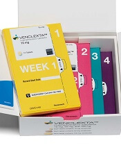
venetoclax (US version)
Photo courtesy of Abbvie
The Australian Therapeutic Goods Administration (TGA) has approved the BCL-2 inhibitor venetoclax (Venclexta™, formerly ABT-199) for use in certain patients with chronic lymphocytic leukemia (CLL).
The drug is now approved to treat Australian patients with relapsed or refractory CLL who have 17p deletion or no other treatment options.
Venetoclax is being developed by AbbVie and Genentech, a member of the Roche Group. The drug is jointly commercialized by the companies in the US and by AbbVie outside of the US.
Now that venetoclax has been approved by the TGA, it can be registered on the Australian Register of Therapeutic Goods and legally marketed and sold in Australia.
To make the drug affordable to the Australian public, the manufacturer can apply to the Pharmaceutical Benefits Advisory Committee to have the cost of the drug subsidized by the Australian government on the Pharmaceutical Benefits Scheme (PBS).
Venetoclax is not listed on the PBS. Historically, the delay between TGA approval and PBS listing ranges from 14 months to 31 months for cancer drugs.
Phase 2 trials
Venetoclax has produced high objective response rates (ORR) in two phase 2 trials of CLL patients.
In one of these trials, researchers tested venetoclax in 107 patients with previously treated CLL and 17p deletion. The results were published in The Lancet Oncology in June 2016.
The ORR in this trial was 79%. At the time of analysis, the median duration of response had not been reached. The same was true for progression-free survival and overall survival.
The progression-free survival estimate for 12 months was 72%, and the overall survival estimate was 87%.
The incidence of treatment-emergent adverse events was 96%, and the incidence of serious adverse events was 55%.
Grade 3 laboratory tumor lysis syndrome (TLS) was reported in 5 patients. Three of these patients continued on venetoclax, but 2 patients required a dose interruption of 1 day each.
In the second trial, researchers tested venetoclax in 64 patients with CLL who had failed treatment with ibrutinib and/or idelalisib. Results from this trial were presented at the 2016 ASH Annual Meeting.
The ORR was 67%. At 11.8 months of follow-up, the median duration of response, progression-free survival, and overall survival had not been reached. The estimated 12-month progression-free survival was 80%.
The incidence of adverse events was 100%, and the incidence of serious adverse events was 53%. No clinical TLS was observed, but 1 patient met Howard criteria for laboratory TLS.
In the past, TLS has caused deaths in patients receiving venetoclax. In response, AbbVie stopped dose-escalation in patients receiving the drug and suspended enrollment in phase 1 trials.
However, researchers subsequently found that a modified dosing schedule, prophylaxis, and patient monitoring can reduce the risk of TLS. ![]()
NCI launches program to expedite drug research

Photo by Bill Branson
The National Cancer Institute (NCI) has launched a new drug formulary designed to provide investigators at NCI-designated cancer centers with quicker access to approved and investigational agents for use in preclinical studies and clinical trials.
The formulary will enable NCI to act as an intermediary between investigators and participating pharmaceutical companies, facilitating the arrangements for access to and use of pharmaceutical agents.
Following company approval, investigators will be able to obtain agents from the formulary and test them in preclinical or clinical studies, including studies combining formulary agents from different companies.
The NCI says having agents available through the formulary will expedite the start of clinical trials by alleviating the lengthy negotiation process—sometimes up to 18 months—that has been required for investigators to access such agents on their own.
“The NCI Formulary will help researchers begin testing promising drug combinations more quickly, potentially helping patients much sooner,” said NCI Acting Director Douglas Lowy, MD.
“Rather than spending time negotiating agreements, investigators will be able to focus on the important research that can ultimately lead to improved cancer care.”
The NCI Formulary includes 15 targeted agents:
- Alectinib (ALK inhibitor, tyrosine kinase inhibitor)
- Atezolizumab (PD-L1 blocking monoclonal antibody)
- Bevacizumab (anti-angiogenesis inhibitor, monoclonal antibody)
- Cobimetinib (MEK1/2 inhibitor)
- Ensartinib (ALK inhibitor)
- Ipilimumab (anti-CTLA-4 monoclonal antibody)
- Larotrectinib (tyrosine kinase inhibitor)
- LY3039478 (Notch inhibitor)
- Nivolumab (PD-1 blocking monoclonal antibody)
- Obinutuzumab (anti-CD20 monoclonal antibody)
- Pertuzumab (anti-HER2 monoclonal antibody)
- Prexasertib (checkpoint kinase 1 inhibitor)
- Trastuzumab (anti-HER2 monoclonal antibody)
- Vemurafenib (BRAF mutant v600 inhibitor)
- Vismodegib (Hedgehog inhibitor)
The agents are products of 6 different pharmaceutical companies: Bristol-Myers Squibb, Eli Lilly and Company, Genentech, Kyowa Hakko Kirin, Loxo Oncology, and Xcovery Holding Company LLC.
“The agreements with these companies demonstrate our shared commitment to expedite cancer clinical trials and improve outcomes for patients,” said James Doroshow, MD, NCI deputy director for clinical and translational research.
“We are very pleased that several additional pharmaceutical companies have already pledged a willingness to participate and are in various stages of negotiation with NCI. By the end of 2017, we expect to have doubled the number of partnerships and drugs available in the NCI Formulary.” ![]()
Intervention may improve warfarin management

Photo courtesy of NIGMS
Researchers have developed an intervention intended to help hospitals and clinicians improve the management of patients on warfarin.
The team implemented the intervention at 8 medical centers in the New England region of the Veterans Health Administration (VA).
These
centers saw an improvement in time in the therapeutic range
(TTR) that was significantly better than the improvement seen in medical
centers without the intervention.
“Insufficient attention has been given in the past to how we can improve the management of warfarin,” said Adam Rose, MD, of Boston University School of Medicine in Massachusetts.
“This study demonstrates that a relatively simple approach can have a large impact.”
Dr Rose and his colleagues described this study in the Annals of Pharmacotherapy.
Intervention
The researchers implemented the intervention at 8 VA sites in New England known as Veterans Integrated Service Network 1 (VISN 1).
The team described the intervention as performance measurement augmented by targeted audit and feedback. Performance was measured via an online reporting system—known as a dashboard—that provided real-time data to clinicians.
The dashboard reported TTR at the patient and site level. The dashboard also reported processes of care that have been linked to TTR, including timely follow-up when the international normalized ratio (INR) is very low or very high, gaps in monitoring, and the proportion of patients with a mean INR value between 2.3 and 2.7.
The dashboard allowed clinicians to call patients who had been lost to follow-up, had low TTR, or were in need of urgent follow-up as a result of extreme INR values.
Results
The researchers examined changes in anticoagulation control, measured as TTR, after the intervention was implemented. They compared changes at VISN 1 sites with 116 other VA sites.
A total of 11,794 patients within VISN 1 and 1,248,782 patients outside of VISN 1 received warfarin for at least part of the study period.
At VISN 1 sites, TTR improved from 66.4% to 69.2%. At the other sites, TTR improved from 65.9% to 66.4% (P<0.001 for the between-group difference).
The researchers said improvement in TTR correlated strongly with the extent of improvement on process-of-care measures, which varied widely across VISN 1 sites.
“Patients who used these measures did better clinically than those in the control group,” Dr Rose said. “This study serves as a model for how other sites and health networks could feasibly approach improving the management of warfarin in their systems.”
“If all anticoagulation clinics in the VA were to achieve this level of improvement, it would prevent 48 strokes and 68 major bleeding events each year, with a savings to the VA system of more than $4 million annually.” ![]()
A new approach to treat MLL-rearranged leukemia?

Investigators may have discovered a new way to treat mixed-lineage leukemia (MLL)-rearranged leukemia, according to research published in Cell.
The team found they could disrupt the balance between wild-type MLL proteins and MLL chimeras.
This impeded MLL leukemia cell proliferation in vitro, delayed disease progression in a mouse model of MLL-AF9 leukemia, and prolonged survival in the mice.
The investigators are now attempting to translate these findings to the clinic.
“We’ve spent the last 20 years in my laboratory trying to molecularly understand how MLL translocations cause this rare and devastating form of leukemia in children so that we can use this information to develop an effective therapy for this cancer,” said lead investigator Ali Shilatifard, PhD, of Northwestern University Feinberg School of Medicine in Chicago, Illinois.
“Now, we’ve made a fundamentally important breakthrough.”
The investigators found that wild-type MLL protein is less stable than the MLL chimeras in MLL leukemia cells. They therefore theorized that stabilizing the wild-type copy of the protein would displace the mutated version that drives MLL-rearranged leukemia.
The team set out to identify factors regulating MLL protein degradation and found the ubiquitin-conjugating enzyme E2O (UBE2O).
The investigators said UBE2O regulates the stability of wild-type MLL in response to interleukin-1 signaling. And inhibiting interleukin-1 receptor-associated kinases (IRAKs) increases the stability and chromatin occupancy of wild-type MLL.
The team also found that IRAK inhibition displaces the MLL chimera and subunits of the super elongation complex at a subset of target genes (LGALS1, LMO2, and GNA15).
To determine the implications of these findings for treatment, the investigators tested an IRAK4 inhibitor in patient-derived cell lines, including MLL leukemia and non-MLL leukemia/lymphoma cells. The inhibitor preferentially impeded the growth of MLL-rearranged leukemia cells.
The team also tested IRAK inhibitors in a murine MLL-AF9 leukemia transplantation model. They injected the animals with IRAK inhibitors on day 19 after transplant, which is just before the mice succumb to leukemia.
The mice received injections with an IRAK1/4 inhibitor (8 mg/kg), an IRAK4 inhibitor (75 mg/kg), or vehicle control every other day for 10 days.
The investigators said both IRAK inhibitors significantly extended survival beyond the 27-day mark, when all of the vehicle-treated mice had succumbed to the disease. Two mice treated with an IRAK inhibitor (1 mouse for each drug) were still alive at day 55.
The team also treated mice with the IRAK inhibitors or vehicle control at 10 days after transplant.
Eight of the 10 mice that received the IRAK1/4 inhibitor had not developed MLL-AF9 leukemia as of day 55. And the same was true for 4 of the 9 mice that received the IRAK4 inhibitor.
However, all of the vehicle-treated mice had succumbed to the disease by the 31-day mark.
The investigators said they are now synthesizing better compounds and hope to eventually launch a phase 1 trial to test these compounds in Chicago. ![]()
ACC releases guidance on anticoagulant use in NVAF

The American College of Cardiology (ACC) has published a guidance document for periprocedural management of anticoagulation in patients with nonvalvular atrial fibrillation (NVAF).
The document includes recommendations on how and when to stop anticoagulants in NVAF patients undergoing surgery, deciding if a substitute medication should be used, and determining when it is safe for patients to resume anticoagulants after surgery.
The document was published in the Journal of the American College of Cardiology.
“With this new decision pathway [document], physicians will be able to make better-informed decisions, and this will contribute to improved patient outcomes,” said John U. Doherty, MD, chair of the document writing committee.
“In North America alone, more than 250,000 nonvalvular atrial fibrillation patients undergo surgery annually, so this document will impact many people.”
The document provides guidance on:
- The overall decision to keep a patient chronically on an anticoagulant by examining whether anticoagulation is warranted based on overall thrombotic risk.
- The decision to take the patient off an anticoagulant temporarily.
- How to temporarily stop the use of vitamin K antagonists and direct-acting oral anticoagulants.
- Deciding if bridging a patient—temporarily discontinuing an oral anticoagulant and replacing it with a subcutaneous or intravenous anticoagulant—before, during, and after surgery is the best choice.
- Deciding how to bridge before, during, and after surgery.
- Deciding how and when to restart the patient’s regular anticoagulant after surgery.
Team uses light to launch drugs from RBCs

Researchers say they’ve developed a technique that uses light to activate a drug stored in circulating red blood cells (RBCs) so the drug is released exactly when and where it’s needed.
The group believes the work could have profound implications for the field of drug delivery.
They say the technique could drastically reduce the amount of drug needed to treat diseases and therefore decrease the risk of side effects.
“Using light to treat a disease site has a lot of benefits beyond the ‘isn’t-that-cool’ factor,” said study author David Lawrence, PhD, of the University of North Carolina at Chapel Hill.
“Those benefits could include avoiding surgery and the risk of infection, making anesthesia unnecessary, and allowing people to treat themselves by shining a light on a problem area, such as an arthritic knee.”
Dr Lawrence and his colleagues described their technique in Angewandte Chemie.
The researchers attached various drug molecules (methotrexate, colchicine, and paclitaxel) to vitamin B12 and loaded the compounds into RBCs, which can circulate for up to 4 months, potentially providing a lasting reservoir of treatment that could be tapped as needed.
The team then demonstrated their ability to overcome a long-time technical hurdle: using long-wavelength light to penetrate deep enough into the body to break molecular bonds; in this case, the drug linked to vitamin B12.
Long-wavelength light can penetrate much more deeply into the body, but it doesn’t carry as much energy as short-wavelength light and cannot typically break molecular bonds.
To activate the drugs with long-wavelength light, the researchers had to determine how to do it in a way that required less energy.
“That’s the trick, and that’s where we’ve been successful,” Dr Lawrence said.
The team solved the energy problem by introducing a weak energy bond between vitamin B12 and the drug and then attaching a fluorescent molecule to the bond.
The fluorescent molecule acts as an antenna, capturing long-wavelength light and using it to cut the bond between the drug and the vitamin carrier.
Dr Lawrence noted that this technique could prove useful in treating cancers for which patients may need to receive a wide array of anticancer agents.
“The problem is, when you start using 4 or 5 very toxic drugs, you’re going to have intolerable side effects,” he said. “However, by focusing powerful drugs at a specific site, it may be possible to significantly reduce or eliminate the side effects that commonly accompany cancer chemotherapy.”
Dr Lawrence has created a company in partnership with the University of North Carolina, Iris BioMed, to further develop the technology to be used in humans. ![]()
Researchers say they’ve developed a technique that uses light to activate a drug stored in circulating red blood cells (RBCs) so the drug is released exactly when and where it’s needed.
The group believes the work could have profound implications for the field of drug delivery.
They say the technique could drastically reduce the amount of drug needed to treat diseases and therefore decrease the risk of side effects.
“Using light to treat a disease site has a lot of benefits beyond the ‘isn’t-that-cool’ factor,” said study author David Lawrence, PhD, of the University of North Carolina at Chapel Hill.
“Those benefits could include avoiding surgery and the risk of infection, making anesthesia unnecessary, and allowing people to treat themselves by shining a light on a problem area, such as an arthritic knee.”
Dr Lawrence and his colleagues described their technique in Angewandte Chemie.
The researchers attached various drug molecules (methotrexate, colchicine, and paclitaxel) to vitamin B12 and loaded the compounds into RBCs, which can circulate for up to 4 months, potentially providing a lasting reservoir of treatment that could be tapped as needed.
The team then demonstrated their ability to overcome a long-time technical hurdle: using long-wavelength light to penetrate deep enough into the body to break molecular bonds; in this case, the drug linked to vitamin B12.
Long-wavelength light can penetrate much more deeply into the body, but it doesn’t carry as much energy as short-wavelength light and cannot typically break molecular bonds.
To activate the drugs with long-wavelength light, the researchers had to determine how to do it in a way that required less energy.
“That’s the trick, and that’s where we’ve been successful,” Dr Lawrence said.
The team solved the energy problem by introducing a weak energy bond between vitamin B12 and the drug and then attaching a fluorescent molecule to the bond.
The fluorescent molecule acts as an antenna, capturing long-wavelength light and using it to cut the bond between the drug and the vitamin carrier.
Dr Lawrence noted that this technique could prove useful in treating cancers for which patients may need to receive a wide array of anticancer agents.
“The problem is, when you start using 4 or 5 very toxic drugs, you’re going to have intolerable side effects,” he said. “However, by focusing powerful drugs at a specific site, it may be possible to significantly reduce or eliminate the side effects that commonly accompany cancer chemotherapy.”
Dr Lawrence has created a company in partnership with the University of North Carolina, Iris BioMed, to further develop the technology to be used in humans.
Transfusing oldest blood may harm patients
Transfusions of blood that has been stored for 6 weeks can release large and potentially harmful amounts of iron into patients’ bloodstreams, a new study suggests.
Based on these findings, researchers are recommending the US Food and Drug Administration (FDA) reduce the maximum storage limit of red blood cells (RBCs) from 6 weeks to 5 weeks, as long as there is a sufficient supply of blood.
“Our recommendation will be controversial, but we think we have real data to support it,” said study author Steven Spitalnik, MD, of Columbia University College of Physicians and Surgeons — New York Presbyterian Hospital in New York, New York.
“Recent studies have concluded that transfusing old blood has no impact on patient outcomes, but those studies didn’t exclusively examine the oldest blood available for transfusions. Our new study found a real problem when transfusing blood that’s older than 5 weeks.”
Dr Spitalnik and his colleagues described their study in The Journal of Clinical Investigation.
The researchers randomly assigned a group of 60 healthy volunteers to receive a unit of RBCs that had been stored for 1, 2, 3, 4, 5, or 6 weeks. The volunteers were then monitored for 20 hours after transfusion.
The researchers found that a longer duration of RBC storage was associated with a progressive increase in extravascular hemolysis, decreasing post-transfusion RBC recovery, decreasing elevations in hematocrit, and increasing serum ferritin.
None of the volunteers were harmed by the transfusions, but subjects who received 6-week-old blood had outcomes associated with an increased risk of harm.
In 7 of the 9 subjects who received the 6-week-old blood (78%), the amount of iron entering the circulation exceeded the iron-uptake capacity of transferrin, producing circulating nontransferrin-bound iron. The same effect occurred in 1 of the 10 subjects who received 5-week-old blood (10%, P=0.003).
The area under the curve of the change in nontransferrin-bound iron was significantly higher for subjects who received the 6-week-old blood than for all other subjects (P<0.001). The same was true for serum iron (P<0.01) and transferrin saturation (P<0.001).
“Based on the amount of iron circulating in the blood of the volunteers who received 6-week-old blood, we’d predict that certain existing infections could be exacerbated,” said study author Eldad Hod, MD, also of Columbia University College of Physicians and Surgeons — New York Presbyterian Hospital.
“Thus, for ill, hospitalized patients, this excess iron could lead to serious complications,” Dr Spitalnik added.
The researchers said the true impact of 6-week-old blood on the rate of complications is likely to be small. However, since millions of Americans receive transfusions each year, even a 1% difference in complications could affect a large number of patients.
“It’s estimated that up to 10% to 20% of blood units used for transfusions have been stored for more than 5 weeks, so the number of patients who are likely to receive a unit of very old blood is substantial,” Dr Hod noted.
“Based on our findings of potential harm, we think the prudent thing to do, at this time, is for the FDA to reduce the maximum storage period,” Dr Spitalnik added.
“The UK, Ireland, the Netherlands, and the National Institutes of Health have limited storage to 35 days, and we think that can be achieved throughout the US without seriously affecting the blood supply.”
This study was supported by grants from the National Institutes of Health (HL115557 and UL1TR000040).
Combo granted orphan designation for CLL

The US Food and Drug Administration (FDA) has granted orphan drug designation for the combination of TG-1101 (ublituximab) and TGR-1202 for the treatment of patients with chronic lymphocytic leukemia (CLL).
Ublituximab is a glycoengineered anti-CD20 monoclonal antibody, and TGR-1202 is a next-generation PI3K delta inhibitor. Both drugs are being developed by TG Therapeutics, Inc.
Researchers have evaluated ublituximab and TGR-1202 in combination in a phase 1 trial of patients with relapsed or refractory CLL/small lymphocytic lymphoma (SLL) and non-Hodgkin lymphomas (NHLs).
Results were presented at the 2015 ASH Annual Meeting.
There was a 3+3 dose-escalation portion of the study and a dose-expansion phase. The patients received TGR-1202 at doses ranging from 400 mg to 1200 mg and 2 different doses of ublituximab—900 mg for patients with NHL and 600 mg or 900 mg for patients with CLL/SLL.
As of ASH, there were 58 patients evaluable for efficacy and 71 evaluable for safety.
There were 10 CLL/SLL patients exposed to higher doses of TGR-1202. Among these patients, the overall response rate was 80%. Seven patients achieved a partial response, 1 achieved a complete response, and the remaining 2 patients had stable disease.
For the entire safety population, the most common adverse events were nausea (46%), diarrhea (44%), fatigue (41%), neutropenia (30%), and infusion-related reactions (25%).
Grade 3/4 adverse events included neutropenia (25%), diarrhea (3%), fatigue (3%), dyspnea (3%), pyrexia (3%), nausea (1%), infusion-related reactions (1%), sinusitis (1%), anemia (1%), hypophosphatemia (1%), and peripheral edema (1%).
Now, the combination of ublituximab and TGR-1202 is being evaluated in the UNITY-CLL phase 3 trial for patients with previously treated or untreated CLL.
“[W]ith enrollment into our UNITY-CLL phase 3 trial currently exceeding our expectations, we expect to be able to commence a regulatory filing for the combination in 2018, and having orphan drug designation will provide certain cost-saving advantages for us during the regulatory approval process,” said Michael S. Weiss, executive chairman and chief executive officer of TG Therapeutics.
The FDA grants orphan designation to drugs and biologics intended to treat, diagnose, or prevent diseases/disorders that affect fewer than 200,000 people in the US.
The designation provides incentives for sponsors to develop products for rare diseases. This may include tax credits toward the cost of clinical trials, prescription drug user fee waivers, and 7 years of market exclusivity if the product is approved.
Iron deficiency anemia protects kids from malaria better than sickle cell trait

Photo by Aurimas Rimsa
New research suggests that, for young children, iron deficiency anemia offers a
greater protective effect against malaria than sickle cell trait.
The
study indicates that iron deficiency anemia can protect children age 2 and
younger from the blood stage of Plasmodium falciparum malaria, and
treating the anemia with iron supplementation removes this protective
effect.
The researchers reported these findings in EBioMedicine.
“This study is elegant in its simplicity yet remains one of the most
substantial and systematic attempts to unveil the cellular-level
relationship between anemia, iron supplementation, and malaria risk,”
said study author Carla Cerami, MD, PhD, of the Medical Research Council
Unit The Gambia in Banjul, Gambia.
For this study, she and her colleagues analyzed the red blood cells of 135 anemic children who were participating in an iron supplementation trial.
The children ranged in age from 6 months to 24 months and lived in a malaria-endemic region of The Gambia where sickle cell trait was also common.
The children received iron (12 mg/day) through micronutrient powder for 84 days, and the researchers analyzed the children’s red blood cells at baseline, day 49, and day 84.
The team conducted in vitro growth and invasion assays with P falciparum laboratory and field strains. The study’s primary endpoint was in vitro parasite growth in the children’s RBCs.
The researchers found that “anemia substantially reduced the invasion and growth of both laboratory and field strains of P falciparum.” The team noted a roughly 10% growth reduction per standard deviation shift in hemoglobin.
On a population-wide basis, anemia reduced the blood stage of malaria by 15.9%, while the sickle cell trait reduced it by 3.5%.
“Our finding that anemia offers greater natural protection against blood-stage malaria infection than sickle cell trait has led us to formulate the interesting hypothesis that the widespread prevalence of anemia in people of African descent is a genetic signature of malaria,” said study author Morgan Goheen, PhD, of University of North Carolina in Chapel Hill.
The researchers also found that deficits in invasion and growth for blood-stage P falciparum were reversed when anemic children had received 7 weeks of iron supplementation. Parasite growth was 2.4-fold higher after supplementation than it was at baseline (P<0.001).
Prior work by the same research group suggested the increased invasion and growth rates following iron supplementation are caused by the parasites’ strong preference for young red blood cells.
The researchers said these new field results consolidate the evidence that iron supplementation increases the risk of P falciparum malaria and provide support for the use of malaria prophylaxis in
conjunction with iron supplementation, especially during the early
phases of erythroid recovery.