User login
How multiple infections make malaria worse

Image by Ute Frevert
and Margaret Shear
New research suggests that infections with 2 types of malaria parasite lead to greater health risks because 1 species helps the other thrive.
Investigators sought to understand what happens when the 2 most common malaria parasites cause infection at the same time, as they are known to attack the body in different ways.
The team found the first parasite helps provide the second with more of the resources it needs to prosper.
“Immune responses are assumed to determine the outcome of interactions between parasite species, but our study clearly shows that resources can be more important,” said Sarah Reece, of the University of Edinburgh in Scotland.
“Our findings also challenge ideas that 1 species will outcompete the other, which explains why infections involving 2 parasite species can pose a greater health risk to patients.”
Dr Reece and her colleagues recounted these findings in Ecology Letters.
In humans, the malaria parasite Plasmodium falciparum infects red blood cells of all ages, while the Plasmodium vivax parasite attacks only young red blood cells.
The current study, conducted in mice with equivalent malaria parasites (P chabaudi and P yoelii), showed that the body’s response to the first infection produces more of the type of red blood cell the second parasite needs.
In response to the first infection, millions of red blood cells are destroyed. The body responds by replenishing these cells.
The fresh cells then become infected by the second type of parasite, making the infection worse.
The investigators said these results appear to explain why infections with both P falciparum and P vivax often have worse outcomes for patients than infections with a single malaria parasite. ![]()
Study explains link between malignant hyperthermia and bleeding abnormalities

A new study helps explain why some patients with malignant hyperthermia may suffer from excessive bleeding.
The findings suggest a mutation that causes malignant hyperthermia can disrupt calcium signaling in vascular smooth muscle cells, leading to bleeding abnormalities.
What’s more, researchers found that a drug clinically approved to treat muscle-related symptoms in malignant hyperthermia helped stop bleeding.
Rubén Lopez, of Basel University Hospital in Switzerland, and his colleagues conducted this research and reported their findings in Science Signaling.
Patients with malignant hyperthermia experience dangerously high fever and severe muscle contractions when exposed to general anesthesia.
Malignant hyperthermia is often caused by mutations in the RYR1 gene, which encodes a calcium channel in skeletal muscle called ryanodine receptor type 1 (RyR1).
For some patients with these mutations, malignant hyperthermia is accompanied by a mild bleeding disorder, but whether the 2 conditions are connected has not been clear.
Working in a mouse model of malignant hyperthermia, researchers found that vascular smooth muscle cells with mutated RyR1 displayed frequent spikes in calcium levels, known as calcium sparks. These sparks led to excessive vasodilation and prolonged bleeding.
Blocking the receptor with dantrolene, a drug used to treat malignant hyperthermia, helped reduce bleeding in the mice and in a single human patient, pointing to an unexpected benefit from the drug.
The findings suggest that mutations in RyR1, which is also found in other types of smooth muscle cells such as those in the bladder and uterus, may have a wider range of effects than previously thought. ![]()
Team identifies potential therapeutic target for AML

New research suggests that E proteins and their antagonists, Id proteins, can play key roles in acute myeloid leukemia (AML).
The study showed that overexpression of the Id2 protein or knockdown of the E2-2 protein can suppress both mixed-lineage leukemia (MLL)-rearranged AML and t(8;21) AML.
These findings, published in Cancer Cell, suggest the Id2/E-protein axis may be a promising therapeutic target for AML.
“There is a particularly urgent need for new, targeted, drug-based therapies for AML, and with every discovery of what’s driving the cancer, we take a step closer to achieving that,” said study author Ricky Johnstone, PhD, of Peter MacCallum Cancer Centre in Melbourne, Victoria, Australia.
“What we found in this case was the suppression of Id2 protein plays an important, and previously unrecognized, role in allowing MLL re-arranged AML cancer cells to take hold and spread. Drugs that influence levels of this protein, or stop it being suppressed by the cancer, could provide a much-needed new avenue to combatting this disease.”
The researchers first found that Id2 regulates leukemia stem cell (LSC) potential. Specifically, low Id2 expression is associated with LSC enrichment, and Id2 overexpression hinders leukemia development.
Further investigation revealed that the fusion protein MLL-AF9 suppresses Id2 and activates E2-2 expression, while E2-2 depletion phenocopies Id2 overexpression in MLL-AF9-AML cells.
The team also found that Id2’s tumor-suppressive function is conserved in t(8;21) AML. And low expression of Id2 and its associated gene signature are associated with poor prognosis in patients with MLL-rearranged AML or t(8;21) AML. ![]()
Study reveals SNPs that may increase risk of MM

A large study has revealed several genetic variations that may increase a person’s risk of developing multiple myeloma (MM).
The findings, published in Nature Communications, build on existing research that suggests MM can run in families.
“Our study expands our understanding of how inherited risk factors can influence the risk of myeloma,” said Richard Houlston, MD, PhD, of The Institute of Cancer Research in London, UK.
“We know that the inherited risk of myeloma does not come from just one or two major risk genes, as can be the case with breast cancer, but from multiple different genetic variants, each with only a small individual effect on risk. Identifying more of these variants gives us new insights into the potential causes of the disease and open up new strategies for prevention.”
For this study, Dr Houlston and his colleagues compared DNA from 9866 MM patients and 239,188 healthy adults.
This confirmed the association between MM and 9 previously reported single nucleotide polymorphisms (SNPs):
- rs6746082 at 2p23.3
- rs1052501 at 3p22.1
- rs4487645 at 7p15.3
- rs10936599 at 3q26.2
- rs2285803 at 6p21.3
- rs4273077 at 17p11.2
- rs877529 at 22q13.1
- rs56219066 at 5q15
- rs138740 at 22q13.
It also revealed 8 new SNPs that may increase the risk of MM:
- rs34229995 at 6p22.3 (P=1.31 × 10⁻⁸)
- rs9372120 at 6q21 (P=9.09 × 10⁻¹⁵)
- rs7781265 at 7q36.1 (P=9.71 × 10⁻⁹)
- rs1948915 at 8q24.21 (P=4.20 × 10⁻¹¹)
- rs2811710 at 9p21.3 (P=1.72 × 10⁻¹³)
- rs2790457 at 10p12.1 (P=1.77 × 10⁻⁸)
- rs7193541 at 16q23.1 (P=5.00 × 10⁻¹²)
- rs6066835 at 20q13.13 (P=1.36 × 10⁻¹³).
These SNPs are located in regions of the genome involved in regulating genes linked to cell processes known to go wrong in MM development—namely, JARID2, ATG5, SMARCD3, CCAT1, CDKN2A, WAC, RFWD3, and PREX1.
This suggests that subtle effects on the activity of key genes could mean the proper development of plasma cells breaks down, increasing the likelihood of developing MM. However, as the researchers noted, further study is needed to confirm and better understand this phenomenon. ![]()
Efficacy of malaria vaccine declines over time
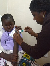
Photo by Caitlin Kleiboer
Results from a phase 2 study of the malaria vaccine RTS,S (also known as RTS,S/AS01 or Mosquirix) suggest its efficacy decreases over time, and this decline is fastest in children living in areas with higher-than-average rates of malaria.
Researchers say the results suggest the benefits of the vaccine are likely to vary across different populations and highlight the need for more research to
determine the most effective way of using RTS,S, which last year became the first malaria vaccine to receive a green light from the European Medicines Agency.
“We found that 3-dose vaccination with RTS,S was initially protective, but this was offset by a rebound in later years among children exposed to higher-than-average levels of malaria-carrying mosquitoes,” said Philip Bejon, PhD, of the Kenya Medical Research Institute–Wellcome Trust Programme in Kilifi, Kenya.
Dr Bejon and his colleagues reported these results in NEJM.
The researchers followed 447 children who had received 3 doses of either RTS,S or a rabies (control) vaccine when they were 5 months to 17 months old.
After 7 years, there were 312 children still involved in the study. During the first year, the risk of getting malaria in the vaccinated children was 35.9% less than in the control group. After 7 years, this protection fell to 3.6%.
And in children exposed to higher-than-average rates of malaria, there were slightly more cases of malaria in the vaccinated group than the control group—1002 and 992 cases, respectively—5 years after vaccination.
This “rebound” effect, which has been seen in previous studies, is thought to occur because children initially protected by the vaccine develop their natural immunity against malaria more slowly than unvaccinated children.
Results from a phase 3 study showed that 3 doses of RTS,S reduced the risk of malaria in young children by 28% over 4 years, but this improved to 36% when children were given a fourth dose 18 months after the first dose. Longer-term follow up of these children is ongoing.
“Overall, our study shows that RTS,S can benefit children but suggests that a fourth dose may be important for sustaining this protection over the long term and to protect against a potential rebound,” said Ally Olotu, PhD, of the Kenya Medical Research Institute–Wellcome Trust Programme.
“Results from 3 sites involved in the original phase 3 study that are continuing follow up, and the WHO’s planned pilot program, will tell us more about the vaccine’s efficacy in different settings and help determine which populations would benefit most from receiving it as part of a wider vaccination strategy.” ![]()
Telomere length linked to neutrophil recovery in AML

Image by Volker Brinkmann
Researchers say they have discovered a way to predict which children with acute myeloid leukemia (AML) are at the highest risk of delayed neutrophil recovery.
The team examined the role of telomeres in neutrophil recovery and found that the length of a patient’s telomeres can indicate the rate of recovery following chemotherapy.
The group reported their findings in the Journal of Clinical Oncology.
“We were interested in telomere length as a marker of blood count recovery because defects in telomere maintenance are known risks for bone marrow failure and aplastic anemia,” said study author Maria Monica Gramatges, MD, PhD, of Baylor College of Medicine in Houston, Texas.
“We know that up to 15% to 20% of children can take 2 months or longer to recover their blood counts after a course of AML chemotherapy. Our goal was to understand if these children had an underlying genetic predisposition associated with an impaired capacity for recovery.”
Dr Gramatges and her colleagues hypothesized that short telomere length could be associated with a delay in neutrophil recovery.
So they obtained bone marrow samples from AML patients who recovered as expected (within 30 days) after each chemotherapy course (n=62), and from AML patients who experienced significant delays in recovery after chemotherapy (n=53).
The team then measured telomere length on each subject and categorized the group by quartile, from shortest to longest.
Subjects in the quartile with the shortest telomere lengths took the longest to recover, especially during the last 2 courses of chemotherapy. In an adjusted analysis, lower telomere content was significantly associated with prolonged neutropenia after the fourth (P=0.002) and fifth courses of chemotherapy (P=0.009).
The researchers said these results support the hypothesis that telomeres are an indicator of capacity for neutrophil recovery following chemotherapy.
Dr Gramatges hopes the results of this study will be helpful in further understanding which children are at a higher risk for prolonged myelosuppression and how to target those children with modified treatments, improved supportive care, and closer monitoring in order to prevent potential complications such as severe infections.
“A significant proportion of children with AML suffer from treatment-related toxicities, with some succumbing to complications of the therapies we give, rather than from the actual cancer itself,” Dr Gramatges said.
“We hope this research will help us identify those who are at a higher risk for delayed recovery and use this knowledge to reduce the morbidity and mortality associated with AML treatment.” ![]()
Old drug could treat CMV

Image by Ed Uthman
Research published in PLOS Pathogens suggests that emetine, a drug once used to treat amebiasis and induce vomiting in cases of poisoning, may also halt replication of cytomegalovirus (CMV).
Although there are drugs available to treat CMV, they can cause serious toxicity.
Furthermore, resistant strains of CMV can emerge, creating “a desperate need for other ways to control this virus,” according to Ravit Boger, MD, of the Johns Hopkins University School of Medicine in Baltimore, Maryland.
Dr Boger and her colleagues decided to search for drugs to treat CMV by screening 1280 pharmacologically active compounds already approved by the US Food and Drug Administration.
The researchers found a “hit” with emetine, a drug that was used in the past to treat amebiasis before other drugs took its place decades ago.
Results showed that emetine can inhibit CMV at much lower doses than those used for amebiasis, and less frequent doses might be effective for CMV inhibition as well.
In vitro and in vivo tests showed that very low doses of emetine significantly reduced viral replication—75 nm in vitro and 0.1 mg/kg in mice.
Furthermore, with a half-life of 35 hours, the drug exerted its effects over a sustained period, effectively inhibiting virus replication at 14 days after 3 doses.
Additional investigation revealed that emetine’s action against CMV was due to its effects on proteins that control the cell cycle.
Dr Boger cautioned that there is a long way to go before emetine or similar agents can be considered for the treatment of CMV.
“But if further research affirms its potential value, emetine might eventually be used in patients who don’t respond to approved anti-CMV drugs, alone or in combination with these,” she said.
Dr Boger and her colleagues also believe that gaining a better understanding of emetine’s activity in cells could lead to the discovery of new drugs that take advantage of the same or similar pathways. ![]()
Image by Ed Uthman
Research published in PLOS Pathogens suggests that emetine, a drug once used to treat amebiasis and induce vomiting in cases of poisoning, may also halt replication of cytomegalovirus (CMV).
Although there are drugs available to treat CMV, they can cause serious toxicity.
Furthermore, resistant strains of CMV can emerge, creating “a desperate need for other ways to control this virus,” according to Ravit Boger, MD, of the Johns Hopkins University School of Medicine in Baltimore, Maryland.
Dr Boger and her colleagues decided to search for drugs to treat CMV by screening 1280 pharmacologically active compounds already approved by the US Food and Drug Administration.
The researchers found a “hit” with emetine, a drug that was used in the past to treat amebiasis before other drugs took its place decades ago.
Results showed that emetine can inhibit CMV at much lower doses than those used for amebiasis, and less frequent doses might be effective for CMV inhibition as well.
In vitro and in vivo tests showed that very low doses of emetine significantly reduced viral replication—75 nm in vitro and 0.1 mg/kg in mice.
Furthermore, with a half-life of 35 hours, the drug exerted its effects over a sustained period, effectively inhibiting virus replication at 14 days after 3 doses.
Additional investigation revealed that emetine’s action against CMV was due to its effects on proteins that control the cell cycle.
Dr Boger cautioned that there is a long way to go before emetine or similar agents can be considered for the treatment of CMV.
“But if further research affirms its potential value, emetine might eventually be used in patients who don’t respond to approved anti-CMV drugs, alone or in combination with these,” she said.
Dr Boger and her colleagues also believe that gaining a better understanding of emetine’s activity in cells could lead to the discovery of new drugs that take advantage of the same or similar pathways.
Computer model shows how spleen filters blood

Researchers have created a computer model that shows how tiny slits in the spleen prevent old, diseased, or misshapen red blood cells from re-entering the bloodstream.
The team says the model can be used to study the spleen’s role in controlling diseases that affect the size and structure of red blood cells—such as sickle cell anemia, thalassemia, and malaria—and to develop new diagnostics and therapeutics for these diseases.
The researchers described the model in PNAS.
Previous studies have shown that part of the spleen’s filtration process relies on having red blood cells squeeze through tiny slits between the endothelial cells that line the spleen’s blood vessels. These “interendothelial slits” are no larger than 1.2 µm tall, 4 µm wide, and 1.9 µm deep.
More rigid and misshapen blood cells might not be able to pass through these narrow passages. And this process cannot be observed in vivo because of the minute size of the slits.
In order to “see” how the interendothelial slits regulate red blood cell circulation, the researchers created a computer simulation based on dissipative particle dynamics, a modeling method.
Their model allowed the team to determine the range of cell sizes and shapes that could fit through the slits. The range closely mirrored the range of sizes and shapes for healthy red blood cells, indicating that only healthy cells should be able to pass through the slits.
“The computational and analytical models from this work, along with a variety of experimental observations, point to a more detailed picture of how the physiology of the human spleen likely influences several key geometrical characteristics of red blood cells,” said study author Subra Suresh, ScD, of Carnegie Mellon University in Pittsburgh, Pennsylvania.
“They also offer better understanding of how the circulatory bottleneck for the red blood cell in the spleen could affect a variety of acute and chronic disease states arising from hereditary disorders, human cancers, and infectious diseases, with implications for therapeutic interventions and drug efficacy assays.”
In addition to giving researchers a better picture of how the spleen functions, the findings provide new insights into drug treatments.
A class of drugs currently in development for treating malaria alters the shape of red blood cells infected with malaria, theoretically preventing them from passing through interendothelial slits. One such drug, spiroindoline KAE609, is in clinical trials.
The researchers’ results might also explain why artemisinin-based antimalarial drugs, which stiffen healthy and malaria-infected red blood cells, can lead to severe anemia.
New type of CAR T cells can produce responses in NHL

Image courtesy of NIAID
Results of a phase 1 study suggest that chimeric antigen receptor T cells specific for the κ light chain (κ.CAR T cells) can produce responses in patients with relapsed or refractory B-cell malignancies, largely without side effects.
The therapy induced complete and partial responses in some patients with non-Hodgkin lymphoma (NHL), and it allowed other patients with chronic lymphocytic leukemia (CLL) or multiple myeloma (MM) to maintain stable disease.
There was 1 adverse event considered possibly related to the treatment.
The researchers reported these results in The Journal of Clinical Investigation.
The κ.CAR T cells were designed to recognize κ-restricted cells and spare normal B cells expressing the nontargeted λ light chain.
“We reasoned that targeting the light chain expressed by malignant B cells should efficiently kill tumor cells while sparing normal B cells expressing the other type of light chain,” said study author Carlos Ramos, MD, of Baylor College of Medicine in Houston, Texas.
He and his colleagues tested the κ.CAR T cells in 16 patients with relapsed or refractory κ+ NHL (n=7), CLL (n=2), or MM (n=7).
The team isolated T cells from these patients and modified the cells so they could target the κ light chain on the surface of malignant B cells. The modified T cells were infused back into the patients, and each patient was monitored for disease progression and side effects.
Eleven patients stopped receiving other treatments at least 4 weeks prior to T-cell infusion. Six patients without lymphopenia received cyclophosphamide at 12.5 mg/kg 4 days before κ.CAR T-cell infusion (0.2×10⁸ to 2×10⁸ κ.CAR T cells/m2).
“We found the treatment to be feasible and safe at all the dose levels studied,” Dr Ramos said.
One MM patient had grade 3 lymphopenia that was deemed possibly related to treatment, but none of the other adverse events were thought to result from the κ.CAR T cells.
Two patients with NHL achieved a complete response to treatment, 1 lasting more than 32 months and the other lasting 6 weeks. A third NHL patient had a partial response lasting 3 months, and a CLL patient had stable disease lasting 6 weeks.
Five of the 7 MM patients had stable disease, 3 lasting 6 weeks, 1 lasting 17 months, and 1 lasting 24 months.
“Our approach, although we are still optimizing it, offers a new possibility for patients in whom other treatments have not been successful,” Dr Ramos concluded.
Health Canada approves mAb for MM

Photo courtesy of Janssen
Health Canada has granted conditional approval, or a Notice of Compliance with Conditions (NOC/c), for daratumumab (Darzalex), a monoclonal antibody (mAb) targeting CD38.
The mAb is now approved to treat patients with multiple myeloma (MM) who have received at least 3 prior lines of therapy, including a proteasome inhibitor (PI) and an immunomodulatory drug (IMiD), or MM patients who are refractory to both a PI and an IMiD.
An NOC/c is authorization to market a drug with the condition that the sponsor—in this case, Janssen Inc.—undertake additional studies to verify a clinical benefit.
The NOC/c policy is designed to provide access to:
- Drugs that can treat serious, life-threatening, or severely debilitating diseases
- Drugs that can treat conditions for which no drug is currently marketed in Canada
- Drugs that provide a significant increase in efficacy or significant decrease in risk when compared to existing drugs marketed in Canada.
Studies of daratumumab
The NOC/c for daratumumab was based on a review of data from the phase 2 SIRIUS study, the phase 1/2 GEN501 study, and additional supportive studies.
The GEN501 study enrolled 102 patients with relapsed MM or relapsed MM that was refractory to 2 or more prior lines of therapy. The patients received daratumumab at a range of doses and on a number of different schedules.
The results suggested daratumumab is most effective at a dose of 16 mg/kg. At this dose, the overall response rate was 36%. Most adverse events in this study were grade 1 or 2, although serious events did occur.
The SIRIUS study enrolled 124 MM patients who had received 3 or more prior lines of therapy. They received daratumumab at different doses and on different schedules, but 106 patients received the drug at 16 mg/kg.
Twenty-nine percent of the 106 patients responded to treatment, and the median duration of response was 7 months. Thirty percent of patients experienced serious adverse events.
Findings from a combined efficacy analysis of the GEN501 and SIRIUS trials demonstrated that, after a mean follow-up of 14.8 months, the estimated median overall survival for patients who received single-agent daratumumab at 16 mg/kg was 20 months.
Five phase 3 clinical studies with daratumumab in MM patients—in relapsed and frontline settings—are ongoing. Additional studies are ongoing or planned to assess the mAb’s potential in other malignant and pre-malignant diseases in which CD38 is expressed.
Janssen has exclusive worldwide rights to the development, manufacturing, and commercialization of daratumumab for all potential indications. The company licensed daratumumab from Genmab A/S in August 2012.