User login
Hybrid drug could treat resistant malaria

Photo by James Gathany
A newly developed hybrid drug can treat malaria that is resistant to other therapies, according to preclinical research.
With previous work, researchers found that chemoreversal agents can re-sensitize resistant malaria parasites to the antimalarial agent chloroquine.
For the current study, the team created hybrid compounds that combine chloroquine and a chemoreversal agent.
One of these compounds, 35, proved particularly active, killing malaria parasites that were resistant to chloroquine and/or artemisinin.
Compound 35 was significantly more effective than chloroquine at killing these resistant strains, which included Hb3 (P<0.001), Dd2 (P<0.001), ARS-233 (P<0.001), ARS-272 (P<0.01), NHP-04559 (P<0.01), NHP04773 (P<0.001), and 7G8 (P<0.01).
In addition, the researchers said compound 35 has a “good therapeutic window” and “favorable drug-like properties,” but they are continuing to refine the compound to make it more effective.
The team noted that malaria drugs and chemoreversal agents have been used to treat drug-resistant malaria before. But this is the first time a hybrid of chloroquine and a newly discovered chemoreversal factor has been used in a single novel molecule for this purpose.
The researchers said a single therapy has several advantages over combination therapy. Besides being more convenient to take, it has less risk of drug-drug interactions, may be better absorbed and distributed in the body, and could result in slower development of new resistant strains of malaria.
Kevin S. W. Tan, PhD, of the National University of Singapore, and his colleagues described this research in Antimicrobial Agents and Chemotherapy. ![]()
Patients may be uninformed about risks of warfarin

ATHENS—A study of patients taking warfarin suggests many do not fully understand the risks associated with the drug.
Researchers asked patients to complete a questionnaire on warfarin use and found that, on average, patients answered 64% of the questions correctly.
The patients tended to be the least informed about food and drug interactions and which side effects necessitate a call or visit to the doctor.
Kjersti Oterhals, RN, PhD, of Haukeland University Hospital in Bergen, Norway, and her colleagues presented these findings at EuroHeartCare 2016 (abstract 36).
“The stroke and bleeding complications from warfarin can be fatal,” Dr Oterhals noted. “Worldwide, warfarin causes the most deaths from drug-related side effects. Patients need to know what foods and drugs have an impact on how warfarin works and what to do if they have symptoms of an overdose or underdose.”
Dr Oterhals and her colleagues evaluated warfarin knowledge in 404 patients with aortic stenosis. The patients’ mean age was 68, and 70% were male.
Nearly two-thirds of the patients (63%) were taking warfarin because they had a mechanical valve, and 24% were taking the drug because they had atrial fibrillation. The remaining patients were taking the drug for unknown reasons (6%) or “other” reasons (7%).
The patients received a postal questionnaire with 28 multiple-choice questions about warfarin. On average, patients answered 18 of the 28 questions correctly. However, 22% of patients answered less than half of the questions correctly.
The questions that were most often answered incorrectly were those concerning food and drug interactions and when to call or see a doctor.
For example, patients were asked which of the following foods would interfere with warfarin: celery, carrots, coleslaw, or green beans. Only 25% correctly said coleslaw. Most patients answered green beans.
“Patients often think green vegetables have the most vitamin K, but that’s not true,” Dr Oterhals said. “Brassica vegetables such as cabbage, broccoli, and cauliflower are rich sources.”
“Patients do not have to avoid these foods, but they should eat an equal amount every week because the vitamin K will decrease their INR and put them at increased risk of thrombosis or embolism. Patients who like to eat a lot of vitamin K-containing foods can take a higher warfarin dosage, but they need to be consistent.”
Dr Oterhals and her colleagues also found that 80% of patients knew they should go directly to the emergency room if they had nose bleeding that would not stop. However, only 45% of patients correctly said diarrhea for more than one day necessitates a visit to the doctor.
The study also showed that increased age was associated with a decrease in correct answers.
“We can only speculate why,” Dr Oterhals said. “Younger people tend to seek out information about how to manage their disease, while the older generation wants the doctor to tell them what to do.”
“Motivated patients should be offered an INR testing kit so that they can monitor their levels and adjust the warfarin dose themselves, just as patients with diabetes who use insulin do. It enables patients to travel and try new foods without having to find a clinic to get tested. Patients tell me that hot weather increases their INR, while another found out while in Asia that nori decreased his INR.”
“Warfarin is a life-saving drug but can be deadly if not used carefully. Health professionals have a responsibility to educate patients, but, unfortunately, even cardiac nurses do not know enough. There is an urgent need to improve health professionals’ warfarin knowledge so they can educate patients.” ![]()
Patients with ASD may have lower cancer risk

Photo by Darren Baker
New research suggests that patients diagnosed with an autism spectrum disorder (ASD) have a higher burden of mutations in oncogenes but lower rates of cancer than the rest of the population.
Investigators analyzed large, publicly available genomic databases of patients with ASD and found that, compared to a set of control subjects, ASD patients had significantly higher rates of DNA variation in oncogenes.
The team followed up this result with an analysis of the University of Iowa Hospitals and Clinics’ electronic medical record and discovered that ASD patients were also significantly less likely to have a co-occurring diagnosis of cancer.
“It’s a very provocative result that makes sense on one level and is extremely perplexing on another,” said Benjamin Darbro, MD, PhD, of the University of Iowa Carver College of Medicine in Iowa City.
Dr Darbro and his colleagues discussed the result, and the research that led to it, in a paper published in PLOS ONE.
The investigators used exome sequencing data from the ARRA Autism Sequencing Collaboration and compared that data to a control cohort from the Exome Variant Server database.
This revealed that rare, coding variants within oncogenes were greatly enriched in the ARRA ASD cohort. By comparison, variants were not significantly enriched in tumor suppressor genes.
To ensure the genetic differences were not technical artifacts but actually bona fide differences in genetic architecture in ASD, the investigators ran numerous controls.
As expected, they found that individuals with ASD had many more DNA variations in genes previously associated with autism, epilepsy, and intellectual disability compared to control individuals.
However, there was no difference between the ASD and control groups when it came to genes involved in other, unrelated conditions such as skeletal dysplasia, retinitis pigmentosa, dilated cardiomyopathy, and non-syndromic hearing loss.
The investigators then turned their attention to the electronic medical record at the University of Iowa Hospitals and Clinics and conducted a retrospective case-control analysis comparing 1837 patients with ASD to 9336 patients with any other diagnosis, and determined what proportion of each group carried a cancer diagnosis.
The team found that, for children and adults with ASD, there appeared to be a protective effect against cancer. The cancer incidence was 1.3% for patients with ASD and 3.9% for controls.
The protective effect was evident in both males and females with ASD, but it was strongest for the youngest group of patients and decreased with age. For ASD patients who were under 14 years of age, the odds of having cancer were reduced by 94% compared to controls.
When the investigators determined the rates of other systemic diseases—such as high blood pressure and diabetes—in the ASD population, they found no relationship.
Furthermore, the team found no relationship with cancer when they examined the rates of other common conditions such as esophageal reflux, allergic rhinitis, atopic dermatitis, and short stature. They said this demonstrated that the inverse relationship observed between ASD and cancer is not due to a technical artifact. ![]()
Short transfusion delays can increase risk of death
![]()
Photo courtesy of UAB Hospital
Even a short delay in the administration of packed red blood cells (pRBCs) can increase the risk of death for some traumatically injured patients, according to research published in the Journal of Trauma and Acute Care Surgery.
The study showed that a delay of 10 minutes was associated with a higher risk of death among patients who required pRBCs early.
However, a 10-minute delay did not increase the risk of death for the entire study cohort.
For this study, researchers tracked trauma patients taken from the scene of their injury to the University of Cincinnati Medical Center by a helicopter service known as Air Care. The service carries 2 units of pRBCs for protocol-driven prehospital transfusion.
“Air Care is the only helicopter in the area to carry blood (and plasma), so we had the research platform to study how early blood transfusions impact outcomes,” said study author Elizabeth Powell, MD, of the University of Cincinnati Medical Center in Ohio.
Dr Powell and her colleagues studied 94 patients who had received at least 1 unit of pRBCs within 24 hours of arriving at the hospital. Ninety-three percent of patients (n=87) were Caucasian, 70% (n=66) were male, and they had a mean age of 43.
Ninety-four percent of patients (n=88) had sustained blunt force injuries, and 33% (n=31) died within 30 days of hospital arrival.
Thirty-three percent of patients (n=31) received a transfusion during transport, 54% (n=51) were transfused within an hour of hospital arrival, and 13% (n=12) were transfused after the first hour but within 24 hours of hospital arrival.
When considering all 94 patients together, the researchers found that a 10-minute increase in time to pRBC administration did not significantly affect the odds of death, even when adjusting for injury severity. The odds ratio was 1.00 (P=0.575).
However, among the 82 patients who received their first pRBC transfusion during transport or within an hour of hospital arrival, each 10 minute increase in time to transfusion increased the odds of death. When the researchers controlled for Trauma Injury Severity Score, the odds ratio was 1.27 (P=0.044).
“Delays in the time to blood transfusion are associated with increased chances of dying,” Dr Powell said. “Shortening the time to transfusion, including having blood available in the prehospital setting, may improve outcomes.” ![]()
Antimalarial resistance can’t be passed on, team says

in mosquito gut
Image by Antoine Nicot
and Jacques Denoyelle
Parasites that develop resistance to the antimalarial drug atovaquone cannot pass this resistance on to their offspring, a new study suggests.
Researchers found that malaria parasites develop resistance to atovaquone via mutations in the mitochondrial cytochrome b complex.
However, these mutations also prevent female parasites from reproducing, so the resistance cannot be passed on to future generations.
Geoff McFadden, PhD, of the University of Melbourne in Victoria, Australia, and his colleagues reported these findings in Science.
“These results are very exciting because the spread of drug resistance is currently destroying our ability to control malaria,” Dr McFadden said.
“We now understand the particular genetic mutation that gave rise to drug resistance in some malaria parasite populations and how it eventually kills them in the mosquito, providing new targets for the development of drugs. So the development of drug resistance may not be a major problem if the resistance cannot spread, meaning the drug atovaquone could be more widely used in malaria control.”
To conduct this study, Dr McFadden and his colleagues analyzed 3 atovaquone-resistant strains of Plasmodium berghei, a malaria parasite that infects rodents. Each strain contained a different mutation in cytochrome b.
The researchers found that 2 of the mutations resulted in developmental defects in the parasite zygotes, and the third mutation resulted in complete infertility in the parasites due to severely impaired female germ cells.
Cross breeding parasites with and without these mutations showed that the mutations are not passed on to offspring. From 44 separate transmission attempts involving 750 mosquito bites, transmission of atovaquone resistance was only observed once, and this mutant was unable to transmit further, despite 7 attempts.
The researchers said it appears that atovaquone-resistant mutations severely impair the lifecycle of the parasites when they are living in mosquito hosts, so these mutations cannot be passed on.
In the human malaria parasite Plasmodium falciparum, the researchers identified similar mutations that impaired the ability of the parasites to infect mosquitos, as well as the number of oocysts produced when infection did occur. ![]()
Gut bacteria could help prevent lymphoma, other cancers

New research published in PLOS ONE suggests certain intestinal bacteria could potentially be used to reduce the risk of lymphomas and other cancers.
Researchers believe doctors might be able to reduce a person’s risk of these cancers by analyzing the levels and types of intestinal bacteria in the body and then prescribing probiotics to replace or bolster the amount of bacteria with anti-inflammatory properties.
“It is not invasive and rather easy to do,” said study author Robert Schiestl, PhD, of the University of California, Los Angeles.
Dr Schiestl and his colleagues isolated a bacterium called Lactobacillus johnsonii 456, which is the most abundant of the beneficial bacteria.
The team found this bacterium reduced gene damage and inflammation, which, as they pointed out, plays a key role in cancers and other diseases.
Previous research led by Dr Schiestl presented the first evidence of a relationship between intestinal microbiota and the onset of lymphoma.
The new study explains how this microbiota might delay the onset of cancer and suggests that probiotic supplements could help keep cancer from forming.
For both studies, Dr Schiestl and his team used mice that had mutations in the gene ATM, which made them susceptible to a neurologic disorder called ataxia telangiectasia. The disorder is associated with a high incidence of leukemias, lymphomas, and other cancers.
The mice were divided into two groups—one that was given only anti-inflammatory bacteria and another that received a mix of inflammatory and anti-inflammatory microbes that typically co-exist in the intestines.
With their previous study, Dr Schiestl and his team showed that, in the mice with more of the beneficial bacteria, the lymphoma took significantly longer to form.
For the new study, the researchers analyzed metabolites in the mice’s urine and feces and found the mice that were receiving only the beneficial microbiota produced metabolites that are known to prevent cancer.
Those mice also had more efficient fat and oxidative metabolism, which the researchers believe might also lower the risk for cancer.
Among the other results, in the mice receiving only the good bacteria, lymphoma formed only half as quickly as it did in the other mice. In addition, mice with the good bacteria lived 4 times longer and had less DNA damage and inflammation.
The researchers said these findings lend credence to the idea that manipulating microbial composition could be used to prevent or alleviate cancer susceptibility. They hope that, in the future, probiotic supplements could be chemopreventive for the average person and decrease tumor incidence in cancer-susceptible populations.
The University of California, Los Angeles has a patent pending on the use of Lactobacillus johnsonii 456 as an anti-inflammatory agent. ![]()
PD-1 inhibitor granted priority review for cHL
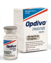
Photo courtesy of Business Wire
The US Food and Drug Administration (FDA) has granted priority review to a supplemental biologics license application seeking to expand use of the PD-1 inhibitor nivolumab (Opdivo) to patients with previously treated classical Hodgkin lymphoma (cHL).
A priority review designation means the FDA’s goal is to take action on an application within 6 months, rather than the 10 months typically taken for a standard review.
To grant an application priority review, the FDA must believe the drug would provide a significant improvement in the treatment, diagnosis, or prevention of a serious condition.
About nivolumab
Nivolumab is an inhibitor that binds to the checkpoint receptor PD-1, which is expressed on activated T cells. The drug prevents PD-L1 and PD-L2 from binding, thereby preventing the PD-1 pathway’s suppressive signaling on the immune system, including interference with an anti-tumor immune response.
Nivolumab is being developed by Bristol-Myers Squibb. The drug currently has regulatory approval in 48 countries.
In the US, nivolumab is approved—both as a single agent and in combination—to treat certain patients with melanoma, non-small-cell lung cancer, or advanced renal cell carcinoma.
According to Bristol-Myers Squibb, nivolumab has the potential to become first PD-1 inhibitor approved for a hematologic malignancy in the US.
The supplemental biologics license application for nivolumab included data from the phase 2 trial CheckMate 205. In this ongoing trial, researchers are evaluating nivolumab in patients with relapsed or refractory cHL who have received an autologous stem cell transplant and brentuximab vedotin.
Data from this trial are expected to be presented at a medical meeting later this year.
The FDA previously granted nivolumab breakthrough therapy designation for cHL. The FDA’s breakthrough therapy designation is intended to expedite the development and review of drugs for serious or life-threatening conditions. ![]()
Photo courtesy of Business Wire
The US Food and Drug Administration (FDA) has granted priority review to a supplemental biologics license application seeking to expand use of the PD-1 inhibitor nivolumab (Opdivo) to patients with previously treated classical Hodgkin lymphoma (cHL).
A priority review designation means the FDA’s goal is to take action on an application within 6 months, rather than the 10 months typically taken for a standard review.
To grant an application priority review, the FDA must believe the drug would provide a significant improvement in the treatment, diagnosis, or prevention of a serious condition.
About nivolumab
Nivolumab is an inhibitor that binds to the checkpoint receptor PD-1, which is expressed on activated T cells. The drug prevents PD-L1 and PD-L2 from binding, thereby preventing the PD-1 pathway’s suppressive signaling on the immune system, including interference with an anti-tumor immune response.
Nivolumab is being developed by Bristol-Myers Squibb. The drug currently has regulatory approval in 48 countries.
In the US, nivolumab is approved—both as a single agent and in combination—to treat certain patients with melanoma, non-small-cell lung cancer, or advanced renal cell carcinoma.
According to Bristol-Myers Squibb, nivolumab has the potential to become first PD-1 inhibitor approved for a hematologic malignancy in the US.
The supplemental biologics license application for nivolumab included data from the phase 2 trial CheckMate 205. In this ongoing trial, researchers are evaluating nivolumab in patients with relapsed or refractory cHL who have received an autologous stem cell transplant and brentuximab vedotin.
Data from this trial are expected to be presented at a medical meeting later this year.
The FDA previously granted nivolumab breakthrough therapy designation for cHL. The FDA’s breakthrough therapy designation is intended to expedite the development and review of drugs for serious or life-threatening conditions.
Study provides new insight into malaria transmission

infecting a red blood cell
Image courtesy of St. Jude
Children’s Research Hospital
Research published in PNAS helps explain how the malaria parasite Plasmodium falciparum undergoes the changes that enable transmission of the parasite from humans to mosquitoes.
Investigators determined how the parasite transforms its own structure and the structure of a host red blood cell so the parasite can hide from the body’s normal defenses and later re-enter the bloodstream for transmission via mosquito bite.
The team believes that, by understanding this process, it may be possible to inhibit the blood cell’s transformation.
“Once you understand the molecular mechanisms, it becomes easier to find drugs to target them,” said Sulin Zhang, PhD, of Pennsylvania State University in University Park.
Dr Zhang developed the computational methods used to understand the physical transformations in the infected red blood cells that allow them to avoid removal in the spleen and prepare for transmission to a mosquito host.
He and his colleagues knew that healthy red blood cells are able to squeeze through small slits in the spleen, but damaged and aging red blood cells cannot and are filtered out and removed from the circulation.
To avoid this fate, the sexual stage malaria parasite first makes the red blood cell rigid and hides out in deep tissue. Then, when the parasite is mature, the infected red blood cells become flexible and elastic, ready to be picked up by a mosquito for disease transmission.
To understand these changes, the investigators prepared samples of parasites at each stage and studied the changing microstructure using atomic force microscopy.
This revealed changes in the organization of a meshwork of tiny spring-like proteins in the blood cell membrane. When the parasite is ready for transmission, it reverses the structural changes.
The team then turned to Dr Zhang, who developed a model to explain how subtle changes to the molecular structure of the spring-like proteins were sufficient to make the red blood cell either rigid or flexible.
The investigators are continuing to use Dr Zhang’s model to simulate the overall shapes and the flow dynamics of infected red blood cells in the bloodstream, providing information that could aid researchers looking to inhibit the malaria parasite’s spread.
TKI trial leaves questions unanswered

Image by Difu Wu
The phase 3 EPIC trial, a comparison of tyrosine kinase inhibitors (TKIs), has left some questions unanswered.
The trial did not determine whether the third-generation TKI ponatinib is more effective than the first-generation TKI imatinib for patients with previously untreated chronic myeloid leukemia (CML).
The study was terminated early due to safety concerns associated with ponatinib, so the primary endpoint could only be analyzed in a small number of patients.
Results in these patients showed no significant difference in that endpoint—major molecular response (MMR) at 12 months—between the imatinib and ponatinib arms.
Results in the entire study cohort suggested that, overall, ponatinib was more toxic than imatinib. In particular, ponatinib produced more arterial occlusive events.
However, the trial’s investigators have questioned whether reducing the dose of ponatinib might change that.
Jeffrey H. Lipton, MD, of Princess Margaret Cancer Centre in Toronto, Ontario, Canada, and his colleagues reported results from the EPIC trial in The Lancet Oncology. The trial was supported by Ariad Pharmaceuticals.
Problems with ponatinib
Ponatinib was approved by the US Food and Drug Administration (FDA) in December 2012 to treat adults with CML or Philadelphia chromosome-positive acute lymphoblastic leukemia that is resistant to or intolerant of other TKIs.
In October 2013, follow-up results from the phase 2 PACE trial suggested ponatinib can increase a patient’s risk of arterial and venous thrombotic events. So all trials of the drug were placed on partial clinical hold, with the exception of the EPIC trial, which was terminated.
That November, the FDA suspended sales and marketing of ponatinib, pending results of a safety evaluation. In December, the agency decided ponatinib could return to the market if new safety measures were implemented. In January 2014, ponatinib was put back on the market in the US.
EPIC trial
The trial enrolled 307 patients with newly diagnosed, chronic-phase CML. Patients were randomized to receive ponatinib at 45 mg (n=155) or imatinib at 400 mg (n=152) once daily until progression, unacceptable toxicity, or other criteria for withdrawal were met.
The median age was 55 (range, 18-89) in the ponatinib arm and 52 (range, 18-86) in the imatinib arm. Most patients were male—63% and 61%, respectively—and most had an ECOG performance status of 0—75% and 78%, respectively.
Patients were randomized between August 14, 2012, and October 9, 2013, and the trial was terminated on October 17, 2013.
Because of the early termination, only 10 patients in the ponatinib arm and 13 in the imatinib arm were evaluable for the primary endpoint—MMR at 12 months. Eighty percent (8/10) of the evaluable patients in the ponatinib arm and 38% (5/13) of those in the imatinib arm achieved an MMR at 12 months (P=0.074).
The investigators also evaluated the incidence of MMR at any time in patients with any post-baseline molecular response assessment. This time, the incidence of MMR was significantly higher in the ponatinib arm than the imatinib arm—41% (61/149) and 18% (25/142), respectively (P<0.0001).
All of the patients were evaluable for safety—154 in the ponatinib arm and 152 in the imatinib arm.
Arterial occlusive events occurred in 7% (n=11) of patients in the ponatinib arm and 2% (n=3) in the imatinib arm (P=0.052). These events were considered serious in 6% (n=10) and 1% (n=1), respectively (P=0.010).
Common grade 3/4 adverse events—in the ponatinib and imatinib arms, respectively—were increased lipase (14% vs 2%), thrombocytopenia (12% vs 7%), rash (6% vs 1%), and neutropenia (3% vs 8%).
Serious adverse events that occurred in 3 or more patients in the ponatinib arm were pancreatitis (n=5), atrial fibrillation (n=3), and thrombocytopenia (n=3). There were no serious adverse events that occurred in 3 or more patients in the imatinib arm.
Dr Lipton and his colleagues said the premature termination of the EPIC trial restricts the interpretation of its results, but the available data provide some insight into the activity and safety of ponatinib in previously untreated CML.
The investigators also noted that data from this trial and the clinical development program for ponatinib suggest that lowering doses of the drug could improve its vascular safety profile and, therefore, the benefit-risk balance.
Two ongoing trials (NCT02467270 and NCT02627677) may provide more insight. Both are investigating starting doses of ponatinib at 15 mg or 30 mg.
CDC says Zika causes microcephaly, other birth defects

Photo by Nina Matthews
After reviewing existing evidence, scientists from the US Centers for Disease Control and Prevention (CDC) have concluded that Zika virus causes
microcephaly and other severe fetal brain defects.
“This study marks a turning point in the Zika outbreak,” said Tom Frieden, MD, director of the CDC.
“It is now clear that the virus causes microcephaly. We’ve now confirmed what mounting evidence has suggested.”
Details on the CDC’s review were published in NEJM.
The report notes that no single piece of evidence provides conclusive proof that Zika virus infection is a cause of microcephaly and other fetal brain defects. Rather, increasing evidence from a number of recently published studies and a careful evaluation using established scientific criteria support the authors’ conclusions.
The finding that Zika virus infection can cause microcephaly and other severe fetal brain defects means that a woman who is infected with Zika during pregnancy has an increased risk of having a baby with these health problems.
However, as we’ve seen during the current Zika outbreak, some infected women deliver babies that appear to be healthy.
The CDC said establishing the causal relationship between Zika and fetal brain defects is an important step in driving additional prevention efforts, focusing research activities, and reinforcing the need for direct communication about the risks of Zika.
However, the agency noted that many questions about the Zika virus remain. And answering these questions will be the focus of ongoing research.
The CDC also said it is not changing its current recommendations regarding the Zika virus. Pregnant women should continue to avoid travel to areas where Zika is actively spreading.
If a pregnant woman travels to or lives in an area with active Zika virus transmission, she should talk with her healthcare provider and take steps to prevent mosquito bites and sexual transmission of the virus.
The CDC also encourages women and their partners in areas with active Zika transmission to engage in pregnancy planning and counseling with their healthcare providers so they know the risks and the ways to mitigate them.