User login
Long-term risk of hospitalization in cancer survivors

patient and her father
Photo by Rhoda Baer
Results of a large study suggest that adolescent and young adult cancer survivors have an increased risk of hospitalization up to 34 years after their diagnosis.
Cancer survivors with the highest risk of hospitalization were those who had been diagnosed with leukemia, brain cancer, or Hodgkin lymphoma.
Kathrine Rugbjerg, PhD, and Jørgen H. Olsen, MD, of the Danish Cancer Society Research Center in Copenhagen, Denmark, reported these results in JAMA Oncology.
The pair examined the risk of hospitalization in 33,555 subjects who had cancer as adolescents or young adults and survived at least 5 years. The subjects were diagnosed from 1943 through 2004, when they were 15 to 39 years of age.
The researchers compared the cancer survivors to a cohort of 228,447 subjects from the general population who were matched to the cancer survivors by sex and year of birth.
All study subjects were followed up for hospitalizations in the Danish Patient Register through December 2010. The median follow-up was 14 years.
There were 53,032 hospitalizations among the cancer survivors, but only 38,423 were expected. So the standardized hospitalization rate ratio (RR) was 1.38.
The highest risks of hospitalization were for diseases of blood and blood-forming organs (RR=2.00), infectious and parasitic diseases (RR=1.69), and malignant neoplasms (RR=1.63).
The overall absolute excess risk of hospitalization for the cancer survivors was 2803 per 100,000 person-years. The highest absolute excess risks were for malignant neoplasms (18%), diseases of digestive organs (15%), and diseases of the circulatory system (14%).
The researchers said these results suggest that survivors of adolescent and young adult cancers face persistent risks for a broad range of somatic diseases that require hospitalization. And the morbidity pattern is highly dependent on the type of cancer being treated. ![]()
Unexpected findings in young cancer patients

Photo by Bill Branson
Many young cancer patients—not just those with a family history of cancer—may benefit from comprehensive genomic screening, according to a study published in NEJM.
The research revealed germline mutations in cancer-predisposing genes in 8.5% of the children and adolescents studied.
Information on family history was available for roughly 60% of these cancer patients, and, in this group, only 40% of patients had a family history of cancer.
Prior to this study, the presence of such germline mutations was thought to be extremely rare and restricted to children in families with strong histories of cancer.
“This paper marks an important turning point in our understanding of pediatric cancer risk and will likely change how patients are evaluated,” said James R. Downing, MD, of St. Jude Children’s Research Hospital in Memphis, Tennessee.
“For many pediatric cancer patients, comprehensive next-generation DNA sequencing of both their tumor and normal tissue may provide valuable information that will not only influence their clinical management but also lead to genetic counseling and testing of their parents and siblings who may be at risk and would benefit from ongoing surveillance.”
To conduct this research, Dr Downing and his colleagues performed next-generation sequencing of both the tumor and normal tissues of 1120 cancer patients younger than 20 years of age.
The investigators sequenced the whole genome in 595 patients, the whole exome in 456 patients, and both in 69 patients.
The team analyzed the DNA sequences of 565 genes, including 60 that have been associated with autosomal dominant cancer-predisposition syndromes, for the presence of germline mutations.
A total of 95 patients, or 8.5%, had germline mutations in 21 of the 60 genes. In comparison, only 1.1% of individuals in a non-cancer cohort had alterations in the same genes.
Fifty-eight of the cancer patients who had a cancer-predisposing mutation also had available information on their family history. Forty percent (n=23) of these patients had a family history of cancer.
The frequency of germline mutations in cancer-predisposition genes varied by the type of cancer a patient had. The highest frequency, 16.7%, was in patients with non-central nervous system (CNS) solid tumors, followed by CNS tumors, at 9%, and leukemia, at 4.4%.
The most commonly mutated genes were TP53 (n=50), APC (n=6), BRCA2 (n=6), NF1 (n=4), PMS2 (n=4), RB1 (n=3), and RUNX1 (n=3).
The investigators were surprised to find mutations in the breast and ovarian cancer genes BRCA1 and BRCA2 in a number of the patients. These genes are not currently included in pediatric cancer genetic screening.
“Another surprising finding to emerge from this study was the prevalence of germline mutations in 6 patients with Ewing sarcoma,” said study author Kim Nichols, MD, also of St. Jude. “[This cancer] was not previously thought to be part of any cancer-predisposition syndrome.” ![]()
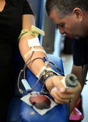
NICE issues guideline for blood transfusions
![]()
Photo courtesy of UAB Hospital
The National Institute for Health and Care Excellence (NICE) has issued its first guideline on blood transfusions.
The agency said that national audits on the use of blood in England have suggested that at least one-fifth of transfusions are unnecessary.
So NICE developed a guideline to provide recommendations on when a transfusion should be used and when alternatives should be considered.
“This guideline will ensure blood products are used safely and efficiently,” said Mark Baker, MD, director of clinical practice at NICE.
“Hundreds of thousands of people receive blood transfusions every year in England and Wales. We must do all we can to ensure that their experience is a safe one. We know that practice is improving, and, with this guideline, we want to drive things even further so we get closer to transfusions being completely risk-free and people are spared from avoidable harm.”
The guideline contains recommendations to help hospitals reduce the need for transfusions wherever possible. This includes encouraging clinicians to prescribe tranexamic acid, which can be given to patients undergoing surgery to stop them losing too much blood.
The guideline also calls for hospitals to consider using a system that electronically identifies patients to make the transfusion process safer and more efficient.
Electronic patient identification systems prompt staff to carry out key steps in the correct order and ensure that transfusions are given to the right patients through scanning of barcodes on patient wristbands and blood component containers.
“Electronic systems make it easy for staff to do the right thing every time and avoid errors,” said Mike Murphy, MD, a consultant hematologist at Oxford University Hospitals and chair of the committee that developed the guideline.
“They can also provide ‘decision support’ for doctors when they are ordering blood and promote the restrictive use of blood. The guideline also, importantly, highlights the benefits of the routine use of tranexamic acid in patients undergoing surgery. Routine implementation of these measures in the [National Health Service] will make best use of a valuable resource, be safer for patients, and save money for hospitals.”
The guideline is available on the NICE website. NICE has also published a guide for members of the public so they know what care they should expect to receive. ![]()
Model enables screening of MM drugs

showing multiple myeloma
Researchers have created a 3-dimensional model that can be used to screen drugs designed to treat multiple myeloma (MM).
The team developed 3D tissue-engineered bone marrow (3DTEBM) cultures that could, ideally, help physicians determine which drug or combination therapy might be most effective for a particular MM patient.
The cultures also provide guidance regarding dosage.
The researchers described this work in Biomaterials.
“Even before the patient completes all of the MRIs, CT scans, and other imaging procedures following diagnosis, we can have a recommendation for which drug and dosage to prescribe,” said study author Kareem Azab, PhD, of Washington University School of Medicine in St Louis, Missouri. “The test results come in 3 to 4 days.”
Dr Azab and his colleagues developed their 3DTEBM cultures using bone marrow samples from MM patients. The team took small samples of a patient’s cells and remodeled them in the lab.
This tumor microenvironment includes the cancer cells and neighboring blood vessels, immune cells, and other components whose interaction can help or inhibit the tumor cells’ growth. Drugs are tested on the remodeled patient cells to determine which treatment is likely to be most effective.
Dr Azab’s method gauges the sensitivity of a patient’s cells to different drugs at any time in the course of the disease.
Therefore, as a patient’s disease becomes more resistant to particular drugs, continued drug screening could suggest when to change therapies. This could save valuable time, Dr Azab said.
“Now, we have a drug test that closely replicates what’s going on with a patient at any given moment,” he noted. “We think this method has a better chance of working than existing options.”
The 3DTEBM cultures are currently being tested in a clinical trial of MM patients.
To further the technology, Dr Azab and his colleagues have launched a company, Cellatrix, in coordination with Washington University’s Office of Technology Management and BioGenerator, a nonprofit organization that helps launch bioscience companies.
Cellatrix is scheduled to begin testing potential therapies on behalf of pharmaceutical companies soon. Dr Azab’s team is also studying how well the screening method works for patients with leukemia or lymphoma. ![]()
Finger-prick blood tests may need repeating
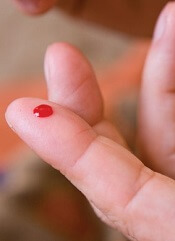
Finger-prick blood tests can deliver inaccurate results, according to a study published in the American Journal of Clinical Pathology.
Investigators found that results varied greatly when they performed multiple finger-prick tests on a single subject.
However, averaging the results of multiple droplet tests allowed the investigators to achieve results on par with venous blood tests.
They required 6 to 9 drops of blood to achieve consistent results.
“We began looking at this after we got some surprising results from our controls in an earlier study,” explained lead investigator Rebecca Richards-Kortum, PhD, of Rice University in Houston, Texas.
“Students in my lab are developing novel, low-cost platforms for anemia, platelet, and white blood cell testing in low-resource settings, and one of my students, Meaghan Bond, noticed there was wide variation in some of the benchmark tests that she was performing on hospital-grade blood analyzers.”
The benchmark controls are used to gauge the accuracy of test results from the new technology under study, so the variation among the control data was a sign that something was amiss.
What wasn’t immediately clear was whether the readings resulted from a problem with the current experiments or actual variations in the amount of hemoglobin, platelets, and white blood cells in the different drops of blood.
So Dr Richards-Kortum and Bond designed a simple protocol to test whether there was actual variation and, if so, how much.
They drew 6 successive 20 µL droplets of blood from 11 donors. As an additional test to determine whether minimum droplet size might also affect the results, the pair drew 10 successive 10 µL droplets from 7 additional donors.
All droplets were drawn from the same finger prick, and the investigators followed best practices in obtaining the droplets. The first drop was wiped away to remove contamination from disinfectants, and the finger was not squeezed or “milked,” which can lead to inaccurate results.
For experimental controls, the investigators used venipuncture to draw tubes of blood from an arm vein.
Each 20 µL droplet was analyzed with a hospital-grade blood analyzer for hemoglobin concentration, total white blood cell count, platelet count, and 3-part white blood cell differential. Each 10 µL droplet was tested for hemoglobin concentration with a popular point-of-care blood analyzer.
“A growing number of clinically important tests are performed using finger-prick blood, and this is especially true in low-resource settings,” Bond noted.
“It is important to understand how variations in finger-prick blood collection protocols can affect point-of-care test accuracy as well as how results might vary between different kinds of point-of-care tests that use finger-prick blood from the same patient.”
She and Dr Richards-Kortum found that hemoglobin content, platelet count, and white blood cell count each varied significantly from blood drop to blood drop.
“Some of the differences were surprising,” Bond said. “For example, in some donors, the hemoglobin concentration changed by more than 2 g/dL in the span of 2 successive drops of blood.”
Fortunately, the investigators found that averaging the results of the droplet tests could produce results that were on par with venous blood tests, but tests on 6 to 9 drops of blood were needed to achieve consistent results.
“Finger-prick blood tests can be accurate, and they are an important tool for healthcare providers, particularly in point-of-care and low-resource settings,” Bond said.
“Our results show that people need to take care to administer finger-prick tests in a way that produces accurate results because accuracy in these tests is increasingly important for diagnosing conditions like anemia, infections, and sickle cell anemia, malaria, HIV, and other diseases.” ![]()
Device linked to anemia in blood donors
A device used to measure hemoglobin can deliver inaccurate results and may have prompted an increase in anemia among blood donors in Ireland, according to the Irish Blood Transfusion Service (IBTS).
The IBTS began using the device—called haemospect—in July 2014 but recently discovered that it cannot detect anemia accurately in all cases.
This may have resulted in anemic individuals donating blood or caused individuals to become anemic by donating.
The IBTS said this issue likely affected female donors more than males, so the organization has decided to temporarily stop taking blood donations from women who have donated in the last 18 months.
The IBTS also said it will perform a full blood count on all male and female donors going forward, until this issue has been resolved.
“Until we have a resolution to the problem that has arisen, we are asking male donors to attend our clinics and give blood,” said IBTS Medical and Scientific Director William Murphy, MD.
“We need male donors to make an extra effort to donate and maintain the blood supply. We will be double-checking all hemoglobin results on these donors.”
Discovering the problem
Haemospect is a noninvasive device that uses white light to detect hemoglobin levels. The device is made by the German company MBR Optical Systems.
IBTS learned of the potential for inaccuracies with haemospect after a blood donor contacted the organization. She had been diagnosed as severely anemic by her doctor but had given blood the week before.
“We have now discovered that the device gives inaccurate results in some individuals with anemia down to, and probably below, 8.4 g/dL,” Dr Murphy said. “As a result of the issue . . . , some women, and probably a much smaller number of men, could have been rendered iron-deficient and anemic from blood donation in the past 18 months.”
Steps to resolution
Since the issue was discovered, more than 30 women with anemia have been identified and contacted. The IBTS said it will contact all donors who are found to be anemic.
“Over the next few weeks, we will introduce new software to reanalyze all the electronic results from all donors who have been tested and accepted for donation since we introduced this device,” Dr Murphy said. “Any discrepant results will be notified to the donors involved.”
Dr Murphy also said donors who think they might be anemic or iron-deficient should visit their doctors for testing, and the IBTS will foot the bill. Concerned donors can contact the IBTS at 1850 731137.
The IBTS plans to replace haemospect with an alternative device as soon as possible. The organization has contacted the Health Products Regularity Authority about the issue with haemospect, which is used in Austria and Germany as well.
“We are determined to have this matter resolved as soon as possible,” Dr Murphy said. “We are confident that this issue . . . has not had any impact on blood received by patients.” ![]()
Group quantifies cardiotoxicity risk with HL treatment

Photo by Rhoda Baer
European researchers say they have quantified the risk of cardiovascular disease associated with treatments for Hodgkin lymphoma (HL).
The group analyzed the risks associated with specific doses of radiation and anthracycline exposure.
They believe their results, published in The Lancet Haematology, could help clinicians identify the optimal treatment regimen for each individual HL patient.
“These study results are exciting,” said Maja V. Maraldo, PhD, of Rigshospitalet in Copenhagen, Denmark.
“They should allow physicians to optimize the combination of systemic therapy and radiation and thereby balance the risks and benefits of different regimens in individual patients.”
Study details
Dr Maraldo and her colleagues analyzed data from patients who participated in 9 trials conducted by the European Organisation for Research and Treatment of Cancer (EORTC) and the Groupe d’Etude des Lymphomes de l’Adulte (GELA, now renamed LYSA) between 1964 and 2004.
In 2009 and 2010, the researchers mailed a Life Situation Questionnaire (LSQ) to the trial participants. The goal was to determine late-onset effects of HL and its treatment.
The team also reconstructed patients’ mean radiation doses to the heart and carotid arteries and the cumulative doses of anthracyclines and vinca-alkaloids they received. The incidence of cardiovascular disease was reported during follow-up and updated through the LSQ.
Patient data
The researchers were able to collect complete information on primary treatment for 6039 HL survivors. Of these patients, 2923 received the LSQ, and 1919 responded. The median follow-up was 9 years, and the patients’ median age at diagnosis was 30.
There were 1238 cardiovascular events in 703 patients, including 46 patients who died from such an event.
The events included ischemic heart disease (24%), congestive heart failure (21%), arrhythmia (17%), valvular disease (14%), disease of the arterial vessels (9%), stroke (6%), venous thromboembolism (5%), pericarditis (3%), peripheral vasculopathy (1%), other vascular events (1%), and other cardiac events (<1%).
Predictors of risk
The researchers found that the mean radiation dose to the heart, per 1 Gy increase, was a significant predictor of cardiovascular disease, with a hazard ratio of 1.015 (P=0.0014).
However, the mean radiation doses to the left internal carotid artery and the right internal carotid artery were not significant predictors of cardiovascular events (P=0.41 and 0.70, respectively).
The dose of anthracyclines, per 50 mg/m2 increase in cumulative dose, was a significant predictor of cardiovascular disease, with a hazard ratio of 1.077 (P=0.0064).
But the cumulative dose of vinblastine or vincristine was not (P=0.77 and 0.36, respectively).
The researchers said a limitation of this study is that they were not able to assess the impact of cardiovascular risk factors such as smoking, hypertension, and diabetes because that information was not consistently available.
Still, the team believes their analyses quantified the effect of radiotherapy and anthracyclines on the risk of cardiovascular disease, and the findings should aid treatment decisions in HL. ![]()
Photo by Rhoda Baer
European researchers say they have quantified the risk of cardiovascular disease associated with treatments for Hodgkin lymphoma (HL).
The group analyzed the risks associated with specific doses of radiation and anthracycline exposure.
They believe their results, published in The Lancet Haematology, could help clinicians identify the optimal treatment regimen for each individual HL patient.
“These study results are exciting,” said Maja V. Maraldo, PhD, of Rigshospitalet in Copenhagen, Denmark.
“They should allow physicians to optimize the combination of systemic therapy and radiation and thereby balance the risks and benefits of different regimens in individual patients.”
Study details
Dr Maraldo and her colleagues analyzed data from patients who participated in 9 trials conducted by the European Organisation for Research and Treatment of Cancer (EORTC) and the Groupe d’Etude des Lymphomes de l’Adulte (GELA, now renamed LYSA) between 1964 and 2004.
In 2009 and 2010, the researchers mailed a Life Situation Questionnaire (LSQ) to the trial participants. The goal was to determine late-onset effects of HL and its treatment.
The team also reconstructed patients’ mean radiation doses to the heart and carotid arteries and the cumulative doses of anthracyclines and vinca-alkaloids they received. The incidence of cardiovascular disease was reported during follow-up and updated through the LSQ.
Patient data
The researchers were able to collect complete information on primary treatment for 6039 HL survivors. Of these patients, 2923 received the LSQ, and 1919 responded. The median follow-up was 9 years, and the patients’ median age at diagnosis was 30.
There were 1238 cardiovascular events in 703 patients, including 46 patients who died from such an event.
The events included ischemic heart disease (24%), congestive heart failure (21%), arrhythmia (17%), valvular disease (14%), disease of the arterial vessels (9%), stroke (6%), venous thromboembolism (5%), pericarditis (3%), peripheral vasculopathy (1%), other vascular events (1%), and other cardiac events (<1%).
Predictors of risk
The researchers found that the mean radiation dose to the heart, per 1 Gy increase, was a significant predictor of cardiovascular disease, with a hazard ratio of 1.015 (P=0.0014).
However, the mean radiation doses to the left internal carotid artery and the right internal carotid artery were not significant predictors of cardiovascular events (P=0.41 and 0.70, respectively).
The dose of anthracyclines, per 50 mg/m2 increase in cumulative dose, was a significant predictor of cardiovascular disease, with a hazard ratio of 1.077 (P=0.0064).
But the cumulative dose of vinblastine or vincristine was not (P=0.77 and 0.36, respectively).
The researchers said a limitation of this study is that they were not able to assess the impact of cardiovascular risk factors such as smoking, hypertension, and diabetes because that information was not consistently available.
Still, the team believes their analyses quantified the effect of radiotherapy and anthracyclines on the risk of cardiovascular disease, and the findings should aid treatment decisions in HL.
Coconut oil may prevent bloodstream infection

Coconut oil may combat infection with Candida albicans, according to preclinical research published in mSphere.
Mice on a diet that included coconut oil had significantly lower gastrointestinal colonization by C albicans than mice that were fed high-fat diets without coconut oil or mice that received a standard diet.
“We found that diet can be an effective way to reduce the amount of Candida in the mouse,” said study author Carol Kumamoto, PhD, of Tufts University School of Medicine in Boston, Massachusetts.
“The extension of this finding to the human population is something that needs to be addressed in the future.”
Previous research showed that changes to diet, including changes in the amount and type of fat, can alter gastrointestinal microbiota. And in vitro studies showed that coconut oil has antifungal properties.
So Dr Kumamoto and her colleagues studied the effect of different diets on C albicans colonization in mice.
The mice received standard diets or high-fat diets containing coconut oil, beef tallow, or soybean oil. The mice were fed these diets for 14 days prior to inoculation with C albicans and 21 days after.
At 21 days post-inoculation, gastrointestinal colonization with C albicans was significantly lower in the stomach contents of mice fed the coconut oil than mice fed the beef tallow (P<0.0001), the soybean oil (P<0.0001), or the standard diet (P<0.0001).
“When you compared a mouse on a high-fat diet that contained either beef fat or soybean oil to mice eating coconut oil, there was about a 10-fold drop in colonization,” Dr Kumamoto said.
In another experiment, the researchers switched mice receiving beef tallow to coconut oil.
“Four days after the change in diet, the colonization changed so it looked almost exactly like what you saw in a mouse who had been on coconut oil the entire time,” Dr Kumamoto said.
“There are 2 directions that we would like to take with this research now,” she added. “One of them is finding out the mechanism of how this works. That is a big question we would like to answer. The second question is whether this can have any impact on humans.”
The researchers are in discussion with Joseph Bliss, MD, PhD, of Women and Infants Hospital of Rhode Island, about launching a clinical trial testing coconut oil in hospitalized infants at high risk of developing systemic candidiasis.
Team characterizes EMH niche

Image by John Perry
Previous studies have shown that hematopoietic stresses—such as myelofibrosis, anemia, and myeloablation—can induce extramedullary hematopoiesis (EMH), in which hematopoietic stem cells (HSCs) are mobilized to sites outside the bone marrow.
The splenic red pulp is known to be a prominent site of EMH in both mice and humans, but not much is known about the EMH niche.
Now, investigators say they have characterized this niche.
They detailed their findings in Nature.
The team used mouse models to examine the expression patterns of 2 known niche cell factors, SCF and CXCL12.
They discovered that the hematopoietic microenvironment in the spleen is found near sinusoidal blood vessels and is created by endothelial cells and perivascular stromal cells, just like the microenvironment in the bone marrow.
“Under emergency conditions, the endothelial cells and perivascular stromal cells that reside in the spleen are induced to proliferate so they can sustain all the new blood-forming stem cells that migrate into the spleen,” explained study author Sean Morrison, PhD, of the University of Texas Southwestern Medical Center, Dallas.
“We determined that this process in the spleen is physiologically important for responding to hematopoietic stress. Without it, the mice we studied could not maintain normal blood cell counts during pregnancy or quickly regenerate blood cell counts after bleeding or chemotherapy.”
The investigators believe these findings could aid the development of therapeutic interventions to enhance blood formation following chemotherapy or HSC transplant and thus accelerate the recovery of blood cell counts.
High-fat diet linked to RBC dysfunction

Preclinical research suggests a high-fat diet is associated with red blood cell (RBC) dysfunction and helps explain how these dysfunctional cells may mediate atherosclerosis.
The researchers believe their findings may have implications for the pathogenesis of atherosclerosis in obesity, but the work may aid the study of other health conditions as well, such as thrombosis in the context of cancer.
The team detailed their findings in Circulation.
“Obesity caused by chronic consumption of a high-calorie, high-fat diet is a worldwide epidemic, representing one of the greatest threats to global health,” said principal investigator Vladimir Bogdanov, PhD, of the University of Cincinnati in Ohio.
“White blood cells play a key role in fueling adipose tissue inflammation and insulin resistance in obesity and also promote the clogging of arteries, or atherosclerosis, setting the stage for heart attack and stroke. While these outcomes linked with a high-fat diet and fat in the blood on white blood cells have been shown in animal models and humans, the impact of high-fat diets on other bone marrow-derived cells, like red blood cells, is not well-defined.”
“Evidence is emerging that red blood cells play an important regulatory role in the development of atherosclerosis, binding pro-inflammatory proteins that cause dysfunction in the inner lining of the blood vessel wall—the endothelium. We explored how a high fat-diet causes red blood cell dysfunction in this study.”
Dr Bogdanov and his team fed a 60% high-fat diet to mice for 12 weeks and compared these animals to control mice that received a normal diet.
There was an increase in the level of chemokines bound to the RBCs of mice that received the high-fat diet. These chemokines were bound to RBCs via the Duffy antigen receptor for chemokines.
The researchers exposed RBCs from mice on the high-fat diet to an endothelial monolayer in vitro, and they observed “significantly enhanced” macrophage transendothelial migration. They said this confirms the functional importance of RBC-bound chemokines in the setting of a high-fat diet.
“In red blood cells from animal models fed a high-fat diet, there was an increase in cholesterol found in the cell membrane and phosphatidylserine levels, promoting inflammatory reactions,” Dr Bogdanov added.
“Phosphatidylserine is a phospholipid membrane component which plays a key role in the cycle of cells. When red blood cells from the animals being fed the high-fat diet were injected into a control group, eating a normal diet, there was a 3-fold increase in their spleens’ uptake of red blood cells. The spleen is involved in the removal of blood cells, as well as systemic inflammation.”
“All of these findings show that the dysfunction of red blood cells, corresponding with dysfunction of the lining of blood vessels, occurs very early in diet-induced obesity and may play a part in the formation of atherosclerosis. Diets high in saturated fat have long been associated with endothelial dysfunction, the precursor to atherosclerosis, but, to our knowledge, the effects of high-fat diet on red blood cells have not been rigorously examined.”
Dr Bogdanov noted that, in humans, high cholesterol is associated with alterations in RBCs that are improved by treatment with statins. But the majority of obese humans do not have severe high cholesterol, as was the case with the animals in this study.
The researchers are now working on translating their findings to humans.