User login
Morphea-like plaques, induration of the extremities, and eosinophilia
A 52-year-old woman who had been diagnosed with morphea 1 year before was referred to our department for evaluation of a 6-week history of myalgia and limited flexion and extension of the wrists and elbows. She had a history of hypertension, which was controlled with enalapril 10 mg daily. She had no fever, weight loss, dyspnea, dysphagia, Raynaud phenomenon, or other symptoms, and she had not traveled recently.
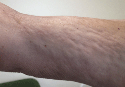
On physical examination, she had multiple dark-brown plaques on the legs (Figure 1), induration of both forearms, puckering of the skin (peau d’orange) of both arms (Figure 1), and marked limitation of motion of the wrists (Figure 2). Sclerodactyly was absent, and the rest of the examination was unremarkable. Laboratory tests showed an elevated serum C-reactive protein (1.97 mg/dL, reference range 0–0.5) and a white blood cell count of 9.59 × 10⁹/L, with 25.2% eosinophils (reference range 0%–7%). Antinuclear antibodies were negative.
Because the patient refused full-thickness biopsy of the skin (ie, down to superficial muscle), magnetic resonance imaging (MRI) of the right arm was ordered. T1-weighted fat-saturated imaging with contrast showed marked contrast enhancement along all muscle fascia. This finding, along with the patient’s presentation and the results of laboratory testing, indicated a diagnosis of eosinophilic fasciitis.
Prednisone was prescribed in an initial dose of 1 mg/kg/day and was subsequently tapered, and this brought a partial response. Methotrexate (15 mg weekly) was added 1 month later. Follow-up visits at 3, 6, and 12 months were scheduled. At 12 months, levels of markers of inflammation were normal, the eosinophilia had completely resolved (< 0.125 × 10⁹/L, < 2%), joint mobility had improved (Figure 2), and the morphea-like lesions had lightened somewhat. MRI repeated 6 months after the start of methotrexate showed complete resolution of the fascia infiltration.
EOSINOPHILIC FASCIITIS
Eosinophilic fasciitis, or Shulman syndrome, is a rare localized fibrosing disorder of the fascia characterized in its early phase by symmetrical limb or trunk erythema and swelling, and later by progressive thickening and induration of the dermis and subcutaneous fascia.1
The cause is unknown, but triggers have been suggested such as vigorous exercise or trauma, Borrelia infection, and drugs (statins, phenytoin, l-tryptophan). It has also been associated with hematologic disease. In our patient, none of these was present.2,3
There are no international diagnostic criteria for eosinophilic fasciitis. Rather, the diagnosis is suspected in a patient with eosinophilia and characteristic cutaneous features, such as pitting edema, induration, hyperpigmentation, and a peau d’orange aspect caused by inflammation and fibrosis of the fascia.1,2 These features have been reported in up to 90% of patients at the time of diagnosis.1–3 Myalgia, muscle weakness, and articular involvement including inflammatory arthralgia and joint contracture, as in our patient, are also common.1,2,4,5 Raynaud phenomenon in these patients is typically absent. Furthermore, pulmonary, cardiac, and renal involvement has also been reported, but this is very infrequent, and if it is present, other diseases such as Churg-Strauss vasculitis and hypereosinophilic syndrome should be excluded.1,2,4
The presence of thickened fascia and inflammatory infiltrates composed of lymphocytes or eosinophils (or both) on histologic examination of the skin-to-muscle biopsy remains the gold standard for diagnosis.2,4 However, when biopsy is nondiagnostic or is not possible, imaging with MRI or ultrasonography has been helpful.6,7 Muscle MRI is considered the best morphologic procedure for diagnosis, as it can specifically indicate the optimal location for biopsy in atypical cases (eg, fasciitis without skin changes and when eosinophilia is absent); it is also used to monitor the response to therapy.7,8 Although nonspecific, muscle MRI typically shows a markedly increased signal intensity within the fascia. And gadolinium administration shows marked fascia enhancement in the acute phase of the disease.1,4,6–8
MORPHEA PREDICTS POOR OUTCOME
Morphea has been reported to be present in 19% to 41% of patients with eosinophilic fasciitis due to dermis infiltration.2,4,5 Although rare, morphea plaques may also be present before the onset of eosinophilic fasciitis.3,4 Morphea has been described as predictive of poor outcome and residual fibrosis, and it has been associated with eosinophilic fasciitis resistant to usual therapies.3,5
Treatment of eosinophilic fasciitis is largely empirical because evidence from controlled trials is lacking. Corticosteroids remain the standard therapy. However, in patients with morphea-like lesions, treatment with an immunosuppressive drug such as methotrexate has been reported to be useful.1,4
- Lebeaux D, Sène D. Eosinophilic fasciitis (Shulman disease). Best Pract Res Clin Rheumatol 2012; 26:449–458.
- Lakhanpal S, Ginsburg WW, Michet CJ, Doyle JA, Moore SB. Eosinophilic fasciitis: clinical spectrum and therapeutic response in 52 cases. Semin Arthritis Rheum 1988; 17:221–231.
- Moulin C, Cavailhes A, Balme B, Skowron F. Morphoea-like plaques revealing an eosinophilic (Shulman) fasciitis. Clin Exp Dermatol 2009; 34:e851–e853.
- Lebeaux D, Francès C, Barete S, et al. Eosinophilic fasciitis (Shulman disease): new insights into the therapeutic management from a series of 34 patients. Rheumatology (Oxford) 2012; 51:557–561.
- Endo Y, Tamura A, Matsushima Y, et al. Eosinophilic fasciitis: report of two cases and a systematic review of the literature dealing with clinical variables that predict outcome. Clin Rheumatol 2007; 26:1445–1451.
- Dybowski F, Neuen-Jacob E, Braun J. Eosinophilic fasciitis and myositis: use of imaging modalities for diagnosis and monitoring. Ann Rheum Dis 2008; 67:572–574.
- Moulton SJ, Kransdorf MJ, Ginsburg WW, Abril A, Persellin S. Eosinophilic fasciitis: spectrum of MRI findings. AJR Am J Roentgenol 2005; 184:975–978.
- Ronneberger M, Janka R, Schett G, Manger B. Can MRI substitute for biopsy in eosinophilic fasciitis? Ann Rheum Dis 2009; 68:1651–1652.
A 52-year-old woman who had been diagnosed with morphea 1 year before was referred to our department for evaluation of a 6-week history of myalgia and limited flexion and extension of the wrists and elbows. She had a history of hypertension, which was controlled with enalapril 10 mg daily. She had no fever, weight loss, dyspnea, dysphagia, Raynaud phenomenon, or other symptoms, and she had not traveled recently.
On physical examination, she had multiple dark-brown plaques on the legs (Figure 1), induration of both forearms, puckering of the skin (peau d’orange) of both arms (Figure 1), and marked limitation of motion of the wrists (Figure 2). Sclerodactyly was absent, and the rest of the examination was unremarkable. Laboratory tests showed an elevated serum C-reactive protein (1.97 mg/dL, reference range 0–0.5) and a white blood cell count of 9.59 × 10⁹/L, with 25.2% eosinophils (reference range 0%–7%). Antinuclear antibodies were negative.
Because the patient refused full-thickness biopsy of the skin (ie, down to superficial muscle), magnetic resonance imaging (MRI) of the right arm was ordered. T1-weighted fat-saturated imaging with contrast showed marked contrast enhancement along all muscle fascia. This finding, along with the patient’s presentation and the results of laboratory testing, indicated a diagnosis of eosinophilic fasciitis.
Prednisone was prescribed in an initial dose of 1 mg/kg/day and was subsequently tapered, and this brought a partial response. Methotrexate (15 mg weekly) was added 1 month later. Follow-up visits at 3, 6, and 12 months were scheduled. At 12 months, levels of markers of inflammation were normal, the eosinophilia had completely resolved (< 0.125 × 10⁹/L, < 2%), joint mobility had improved (Figure 2), and the morphea-like lesions had lightened somewhat. MRI repeated 6 months after the start of methotrexate showed complete resolution of the fascia infiltration.
EOSINOPHILIC FASCIITIS
Eosinophilic fasciitis, or Shulman syndrome, is a rare localized fibrosing disorder of the fascia characterized in its early phase by symmetrical limb or trunk erythema and swelling, and later by progressive thickening and induration of the dermis and subcutaneous fascia.1
The cause is unknown, but triggers have been suggested such as vigorous exercise or trauma, Borrelia infection, and drugs (statins, phenytoin, l-tryptophan). It has also been associated with hematologic disease. In our patient, none of these was present.2,3
There are no international diagnostic criteria for eosinophilic fasciitis. Rather, the diagnosis is suspected in a patient with eosinophilia and characteristic cutaneous features, such as pitting edema, induration, hyperpigmentation, and a peau d’orange aspect caused by inflammation and fibrosis of the fascia.1,2 These features have been reported in up to 90% of patients at the time of diagnosis.1–3 Myalgia, muscle weakness, and articular involvement including inflammatory arthralgia and joint contracture, as in our patient, are also common.1,2,4,5 Raynaud phenomenon in these patients is typically absent. Furthermore, pulmonary, cardiac, and renal involvement has also been reported, but this is very infrequent, and if it is present, other diseases such as Churg-Strauss vasculitis and hypereosinophilic syndrome should be excluded.1,2,4
The presence of thickened fascia and inflammatory infiltrates composed of lymphocytes or eosinophils (or both) on histologic examination of the skin-to-muscle biopsy remains the gold standard for diagnosis.2,4 However, when biopsy is nondiagnostic or is not possible, imaging with MRI or ultrasonography has been helpful.6,7 Muscle MRI is considered the best morphologic procedure for diagnosis, as it can specifically indicate the optimal location for biopsy in atypical cases (eg, fasciitis without skin changes and when eosinophilia is absent); it is also used to monitor the response to therapy.7,8 Although nonspecific, muscle MRI typically shows a markedly increased signal intensity within the fascia. And gadolinium administration shows marked fascia enhancement in the acute phase of the disease.1,4,6–8
MORPHEA PREDICTS POOR OUTCOME
Morphea has been reported to be present in 19% to 41% of patients with eosinophilic fasciitis due to dermis infiltration.2,4,5 Although rare, morphea plaques may also be present before the onset of eosinophilic fasciitis.3,4 Morphea has been described as predictive of poor outcome and residual fibrosis, and it has been associated with eosinophilic fasciitis resistant to usual therapies.3,5
Treatment of eosinophilic fasciitis is largely empirical because evidence from controlled trials is lacking. Corticosteroids remain the standard therapy. However, in patients with morphea-like lesions, treatment with an immunosuppressive drug such as methotrexate has been reported to be useful.1,4
- Lebeaux D, Sène D. Eosinophilic fasciitis (Shulman disease). Best Pract Res Clin Rheumatol 2012; 26:449–458.
- Lakhanpal S, Ginsburg WW, Michet CJ, Doyle JA, Moore SB. Eosinophilic fasciitis: clinical spectrum and therapeutic response in 52 cases. Semin Arthritis Rheum 1988; 17:221–231.
- Moulin C, Cavailhes A, Balme B, Skowron F. Morphoea-like plaques revealing an eosinophilic (Shulman) fasciitis. Clin Exp Dermatol 2009; 34:e851–e853.
- Lebeaux D, Francès C, Barete S, et al. Eosinophilic fasciitis (Shulman disease): new insights into the therapeutic management from a series of 34 patients. Rheumatology (Oxford) 2012; 51:557–561.
- Endo Y, Tamura A, Matsushima Y, et al. Eosinophilic fasciitis: report of two cases and a systematic review of the literature dealing with clinical variables that predict outcome. Clin Rheumatol 2007; 26:1445–1451.
- Dybowski F, Neuen-Jacob E, Braun J. Eosinophilic fasciitis and myositis: use of imaging modalities for diagnosis and monitoring. Ann Rheum Dis 2008; 67:572–574.
- Moulton SJ, Kransdorf MJ, Ginsburg WW, Abril A, Persellin S. Eosinophilic fasciitis: spectrum of MRI findings. AJR Am J Roentgenol 2005; 184:975–978.
- Ronneberger M, Janka R, Schett G, Manger B. Can MRI substitute for biopsy in eosinophilic fasciitis? Ann Rheum Dis 2009; 68:1651–1652.


