User login
Hospital-Acquired and Community-Acquired MRSA: Two Distinct Infections
Two students, 2 tropical beaches, 2 injured feet
A 21-year-old previously healthy male went to his college health center, reporting that he had injured his foot in Columbia while he was on winter break. Two weeks before this clinic visit, he felt a “scrape” on the bottom of his foot while walking on the beach. One week later, the site began to itch and was progressively enlarging. He had no systemic symptoms and no pain at the site. Physical examination showed a 3-cm-wide indurated filamentous/serpiginous eruption with surrounding erythema, and a crusting central “puncture” (FIGURE 1).
The patient seen immediately following the patient above was a 19-year-old female college student. For the last 1 to 2 weeks she had an enlarging, very pruritic eruption on the sole of her foot that she attributed to a bug bite. She had just returned from winter vacation in Brazil, where she recalled spending a lot of time on the beach. The patient was otherwise healthy and did not have fever, other rash, or joint pain. Examination revealed an indurated, erythematous, serpiginous eruption on the plantar aspect of her left foot (FIGURE 2).
FIGURE 1
Patient 1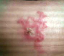
FIGURE 2
Patient 2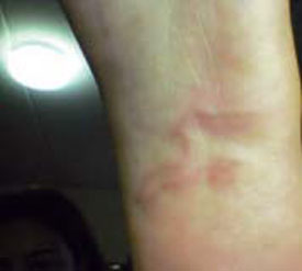
What is your diagnosis for both cases?
How would you treat them, and advise for prevention of future similar problems?
Diagnosis: Cutaneous larva migrans
Cutaneous larva migrans (CLM, sometimes called the “creeping eruption”) occurs when the third-stage larvae of dog (or cat) hookworm (Ancylostoma braziliense and A caninum, most commonly) invade the skin and fail to penetrate the epidermal basement layer. Humans are not the definitive host for this roundworm. Skin manifestations occur due to a hypersensitivity reaction.
The disease is found most commonly in tropical and subtropical climates. Animals defecate on warm, moist sand and the nematode eggs hatch. The larvae quickly invade bare skin in contact with the sand. The highest incidence of infection in the US occurs in Florida and along the Gulf Coast, but the disease is more common in the Caribbean, Central and South America, and Southeast Asia and Africa.
Many patients report a stinging sensation when the larva penetrates, as did patients 1 and 2, who misinterpreted the pain as a puncture (patient 1) or an insect bite (patient 2). Larvae migrate at a rate of several millimeters per day. Migration produces an intensely pruritic, linear or serpiginous track 2 to 4 mm wide, sometimes with vesicles, as the body reacts to the larva or to its excreta. Because each larva leaves an individual track in its wake, lesions may be simple or quite complex, depending upon the number of infecting organisms. A second morphologic variant has been described with follicular papules and pustules accompanying linear burrows.1 Eventually, the larvae die and the condition resolves even without treatment, but treatment accelerates resolution. The lesions may occur anywhere on the body, but most commonly are found on the feet, buttocks, and hands.
Differential diagnosis
The typical lesion is usually a straightforward diagnosis (once the observer is familiar with the infection), but differential diagnosis includes tinea corporis, granuloma annulare, erythema migrans (Lyme disease), ground itch, larva currens, myiasis, and stings by marine invertebrates. In very rare cases, larvae may migrate to the lungs, causing pneumonitis.2
Management of CLM
Among the various treatments, no randomized trials exist comparing their efficacy. Caumes et al1 recommend a single dose of oral ivermectin 12 mg weekly until cure, or oral albendazole 400 mg twice daily for 3 consecutive days. Others recommend ivermectin 200 mcg/kg, repeated once or twice,3 thiabendazole 500 mg 4 times daily for 5 days,4 or even topical thiabendazole 10% cream,5 although creams may be less effective. Topical thiabendazole is a good alternative for young children to avoid the potential side effects of systemic medications.
Folliculitis from the larva may be relatively treatment-resistant, requiring a longer duration of treatment to effect cure (strength of recommendation [SOR]: C).
Outcome and prevention
Both students were treated with ivermectin 15 mg as a single dose, with prompt resolution of symptoms. They were counseled to wear beach shoes and not to sit directly on sand while on the beach.
Additional reading: Brenner MA, Patel MB. Cutaneous larva migrans: the creeping eruption. Cutis 2003; 72:111—115.
1. Caumes E, Ly F, Bricaire F. Cutaneous larva migrans with folliculitis: report of seven cases and review of the literature. Br J Dermatol 2002;146:314-316.
2. Hotez PJ, Brooker S, Bethony JM, Bottazzi ME, Loukas A, Xiao S. Hookworm infection. N Engl J Med 2004;351:799-807.
3. Bouchaud O, Houze S, Schiemann R, et al. Cutaneous larva migrans in travelers: a prospective study, with assessment of therapy with ivermectin. Clin Infect Dis 2001;31:493-498.
4. O’Quinn JC, Dushin R. Cutaneous larva migrans: case report with current recommendations for treatment. J Am Pod Med Assoc 2005;95:291-294.
5. Blackwell V, Vega-Lopez F. Cutaneous larva migrans: clinical features and management of 44 cases presenting in the returning traveler. Br J Dermatol 2001;145:434-437.
A 21-year-old previously healthy male went to his college health center, reporting that he had injured his foot in Columbia while he was on winter break. Two weeks before this clinic visit, he felt a “scrape” on the bottom of his foot while walking on the beach. One week later, the site began to itch and was progressively enlarging. He had no systemic symptoms and no pain at the site. Physical examination showed a 3-cm-wide indurated filamentous/serpiginous eruption with surrounding erythema, and a crusting central “puncture” (FIGURE 1).
The patient seen immediately following the patient above was a 19-year-old female college student. For the last 1 to 2 weeks she had an enlarging, very pruritic eruption on the sole of her foot that she attributed to a bug bite. She had just returned from winter vacation in Brazil, where she recalled spending a lot of time on the beach. The patient was otherwise healthy and did not have fever, other rash, or joint pain. Examination revealed an indurated, erythematous, serpiginous eruption on the plantar aspect of her left foot (FIGURE 2).
FIGURE 1
Patient 1
FIGURE 2
Patient 2
What is your diagnosis for both cases?
How would you treat them, and advise for prevention of future similar problems?
Diagnosis: Cutaneous larva migrans
Cutaneous larva migrans (CLM, sometimes called the “creeping eruption”) occurs when the third-stage larvae of dog (or cat) hookworm (Ancylostoma braziliense and A caninum, most commonly) invade the skin and fail to penetrate the epidermal basement layer. Humans are not the definitive host for this roundworm. Skin manifestations occur due to a hypersensitivity reaction.
The disease is found most commonly in tropical and subtropical climates. Animals defecate on warm, moist sand and the nematode eggs hatch. The larvae quickly invade bare skin in contact with the sand. The highest incidence of infection in the US occurs in Florida and along the Gulf Coast, but the disease is more common in the Caribbean, Central and South America, and Southeast Asia and Africa.
Many patients report a stinging sensation when the larva penetrates, as did patients 1 and 2, who misinterpreted the pain as a puncture (patient 1) or an insect bite (patient 2). Larvae migrate at a rate of several millimeters per day. Migration produces an intensely pruritic, linear or serpiginous track 2 to 4 mm wide, sometimes with vesicles, as the body reacts to the larva or to its excreta. Because each larva leaves an individual track in its wake, lesions may be simple or quite complex, depending upon the number of infecting organisms. A second morphologic variant has been described with follicular papules and pustules accompanying linear burrows.1 Eventually, the larvae die and the condition resolves even without treatment, but treatment accelerates resolution. The lesions may occur anywhere on the body, but most commonly are found on the feet, buttocks, and hands.
Differential diagnosis
The typical lesion is usually a straightforward diagnosis (once the observer is familiar with the infection), but differential diagnosis includes tinea corporis, granuloma annulare, erythema migrans (Lyme disease), ground itch, larva currens, myiasis, and stings by marine invertebrates. In very rare cases, larvae may migrate to the lungs, causing pneumonitis.2
Management of CLM
Among the various treatments, no randomized trials exist comparing their efficacy. Caumes et al1 recommend a single dose of oral ivermectin 12 mg weekly until cure, or oral albendazole 400 mg twice daily for 3 consecutive days. Others recommend ivermectin 200 mcg/kg, repeated once or twice,3 thiabendazole 500 mg 4 times daily for 5 days,4 or even topical thiabendazole 10% cream,5 although creams may be less effective. Topical thiabendazole is a good alternative for young children to avoid the potential side effects of systemic medications.
Folliculitis from the larva may be relatively treatment-resistant, requiring a longer duration of treatment to effect cure (strength of recommendation [SOR]: C).
Outcome and prevention
Both students were treated with ivermectin 15 mg as a single dose, with prompt resolution of symptoms. They were counseled to wear beach shoes and not to sit directly on sand while on the beach.
Additional reading: Brenner MA, Patel MB. Cutaneous larva migrans: the creeping eruption. Cutis 2003; 72:111—115.
1. Caumes E, Ly F, Bricaire F. Cutaneous larva migrans with folliculitis: report of seven cases and review of the literature. Br J Dermatol 2002;146:314-316.
2. Hotez PJ, Brooker S, Bethony JM, Bottazzi ME, Loukas A, Xiao S. Hookworm infection. N Engl J Med 2004;351:799-807.
3. Bouchaud O, Houze S, Schiemann R, et al. Cutaneous larva migrans in travelers: a prospective study, with assessment of therapy with ivermectin. Clin Infect Dis 2001;31:493-498.
4. O’Quinn JC, Dushin R. Cutaneous larva migrans: case report with current recommendations for treatment. J Am Pod Med Assoc 2005;95:291-294.
5. Blackwell V, Vega-Lopez F. Cutaneous larva migrans: clinical features and management of 44 cases presenting in the returning traveler. Br J Dermatol 2001;145:434-437.